. 2010:75:95-102.
doi: 10.1101/sqb.2010.75.036. Epub 2011 Mar 29.

The role of cotranscriptional histone methylations

Affiliations

The role of cotranscriptional histone methylations

S Buratowski et al. Cold Spring Harb Symp Quant Biol. 2010.

Abstract

The carboxy-terminal domain (CTD) of the RNA polymerase II subunit Rpb1 undergoes dynamic phosphorylation, with different phosphorylation sites predominating at different stages of transcription. Our laboratory studies show how various mRNA-processing and chromatin-modifying enzymes interact with the phosphorylated CTD to efficiently produce mRNAs. The H3K36 methyltransferase Set2 interacts with CTD carrying phosphorylations characteristic of downstream elongation complexes, and the resulting cotranscriptional H3K36 methylation targets the Rpd3S histone deacetylase to downstream transcribed regions. Although positively correlated with gene activity, this pathway actually inhibits transcription elongation as well as initiation from cryptic promoters within genes. During early elongation, CTD serine 5 phosphorylation helps recruit the H3K4 methyltransferase complex containing Set1. Within 5' transcribed regions, cotranscriptional H3K4 dimethylation (H3K4me2) by Set1 recruits the deacetylase complex Set3C. Finally, H3K4 trimethylation at the most promoter-proximal nucleosomes is thought to stimulate transcription by promoting histone acetylation by complexes containing the ING/Yng PHD finger proteins. Surprisingly, the Rpd3L histone deacetylase complex, normally a transcription repressor, may also recognize H3K4me3. Together, the cotranscriptional histone methylations appear to function primarily to distinguish active promoter regions, which are marked by high levels of acetylation and nucleosome turnover, from the deacetylated, downstream transcribed regions of genes.


Figures
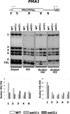
Figure 1
Figure 1
Comparison of histone acetylation on the PMA1 gene in set1Δ and set2Δ cells. (Top) PMA1 locus with transcription start sites (arrows), open reading frames (white rectangles), and positions of polymerase chain reaction (PCR) products (numbered black rectangles) used in chromatin immunoprecipitation (ChIP) analysis. (Middle) Chromatin prepared from wild-type, set1Δ, or set2Δ cells was analyzed by ChIP for histone H3, acetylated H4, or acetylated H3. Middle left panel shows PCR using the Input chromatin. TEL is a primer pair for a non-transcribed sub-telomeric region. Results from triplicate biological repeats are quantitated in bottom panels. Note the strongly increased acetylation at primer sets 1 and 2 and TEL in set1Δ cells. In contrast, set2Δ cells show the largest increase in acetylation at primer sets 3 and 4. Deletion of SET1 also led to a reproducible decrease in overall histone occupancy. Note that this data is adapted with permission from Kim and Buratowski (2009).
Figure 2
Figure 2
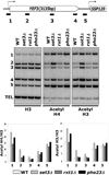
Comparison of histone acetylation in strains deleted for three PHD proteins associated with histone deacetylase complexes. (Top) Schematic of the YEF3 locus with position of ChIP PCR products shown as numbered black boxes. (Middle) Chromatin prepared from wild-type, set3Δ, rxt1Δ, or pho23Δ cells was analyzed by ChIP for histone H3, acetylated H4, or acetylated H3. Results from triplicate biological repeats are quantitated in bottom panels. Note the strongly increased acetylation at primers 1–3 in set3Δ, essentially identical to that seen in set1Δ cells (not shown). Also, pho23Δ cells show increased acetylation only at primer pair 1, which is also the location of H3K4me3 (not shown). Note that this data is adapted with permission from Kim and Buratowski (2009).
Figure 3
Figure 3
Model for the effect of co-transcriptional methylation at H3K4 and H3K36. At the most promoter-proximal nucleosomes, H3K4me3 may interact with Yng1 and Yng2 to target NuA3 and NuA4 HAT complexes. These HATs may also be targeted by activator interactions. However, H3K4me3 can also interact with the Pho23 subunit of the Rpd3L HDAC, perhaps to allow modulation or cycling of acetylation levels near transcription start sites. Slightly further downstream, H3K4me2 targets the activity of Set3C to 5' transcribed regions. In 3' transcribed regions, H3K36me2/3 is necessary for targeting Rpd3S HDAC activity. It should be noted that although these regions are drawn as discrete, there is likely to be some overlap. The combined effect is to leave promoter regions more accessible due to highly acetylated nucleosomes that are subject to rapid remodeling. In contrast, histones at downstream-transcribed regions are methylated and deacetylated such that the nucleosomes are more stably associated with the DNA.

References

    1. Alejandro-Osorio AL, Huebert DJ, Porcaro DT, Sonntag ME, Nillasithanukroh S, Will JL, Gasch AP. The histone deacetylase Rpd3p is required for transient changes in genomic expression in response to stress. Genome Biol. 2009;10:R57. - PMC - PubMed
    1. Berretta J, Pinskaya M, Morillon A. A cryptic unstable transcript mediates transcriptional trans-silencing of the Ty1 retrotransposon in S. cerevisiae. Genes Dev. 2008;22:615–626. - PMC - PubMed
    1. Biswas D, Dutta-Biswas R, Mitra D, Shibata Y, Strahl BD, Formosa T, Stillman DJ. Opposing roles for Set2 and yFACT in regulating TBP binding at promoters. EMBO J. 2006;25:4479–4489. - PMC - PubMed
    1. Buratowski S. The CTD code. Nat Struct Biol. 2003;10:679–680. - PubMed
    1. Buratowski S. Progression through the RNA polymerase II CTD cycle. Mol Cell. 2009;36:541–546. - PMC - PubMed

Publication types

MeSH terms

LinkOut - more resources