Comparative Study
. 2011 May 4;30(9):1804-17.
doi: 10.1038/emboj.2011.93. Epub 2011 Mar 29.

Dissociation by Pelota, Hbs1 and ABCE1 of mammalian vacant 80S ribosomes and stalled elongation complexes

Affiliations
Comparative Study

Dissociation by Pelota, Hbs1 and ABCE1 of mammalian vacant 80S ribosomes and stalled elongation complexes

Vera P Pisareva et al. EMBO J. .

Abstract

No-go decay (NGD) and non-stop decay (NSD) are eukaryotic surveillance mechanisms that target mRNAs on which elongation complexes (ECs) are stalled by, for example, stable secondary structures (NGD) or due to the absence of a stop codon (NSD). Two interacting proteins Dom34(yeast)/Pelota(mammals) and Hbs1, which are paralogues of eRF1 and eRF3, are implicated in these processes. Dom34/Hbs1 were shown to promote dissociation of stalled ECs and release of intact peptidyl-tRNA. Using an in vitro reconstitution approach, we investigated the activities of mammalian Pelota/Hbs1 and report that Pelota/Hbs1 also induced dissociation of ECs and release of peptidyl-tRNA, but only in the presence of ABCE1. Whereas Pelota and ABCE1 were essential, Hbs1 had a stimulatory effect. Importantly, ABCE1/Pelota/Hbs1 dissociated ECs containing only a limited number of mRNA nucleotides downstream of the P-site, which suggests that ABCE1/Pelota/Hbs1 would disassemble NSD complexes stalled at the 3'-end, but not pre-cleavage NGD complexes stalled in the middle of mRNA. ABCE1/Pelota/Hbs1 also dissociated vacant 80S ribosomes, which stimulated 48S complex formation, suggesting that Pelota/Hbs1 have an additional role outside of NGD.


Conflict of interest statement

The authors declare that they have no conflict of interest.

Figures
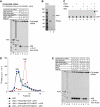
Figure 1
Figure 1
Pelota, Hbs1 and ABCE1 strongly stimulate 48S complex formation from 80S ribosomes by dissociating them into subunits. (A, E) Toe-printing analysis of the influence of different combinations of Pelota, Hbs1, eRF1/eRF3 and ABCE1 on 48S complex formation at 1 mM free Mg2+ on (CAA)nGUS mRNA from 40S subunits or 80S ribosomes. Lanes C, T, A and G depict cDNA sequences corresponding to (CAA)nGUS mRNA. The positions of toe-prints corresponding to ribosomal complexes are indicated. (B) Purified recombinant Pelota and Hbs1 resolved by SDS–PAGE. (C) Thin-layer chromatography analysis of the GTPase activity of Hbs1. (D) Dissociation of vacant 80S ribosomes containing [32P]60S subunits by different combinations of Pelota, Hbs1, ABCE1, eRFs and eIF6 at 1.5 mM free Mg2+, assayed by SDG centrifugation.
Figure 2
Figure 2
Dissociation of vacant 80S ribosomes by Pelota, Hbs1 and ABCE1. (AD) Dissociation of vacant 80S ribosomes containing [32P]60S subunits by incubation with Pelota, Hbs1 and ABCE1 (A, B) in the presence of (A) different concentrations of Pelota and (B) different nucleotides at 1.5 mM free Mg2+, (C) at different free Mg2+ concentrations and (D) after different incubation periods at 1.5 mM free Mg2+, assayed by SDG centrifugation. Upper fractions were omitted for clarity.
Figure 3
Figure 3
Pelota, Hbs1 and ABCE1 cannot dissociate pre-termination and elongation complexes assembled on MVHL-STOP mRNA containing an ∼400-nt-long 3′-UTR. (A) Structure of MVHL-STOP mRNA. (BE) Investigation by (B, C) SDG centrifugation and (D, E) toe-printing analysis of the influence of Pelota, Hbs1 and ABCE1 at 1.5 mM free Mg2+ on (B, D) pre-TCs and (C, E) ECs containing MV-peptidyl-tRNA assembled on MHVL-STOP mRNA. (B, C) Pre-TCs and ECs were assembled using [32P]60S subunits. Upper fractions were omitted for clarity. (D, E) Lanes C, T, A and G depict cDNA sequence corresponding to MVHL-STOP mRNA; the positions of toe-prints corresponding to ribosomal complexes are indicated.
Figure 4
Figure 4
Pelota, Hbs1 and ABCE1 can dissociate pre-termination and elongation complexes assembled on MVHL-STOP mRNA after their treatment with RelE. (A) Primer-extension analysis of A-site mRNA cleavage induced by RelE in 80S initiation and in pre- and post-translocation ECs containing MV-peptidyl-tRNA that were assembled on MVHL-STOP mRNA. (BD) Dissociation by Pelota, Hbs1 and ABCE1 at 1.5 mM free Mg2+ of (B, C) post- and pre-translocation ECs containing MV-peptidyl-tRNA and (D) pre-TCs assembled on MVHL-STOP mRNA using [32P]60S subunits depending on their treatment with RelE, assayed by SDG centrifugation. Upper fractions were omitted for clarity.
Figure 5
Figure 5
Dissociation by Pelota, Hbs1 and ABCE1 of elongation complexes assembled on non-stop mRNAs. (A) Structure of non-stop mRNAs encoding MVHC tetrapeptide and containing 0, 1 or 2 nts after the last codon. (B) Dissociation into subunits of ECs assembled on MVHC-0 mRNA with [32P]60S subunits and containing MVHC-peptidyl-tRNA after their incubation with Pelota, Hbs1 and ABCE1 at 1.5 mM free Mg2+, assayed by SDG centrifugation. (C) Ribosomal association of [35S]MVHC-peptidyl tRNA after incubation with Pelota, Hbs1 and ABCE1 at 1.5 mM free Mg2+ of ECs assembled on MVHC-0 mRNA, assayed by (upper panel) native gel electrophoresis and (lower panel) SDG centrifugation. (C, upper panel) Lanes 1 and 6 contain [35S-Cys]MVHC-tRNACys that was phenol extracted from elongation complexes, and [35S]Cys-tRNACys, respectively. (D) Ribosomal association of [32P]MVHL-0 mRNA after incubation of ECs with Pelota, Hbs1 and ABCE1 at 1.5 mM free Mg2+, assayed by (upper panel) SDG centrifugation and (lower panel) native gel electrophoresis. (E) Ribosomal association of [35S]MVHC-peptidyl tRNA after incubation with Pelota, Hbs1 and ABCE1 at 1.5 mM free Mg2+ of ECs assembled on MVHC-1 and MVHC-2 mRNAs, assayed by SDG centrifugation.
Figure 6
Figure 6
Dissociation by Pelota, Hbs1 and ABCE1 of pre-termination complexes containing different numbers of mRNA nucleotides downstream of the P-site. (A) Structure of mRNAs encoding MVHC tetrapeptide followed by the UAG stop codon and containing 3, 6, 9, 13 or 17 nts after the last sense codon. (B, F) Ribosomal association of [35S-Cys]MVHC-peptidyl tRNA after incubation of pre-TCs assembled on mRNAs shown in panel (A) with (B) recombinant and (F) native Pelota, Hbs1 and ABCE1 at 1.5 mM Mg2+, assayed by native gel electrophoresis. (C) Ribosomal association of [32P]MVHC-9 mRNA after incubation of ECs with Pelota, Hbs1 and ABCE1 at 1.5 mM free Mg2+, assayed by native gel electrophoresis. (D) Purified native and recombinant Pelota resolved by SDS–PAGE. (E) Dissociation by native Pelota, Hbs1 and ABCE1 at 1.5 mM free Mg2+ of pre-TCs assembled on MVHL-STOP mRNA using [32P]60S subunits depending on their treatment with RelE, assayed by SDG centrifugation. Upper fractions were omitted for clarity.
Figure 7
Figure 7
Kinetics and Mg2+ dependence of dissociation by Pelota, Hbs1 and ABCE1 of pre-termination complexes containing 3 mRNA nucleotides downstream of the P-site. Ribosomal association of [35S-Cys]MVHC-peptidyl tRNA after incubation of pre-TCs assembled on MVHC-3 mRNA with Pelota, Hbs1 and ABCE1 (A) at different Mg2+ concentrations, and (B) for different time periods at 1.5 mM Mg2+, assayed by SDG centrifugation. Upper fractions were omitted for clarity.

Similar articles

Cited by

References

    1. Acker MG, Kolitz SE, Mitchell SF, Nanda JS, Lorsch JR (2007) Reconstitution of yeast translation initiation. Methods Enzymol 430: 111–145 - PubMed
    1. Adham IM, Sallam MA, Steding G, Korabiowska M, Brinck U, Hoyer-Fender S, Oh C, Engel W (2003) Disruption of the pelota gene causes early embryonic lethality and defects in cell cycle progression. Mol Cell Biol 23: 1470–1476 - PMC - PubMed
    1. Alkalaeva EZ, Pisarev AV, Frolova LY, Kisselev LL, Pestova TV (2006) In vitro reconstitution of eukaryotic translation reveals cooperativity between release factors eRF1 and eRF3. Cell 125: 1125–1136 - PubMed
    1. Andreev D, Hauryliuk V, Terenin I, Dmitriev S, Ehrenberg M, Shatsky I (2008) The bacterial toxin RelE induces specific mRNA cleavage in the A site of the eukaryote ribosome. RNA 14: 233–239 - PMC - PubMed
    1. Atkinson GC, Baldauf SL, Hauryliuk V (2008) Evolution of nonstop, no-go and nonsense-mediated mRNA decay and their termination factor-derived components. BMC Evol Biol 8: 290. - PMC - PubMed

Publication types