. 2011 Mar 23;6(3):e17563.
doi: 10.1371/journal.pone.0017563.

Expression of DDX3 is directly modulated by hypoxia inducible factor-1 alpha in breast epithelial cells

Affiliations

Expression of DDX3 is directly modulated by hypoxia inducible factor-1 alpha in breast epithelial cells

Mahendran Botlagunta et al. PLoS One. .

Abstract

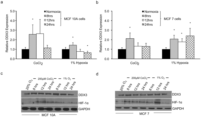
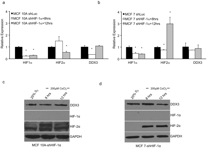
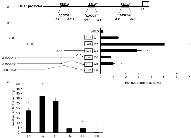
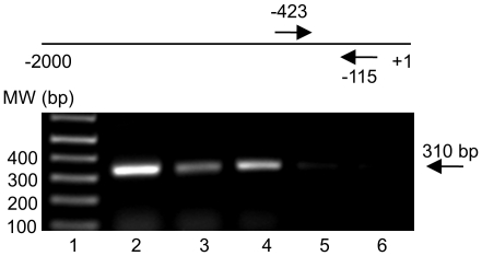
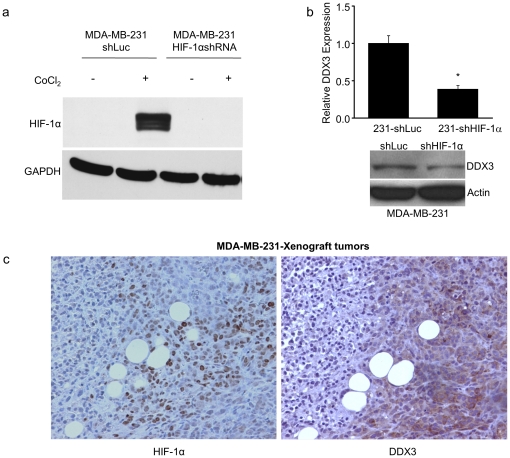
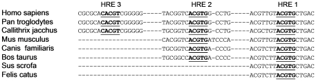
DEAD box protein, DDX3, is aberrantly expressed in breast cancer cells ranging from weakly invasive to aggressive phenotypes and functions as an important regulator of cancer cell growth and survival. Here, we demonstrate that hypoxia inducible factor-1α is a transcriptional activator of DDX3 in breast cancer cells. Within the promoter region of the human DDX3 gene, we identified three putative hypoxia inducible factor-1 responsive elements. By luciferase reporter assays in combination with mutated hypoxia inducible factor-1 responsive elements, we determined that the hypoxia inducible factor-1 responsive element at position -153 relative to the translation start site is essential for transcriptional activation of DDX3 under hypoxic conditions. We also demonstrated that hypoxia inducible factor-1 binds to the DDX3 promoter and that the binding is specific, as revealed by siRNA against hypoxia inducible factor-1 and chromatin immunoprecipitation assays. Thus, the activation of DDX3 expression during hypoxia is due to the direct binding of hypoxia inducible factor-1 to hypoxia responsive elements in the DDX3 promoter. In addition, we observed a significant overlap in the protein expression pattern of hypoxia inducible factor-1α and DDX3 in MDA-MB-231 xenograft tumors. Taken together, our results demonstrate, for the first time, the role of DDX3 as a hypoxia-inducible gene that exhibits enhanced expression through the interaction of hypoxia inducible factor-1 with hypoxia inducible factor-1 responsive elements in its promoter region.


Conflict of interest statement

Competing Interests: The authors have declared that no competing interests exist.

Figures

Figure 1
Figure 1. Effect of hypoxic conditions on the expression of the DDX3 gene in breast epithelial cells.
MCF 10A (a & c) and MCF 7 (b & d) cells were cultured under normoxic conditions (20% O2) or subjected to 200 µM CoCl2 or 1% O2 for 8, 12 and 24 h (a & b) qRT-PCR analysis was performed using specific primers for human DDX3 and HPRT as an internal normalization control. The expression level under normoxia was set to 1. DDX3 protein levels (c & d) in cell lysates of MCF 10A and MCF 7 cells were determined by immunoblots using anti-DDX3 antibodies. In these cases, HIF-1α and GAPDH served as controls indicating hypoxic conditions and equal protein loading respectively. Error bars represent ±SD.
Figure 2
Figure 2. Specificity of HIF-1α dependent activation of the DDX3 gene.
Stable MCF 10A and MCF 7-HIF-1α shRNA clones were generated. (a) MCF 10A-shHIF-1α and (b) MCF 7-shHIF-1α cells were subjected to 200 µM CoCl2 for the indicated times. qRT-PCR analysis was performed using specific primers for human DDX3, HIF-1α and HIF-2α. DDX3, HIF-1α, and HIF-2α protein levels in cell lysates of MCF 10A-shHIF-1α (c) and MCF 7-shHIF-1α (d) cells were examined by immunoblots using protein specific antibodies. GAPDH served as a loading control. Error bars represent ±SD.
Figure 3
Figure 3. Characterization of the putative HRE containing sequences of the human DDX3 promoter.
(a) Schematic representation of putative core HRE sequences in the promoter region of the DDX3 gene. (b) Linear representations of the DDX3 promoter-reporter constructs and the relative firefly luciferase activity of each in transient transfection assays in MCF 7 cells with pRRL-CMV (renilla luciferase expressing vector) as an internal control. After transfection cells were incubated in normoxia for 24 h Relative firefly luciferase activities are presented as a histogram at the right. (c) Effects of over-expression of HIF-1α in MCF 7 cells under normoxia. Cells were co-transfected with the DDX3 promoter-reporter constructs shown in (b), with pCDNA-HIF-1α (constitutive HIF-1α expressing vector), and pRRL-CMV as the internal control. Relative firefly luciferase activities are presented. Mean values from three independent transfections are shown. Error bars represent ±SD.
Figure 4
Figure 4. Identification of functional HRE in the DDX3 promoter.
(a) The regions of the individually mutated HREs-1-3 (Mut1-3) are indicated schematically. MCF 7 and MCF 7-shHIF-1α cells were transfected with wild-type (D2), singly (M1, M2, M3), doubly (M1+M2, M1+M3, M2+M3) or triply (M1+M2+M3) mutated promoter-reporter constructs, as indicated. (b & c) After transfection MCF 7 or MCF 7-shHIF-1α cells were cultured under normoxia. (d) After transfection MCF 7 were cultured in presence of CoCl2. (e) Each transfection of MCF 7 cells included co-transfection with a constitutive HIF-1α expression vector. Histograms show relative firefly luciferase activity. Each experiment was performed in triplicate on at least three separate occasions. Error bars represent ±SD.
Figure 5
Figure 5. ChIP assay: in vivo binding of HIF-1 to the DDX3 promoter in MCF 10A cells.
At the top of the gel is a schematic representation of the DDX3 promoter. Arrows flank the region (-423 to -115) amplified by PCR with DDX3 promoter specific primers. Gel shows: lane 1- molecular weight (MW) marker, lane 2- total input chromatin, lane 3-acetyl histone H3 precipitation, lane 4-anti-HIF-1α precipitation under hypoxic conditions, lane 5-anti-HIF-1α under normoxic conditions, and lane 6-anti-actin precipitation. Identical volumes from each final precipitation were used for PCR (except for the input chromatin, which was diluted 100x).
Figure 6
Figure 6. Correlation between HIF-1α and DDX3 protein expression in MDA-MB-231 breast cancer cells.
(a) Immunoblot showing expression levels of HIF-1α following CoCl2 treatment in HIF-1α knockdown cells. (b) Histogram showing relative fold expression of DDX3 mRNA protein levels of MDA-MB-231 versus MDA-MB-231-shHIF-1α cells. Expression of DDX3 was downregulated in MDA-MB-231-shHIF-1α relative to MDA-MB-231 cells at both the mRNA and protein levels. (c) Immunohistochemical staining for HIF-1α and DDX3 in sequential slices of a MDA-MB-231 tumor xenograft. Tumor sections were stained with antibodies that are specific for HIF-1α and DDX3. Samples were counterstained with hematoxylin to reveal morphology. Error bars represent ±SD.
Figure 7
Figure 7. Comparison of regions of the DDX3 promoter among eutherian mammals.
Conserved core hypoxia response elements (HRE-1, 2, & 3), which are putative HIF-1 binding sites (indicated in bold and underlined) are shown. HRE-1 and surrounding sequences are highly conserved across species.

References

    1. Lahn BT, Page DC. Functional coherence of the human Y chromosome. Science. 1997;278:675–680. - PubMed
    1. Cordin O, Banroques J, Tanner NK, Linder P. The DEAD-box protein family of RNA helicases. Gene. 2006;367:17–37. - PubMed
    1. Linder P. Dead-box proteins: a family affair–active and passive players in RNP-remodeling. Nucleic Acids Res. 2006;34:4168–4180. - PMC - PubMed
    1. Rocak S, Linder P. DEAD-box proteins: the driving forces behind RNA metabolism. Nat Rev Mol Cell Biol. 2004;5:232–241. - PubMed
    1. Leroy P, Alzari P, Sassoon D, Wolgemuth D, Fellous M. The protein encoded by a murine male germ cell-specific transcript is a putative ATP-dependent RNA helicase. Cell. 1989;57:549–559. - PubMed

Publication types

MeSH terms