. 2011 Feb 26;44(1):1-7.
doi: 10.1267/ahc.10028. Epub 2011 Feb 4.

Dynamics of corticosteroid receptors: lessons from live cell imaging

Affiliations

Dynamics of corticosteroid receptors: lessons from live cell imaging

Mayumi Nishi. Acta Histochem Cytochem. .

Abstract

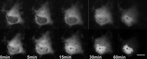
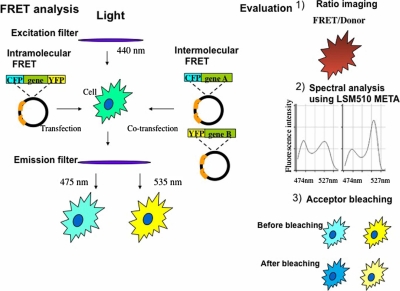
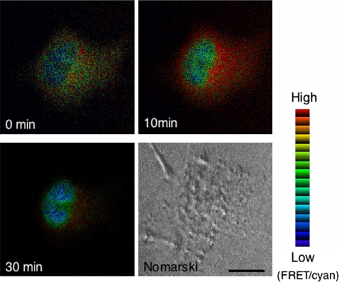
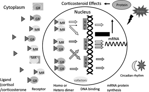
Adrenal corticosteroids (cortisol in humans or corticosterone in rodents) exert numerous effects on the central nervous system that regulates the stress response, mood, learning and memory, and various neuroendocrine functions. Corticosterone (CORT) actions in the brain are mediated via two receptor systems: the glucocorticoid receptor (GR) and the mineralocorticoid receptor (MR). It has been shown that GR and MR are highly colocalized in the hippocampus. These receptors are mainly distributed in the cytoplasm without hormones and translocated into the nucleus after treatment with hormones to act as transcriptional factors. Thus the subcellular dynamics of both receptors are one of the most important issues. Given the differential action of MR and GR in the central nervous system, it is of great consequence to clarify how these receptors are trafficked between cytoplasm and nucleus and their interactions are regulated by hormones and/or other molecules to exert their transcriptional activity. In this review, we focus on the nucleocytoplasmic and subnuclear trafficking of GR and MR in neural cells and non-neural cells analyzed by using molecular imaging techniques with green fluorescent protein (GFP) including fluorescence recovery after photobleaching (FRAP) and fluorescence resonance energy transfer (FRET), and discuss various factors affecting the dynamics of these receptors. Furthermore, we discuss the future directions of in vivo molecular imaging of corticosteroid receptors at the whole brain level.

Keywords: GFP; glucocorticoid receptor; hippocampus; mineralocorticoid receptor; real-time imaging.


Figures

Fig. 1
Fig. 1
Dual-color imaging of GR and MR with GFP color variants in a single COS-1 cell. COS-1 cells co-transfected with CFP-GR and YFP-MR were cultured in the absence of serum and steroids for 24 hr before observation. Upper images were representative time-lapse images of CFP-GR, and bottoms were those of YFP-MR. Note that YFP-MR was accumulated in the nuclear region faster than CFP-GR after treatment with 10−9 M CORT. Bar=10 µm.
Fig. 2
Fig. 2
Procedure and evaluation of fluorescence resonance energy transfer (FRET) experiment using fusion proteins with cyan fluorescent protein (CFP) and yellow fluorescent protein (YFP). In our experiment, FRET is evaluated with three ways: 1) Ratio imaging (FRET image was divided by Donor image). Ratio images were pseudocolored where the red range indicated a high ratio and the blue range indicated a low ratio. 2) For detecting an emission spectral change in FRET imaging, Emission Fingerprinting method using confocal laser-scanning microscope LSM 510 META (Zeiss) was employed. First, spectral signatures of the fluorescence within the specimen were captured by means of lambda stack acquisition with excitation at 458 nm and detection at 10 nm-intervals from 458 through 596 nm using an HFT 458/543 dichroic mirror. Several regions of interest (ROIs) with a diameter of 2 µm were then randomly selected for obtaining emission spectral patterns, and the mean ratio of fluorescence intensity of 527 nm and 474 nm was calculated from selected ROIs at each time point after ligand addition. 3) For acceptor photobleaching, we used the confocal laser-scanning microscope. Energy transfer was detected as an increase in donor fluorescence (CFP) after photobleaching of the acceptor molecules (YFP). The acceptor was photobleached by using a 514 nm laser for 1 min at maximum power (25 mW).
Fig. 3
Fig. 3
Ratio images of the cell co-expressing CFP-GR and YFP-importin α detected by FRET. COS-1 cells were co-expressed with CFP-GR and YFP-importin α and cultured in the absence of serum and steroids for at least 15 hr before observation. Fluorescent images of CFP-GR and YFP-importin α were captured using a filter set of CFP (440AF21 excitation, 480AF30 emission, and 455DRLP dichroic mirror) and YFP (500AF25 excitation, 545AF35 emission, and 525DRLB dichroic mirror), respectively. FRET image was detected using a filter set with 440AF21 excitation and 535AF26 emission, and 455DRPL dichroic mirror at 0, 10, and 30 min after treatment with 10−6 M CORT. Filter sets were purchased from Omega Optical Inc. The ratio of the FRET image was divided by donor image to obtain the ratio images using MetaMorph software (Universal Imaging Corp.). The ratio images were pseudo colored. The red range showed high ratio and blue range showed low ratio. High ratio was observed in the cytoplasm, indicating an interaction of CFP-GR and YFP-importin, whereas low ratio was observed in the nucleus, indicating a dissociation of these two molecules.
Fig. 4
Fig. 4
A schematic model for dimer formation of corticosteroid receptor. A variety of corticosteroid receptor dimers including homodimers and heterodimers may give the potential to provide a more finely tuned regulation of corticosteroid-regulated gene for responding to fluctuations in plasma cortisol/corticosterone level affected by stress responses, circadian rhythm, and so on.

Similar articles

Cited by

References

    1. Arriza J. L., Simerly R. B., Swanson L. W., Evans R. M. The neuronal mineralocorticoid receptor as a mediator of glucocorticoid response. Neuron. 1988;1:887–900. - PubMed
    1. Bohn M. C., O’Banion M. K., Young D. A., Giuliano R., Hussain S., Dean D. O., Cunningham L. A. In vitro studies of glucocorticoid effects on neurons and astrocytes. Annl. N.Y. Acad. Sci. 1994;746:243–259. - PubMed
    1. Collins G. A., Tansey W. P. The proteasome: A utility tool for transcription? Curr. Opin. Genet. Dev. 2006;16:197–202. - PubMed
    1. Dallman M. F. Fast glucocorticoid actions on brain: back to the future. Front. Neuroendocrinol. 2005;26:103–108. - PubMed
    1. Davis L. I. The nuclear pore complex. Annu. Rev. Biochem. 1995;64:865–896. - PubMed