. 2011 Apr;24(2):119-29.
doi: 10.1089/vim.2010.0077.

Viral interactions with B-cells contribute to increased regulatory T-cells during chronic HCV infection

Affiliations

Viral interactions with B-cells contribute to increased regulatory T-cells during chronic HCV infection

Chris L Ayers et al. Viral Immunol. 2011 Apr.

Abstract

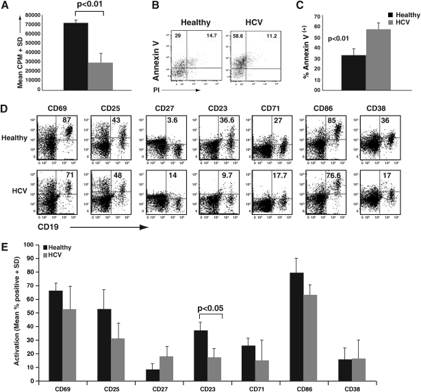
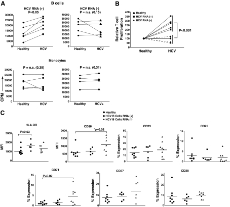
Hepatitis C virus (HCV) has a propensity to establish chronic infection that is characterized by attenuated virus-specific T-cell responses. Mechanisms leading to T-cell attenuation are poorly understood and likely involve dysfunctional interactions between antigen-presenting cells (APC) and effector/regulatory T-cells. Reports on dendritic cells (DC) have described only minor dysfunction during HCV infection. However, there is a paucity of reports regarding B-cell function, despite clear associations with B-cell-related secondary sequelae. In this study we evaluated the state of B-cells during chronic HCV infection, and observed a diminished ability to respond to mitogenic stimuli, correlating with increased apoptosis. This was in contrast to their ex vivo phenotype, which indicated ongoing chronic activation in vivo. There was a high association of HCV-positive strand RNA with B-cells in a subset of HCV patients. Interestingly, ex-vivo-derived HCV RNA-positive B-cells induced significantly greater proliferation in allogeneic T-cells than in HCV-negative B-cells, correlating with an increased generation of CD4(+)CD25(+)FOXP3(+) regulatory T-cells (Tregs). In-vitro exposure of healthy peripheral blood mononuclear cells (PBMC) to HCV resulted in robust activation of resting B-cells. These HCV-exposed B-cells also showed an enhanced ability to generate Tregs. Our results provide strong evidence for a novel and paradoxical link between HCV-induced enhanced APC function and the generation of Tregs.


Figures

FIG. 1.
FIG. 1.
B-cells from HCV patients have an attenuated activation potential. (A) B-cells from chronic HCV patients or healthy donors were isolated using CD19 magnetic microbeads. First, 2 × 105 B-cells (triplicate cultures in 96-well plates) were stimulated for 4 d with a 1:1000 dilution of pansorbin (protein A). 3H-thymidine was added in the last 16 h of culture and the scintillation counts were measured. The data are representative of replicate experiments from three separate donor HCV pairs. (B) CD19+ B-cells from chronic HCV patients or healthy donors were incubated for 2 d with a 1:5000 dilution of pansorbin (protein A). The cells were then stained with annexin V-FITC, propidium iodide (PI), and CD19-APC. Flow cytometry dotplots indicate the percent expression of annexin V versus PI, and are representative of five separate experiments. (C) Cumulative data of pansorbin-stimulated CD19+ B-cells, depicting percentages of annexin V+/PI B-cells, and indicating cells undergoing early apoptosis, showing significant differences in healthy versus HCV B-cells (p < 0.01). (D) Bulk PBMC from chronic HCV patients or healthy donors were incubated for 24 h with 5 μg/mL of pokeweed mitogen. The cells were subsequently stained with the following three panels of activation markers: (1) CD19-PECy7, CD25-APC, and CD69-PE; (2) CD19-PECy7, CD27-FITC, and CD23-PE; and (3) CD19-PECy7, CD71-FITC, CD86-PE, and CD38-APC. Representative dotplots from a single healthy donor and an HCV patient are shown. Numbers indicate percentages of expression of each marker on gated B-cells. (E) Cumulative data from nine HCV patients and four healthy control subjects, depicting percentage expression of the indicated markers. There was an overall trend toward lower induced activation on HCV B-cells. Statistical significance was observed for CD23 differences between healthy subjects and HCV patients (p < 0.05).
FIG. 2.
FIG. 2.
HCV RNA+ B-cells induce significantly greater MLR responses and exhibit an activated phenotype. (A) Microbead-purified B-cells (top panels), and monocytes (bottom panels), from up to 13 healthy donors and 13 HCV-infected patients were used to stimulate purified allogeneic CD3+ T-cells from one of two healthy third-party subjects. Every experimental set-up consisted of at least one healthy-HCV pair against the same third-party donor. Allogeneic cultures were pulsed with 3H-thymidine on day 5 and harvested on day 6. Responses of T-cells at a stimulator:responder ratio of 1:10 are represented as CPM on the y-axis. Data are separated based on HCV RNA status of the patients' B-cells: patients with HCV RNA+ B-cells are represented in the left two panels, whereas those with HCV RNA B-cells are represented in the right two panels. (B) B-cell data from panel A were normalized for responses generated from healthy control subjects (assigned as 100). Responses induced by HCV RNA+ B-cells were significantly greater than those from HCV RNA B-cells (p < 0.001). (C) PBMC from 14 HCV patients (6 B-cell HCV RNA+ and 8 HCV RNA), and 8 healthy donors were stained ex-vivo for HLA-DR, CD23, CD25, CD27, CD38, CD71, and CD86. The expression level of each marker was evaluated on a gated CD19+ B-cell population. Isotypic controls were used to determine cutoffs. Mean fluorescence intensity (MFI) levels were used for HLA-DR and CD86, whereas percentage expression was determined for the remaining markers.
FIG. 3.
FIG. 3.
Exposure of healthy PBMC to HCV results in robust B-cell activation. (A) PBMC from healthy donors were exposed to JFH-1 virus (or mock media) for 3 h, washed, and cultured for 7 d. B-cell activation was monitored at various time points (days 1 and 7 are shown). CD19 is shown on the x-axis, and staining for the indicated activation markers is represented on the corresponding y-axes. The percentages of B-cells that were positive for each marker (i.e., within gated CD19+ cells) is indicated. The data are representative of replicate experiments from five separate donors. (B) PBMC were incubated with viable virus, UV-inactivated virus (UV virus), or were pre-incubated with anti-CD81 (αCD81), or anti-E2 antibody (αE2+), for 1 h prior to the addition of viable virus for 3 h at 37°C, followed by washing and culture for 7 d. PBMC were then harvested and analyzed for B-cell activation (CD71 is shown as a representative activation marker). Data are representative of three replicate experiments, using PBMC from different donors. (C) Bulk PBMC, monocyte-depleted PBMC (PBMC-M), T-cell-depleted PBMC (PBMC-T), purified B-cells + monocytes (B + M), B-cells + T-cells (B + T), and purified B-cells + monocytes + T-cells (B + M + T), were incubated with viable JFH-1 virus at a 0.01 virus:cell ratio. All cell combinations were incubated for 7 d, and gated B-cells were subsequently analyzed for activation (represented by CD71-FITC and CD86-PE expression). Data are representative of three replicate experiments, using PBMC from different donors.
FIG. 4.
FIG. 4.
HCV-modified B-cells are potent inducers of regulatory T-cells. (A) Bead-sorted B-cells from healthy subjects and treatment-naïve HCV patients were used as APC in CFSE-based MLR assays, using sorted third-party CD4+CD25 T-cells as responders. The top panels show representative CFSE histograms obtained from assays with healthy B-cells, HCV RNA+ B-cells, and HCV RNA B-cells as APC. The percentages of proliferation are indicated, demonstrating a significantly greater response from HCV RNA+ B-cells. The bottom panels demonstrate CD25 versus FOXP3 staining on gated CD4+ T-cells from the same cultures. The percentages of CD25+ and FOXP3+ are indicated, showing higher Treg generation in HCV RNA+ MLR. Data are representative of 11 replicate experiments, using different healthy and HCV donors [a total of 11 healthy and 11 HCV (6 HCV+ and 5 HCV) subjects]. (B) Cumulative graph of the CFSE-based MLR assays described in panel A. Results are represented as percentages of CFSElow(proliferating)/CD25+/FOXP3+ cells induced by HCV B-cells relative to corresponding healthy donor B-cells (normalized to 100%). These data confirm a trend toward higher proliferation and increased CD25/FOXP3 induction by HCV+ B-cells (*p = 0.06). (C) B-cell-induced MLR were set up as described for panel A. Activated CD25+ T-cells from these MLR were then tested for suppressor activity, using flow cytometry–based, anti-CD3-stimulated suppression assays. Autologous T-cells were incubated with increasing numbers of putative suppressor T-cells, derived from MLR. The percentage of proliferation was normalized to the proliferation seen in the absence of any suppressors (designated as 100). Both healthy (black bars) and HCV (gray bars) B-cell-induced CD4+CD25+FOXP3+ T-cells demonstrated comparable Treg activity on a per-cell basis. These data are representative of five separate MLR-induced suppressor pair replicate experiments. (D) JFH- or mock-exposed B-cells were used as APC in allogeneic MLR with CD4+25 T-cells. The percentages of CD4+CD25+FOXP3+ Tregs from the indicated cultures are demonstrated. Data are representative of three replicate experiments.

Similar articles

Cited by

References

    1. Altomonte M. Montagner R. Pucillo C. Maio M. Triggering of target of an antiproliferative antibody-1 (TAPA-1/CD81) up-regulates the release of tumour necrosis factor-alpha by the EBV-B lymphoblastoid cell line JY. Scand J Immunol. 1996;43:367–373. - PubMed
    1. Averill L. Lee WM. Karandikar NJ. Differential dysfunction in dendritic cell subsets during chronic HCV infection. Clin Immunol. 2007;123:40–49. - PMC - PubMed
    1. Bain C. Fatmi A. Zoulim F, et al. Impaired allostimulatory function of dendritic cells in chronic hepatitis C infection. Gastroenterology. 2001;120:512–524. - PubMed
    1. Barnes E. Lauer G. Walker B. Klenerman P. T cell failure in hepatitis C virus infection. Viral Immunol. 2002;15:285–293. - PubMed
    1. Bassett SE. Brasky KM. Lanford RE. Analysis of hepatitis C virus-inoculated chimpanzees reveals unexpected clinical profiles. J Virol. 1998;72:2589–2599. - PMC - PubMed

Publication types