. 2011 Aug;163(7):1507-19.
doi: 10.1111/j.1476-5381.2011.01379.x.

Cannabidiol inhibits pathogenic T cells, decreases spinal microglial activation and ameliorates multiple sclerosis-like disease in C57BL/6 mice

Affiliations

Cannabidiol inhibits pathogenic T cells, decreases spinal microglial activation and ameliorates multiple sclerosis-like disease in C57BL/6 mice

Ewa Kozela et al. Br J Pharmacol. 2011 Aug.

Abstract

Background and purpose: Cannabis extracts and several cannabinoids have been shown to exert broad anti-inflammatory activities in experimental models of inflammatory CNS degenerative diseases. Clinical use of many cannabinoids is limited by their psychotropic effects. However, phytocannabinoids like cannabidiol (CBD), devoid of psychoactive activity, are, potentially, safe and effective alternatives for alleviating neuroinflammation and neurodegeneration.

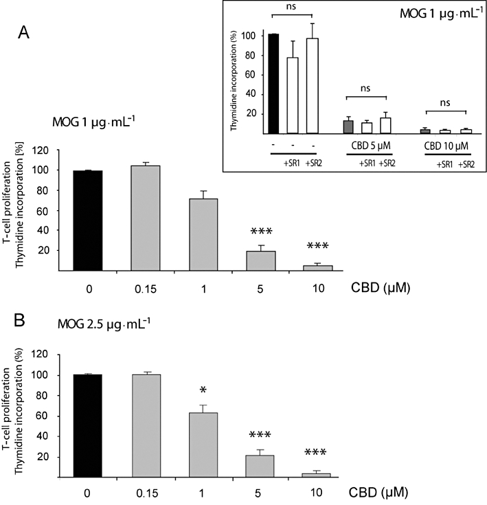
Experimental approach: We used experimental autoimmune encephalomyelitis (EAE) induced by myelin oligodendrocyte glycoprotein (MOG) in C57BL/6 mice, as a model of multiple sclerosis. Using immunocytochemistry and cell proliferation assays we evaluated the effects of CBD on microglial activation in MOG-immunized animals and on MOG-specific T-cell proliferation.

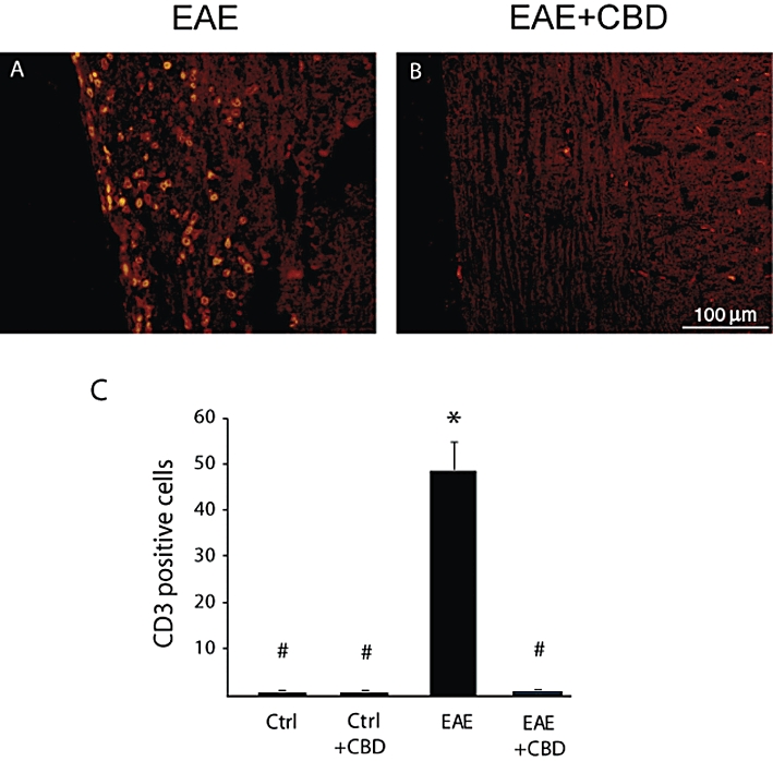
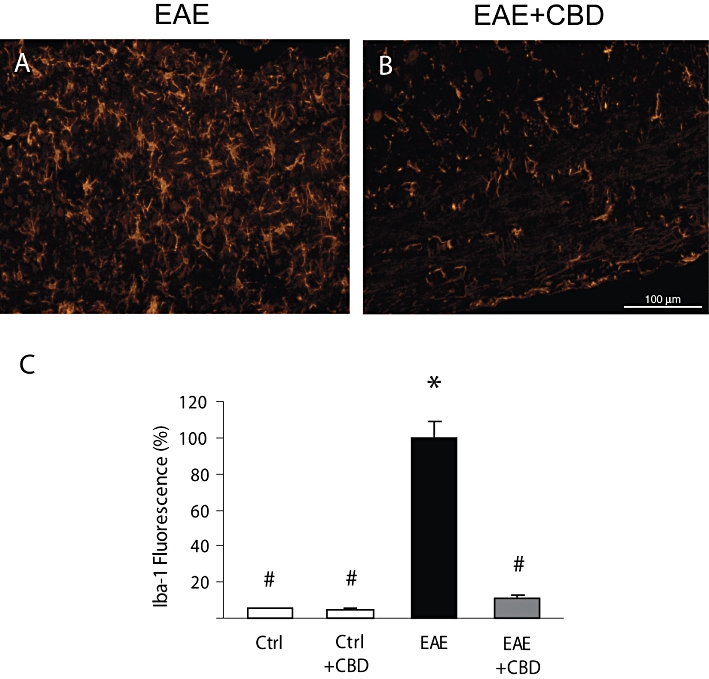
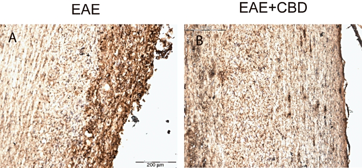
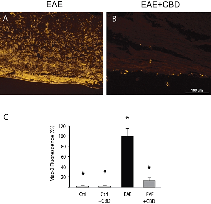
Key results: Treatment with CBD during disease onset ameliorated the severity of the clinical signs of EAE. This effect of CBD was accompanied by diminished axonal damage and inflammation as well as microglial activation and T-cell recruitment in the spinal cord of MOG-injected mice. Moreover, CBD inhibited MOG-induced T-cell proliferation in vitro at both low and high concentrations of the myelin antigen. This effect was not mediated via the known cannabinoid CB(1) and CB(2) receptors.

Conclusions and implications: CBD, a non-psychoactive cannabinoid, ameliorates clinical signs of EAE in mice, immunized against MOG. Suppression of microglial activity and T-cell proliferation by CBD appeared to contribute to these beneficial effects.


Figures

Figure 1
Figure 1
Cannabidiol (CBD) ameliorates the clinical signs and disease progression of EAE induced by myelin oligodendrocyte glycoprotein (MOG). EAE was induced in C57BL/6 mice by flank s.c. immunization with MOG35-55. Clinical disease scores were recorded daily until day 30 after induction. CBD (CBD + EAE) or its vehicle (EAE group), was injected i.p. during the onset of the disease for 3 consecutive days (days 19–21; indicated by arrows). The mean clinical scores ± SD are shown. Each group consisted of 15 mice. Repeated measure anova showed significant differences between the EAE and CBD + EAE groups (P < 0.001).
Figure 2
Figure 2
Cannabidiol (CBD) treatment of control, naïve C57BL/6 mice does not affect spinal cord integrity, basal microglial and T-cell number. The Figure shows representative stainings of spinal cord sections prepared from control healthy mice receiving complete Freund adjuvant either without CBD (Ctrl) or with CBD (Ctrl + CBD) treatment. (A) Haematoxylin and eosin (H&E) staining; (B) Iba-1 staining for the presence of microglial/macrophages; and (C) CD3+ staining for presence of T-cells. Scale bars represent 100 µm. Statistical analysis of the CD3+ and Iba-1 expression is presented in the bar graphs of Figures 5C (CD3+) and 6C (Iba-1).
Figure 5
Figure 5
Cannabidiol (CBD) treatment attenuates EAE-induced T-cell infiltration into spinal cord of mice. CD3+ staining reveals that CBD treatment reduces the infiltration of T cells into the white matter of the spinal cords of EAE mice. (A) EAE mice. (B) EAE mice treated with CBD. Scale bar represents 100 µm. The results are presented as mean ± SEM of CD3+ cells from 9–12 spinal fragments from three to four mice per each experimental group. One-way anovaF(3,43) = 45.3, P < 0.0001, followed by Bonferroni post hoc test. *P < 0.001 versus Ctrl. #P < 0.001 versus EAE mice.
Figure 6
Figure 6
Cannabidiol (CBD) reduces the MOG-up-regulated Iba-1 expression in spinal cords of mice. Representative pictures showing that (A) MOG injections increase Iba-1 staining in spinal cord, and (B) that CBD injections significantly decrease the level of Iba-1 staining. (C) Quantification of Iba-1 fluorescence in sections of control (Ctrl) and of EAE mice with and without CBD treatment. The results are presented as mean ± SEM of total fluorescence from 8–11 spinal fragments from three to four mice per each experimental group. One-way anovaF(3,37) = 30.23, P < 0.0001, followed by Bonferroni post hoc test. *P < 0.001 versus Ctrl. #P < 0.001 versus EAE mice. Scale bar represents 100 µm.
Figure 3
Figure 3
Cannabidiol (CBD) reduces the MOG-induced axonal damage in spinal cords of EAE mice. Mice were killed 30 days after EAE disease induction and sections of spinal cord were analysed for damaged axons using SMI-32 antibody. (A) MOG-induced EAE mice; (B) MOG-induced EAE mice treated with CBD (as described in Figure 1). Scale bar represents 200 µm.
Figure 4
Figure 4
Cannabidiol (CBD) treatment attenuates EAE-induced immune cell infiltration into spinal cord of mice. Haematoxylin and eosin staining revealed that CBD treatment reduces immune cell infiltration into the white matter of the spinal cords of EAE mice. (A) EAE mice and (B) EAE mice treated with CBD. Scale bar represents 100 µm.
Figure 7
Figure 7
Cannabidiol (CBD) reduces the increased Mac-2/Galectin-3 expression in the spinal cords of EAE mice. Representative pictures showing that (A) MOG injections increase Mac-2 expression and (B) that CBD injections during the onset of EAE significantly decrease the level of Mac-2 staining. (C) Quantification of Mac-2 fluorescence in spinal cord sections of control and EAE mice with and without CBD treatment. The results are presented as mean ± SEM of total fluorescence from 8–11 spinal fragments from three to four mice per each experimental group. One-way anovaF(3,34) = 63.25, P < 0.0001, followed by Bonferroni post hoc test. *P < 0.001 versus Ctrl. #P < 0.001 versus EAE mice. Scale bar represents 100 µm.
Figure 8
Figure 8
Cannabidiol (CBD) inhibits MOG-induced T-cell proliferation. MOG-induced T-cell proliferation was determined by [3H]thymidine incorporation. Encephalitogenic T cells were co-cultured with antigen presenting cells and exposed to MOG35-55 at 1 (A) and 2.5 µg·mL−1 (B). CBD was added 5 min before MOG. Assays were carried out each time in triplicate and mean per cent values ± SEM are shown based on four independent experiments. (A) One-way anovaF(4,19) = 124.7, P < 0.0001; (B) One-way anovaF(4,19) = 31.1, P < 0.0001, followed by Bonferroni post hoc test. *P < 0.05, ***P < 0.001 versus MOG alone at the respective concentration. Inset: neither the CB1 receptor antagonist (1 µM of SR141716; SR1) nor the CB2 receptor antagonist (1 µM of SR144528; SR2) applied 30 min before CBD and MOG affected MOG35-55 (1 µg·mL−1) – induced T-cell proliferation either in the absence or presence of CBD; one-way anovaF(8,26) = 26.8, P < 0.05; ns, non-significant.

References

    1. Alexander SPH, Mathie A, Peters JA. Guide to Receptors and Channels (GRAC), 4th edn. Br J Pharmacol. 2009;158:S1–S254. - PMC - PubMed
    1. Arévalo-Martín A, Vela JM, Molina-Holgado E, Borrell J, Guaza C. Therapeutic action of cannabinoids in a murine model of multiple sclerosis. J Neurosci. 2003;23:2511–2516. - PMC - PubMed
    1. Bayewitch M, Avidor-Reiss T, Levy R, Barg J, Mechoulam R, Vogel Z. The peripheral cannabinoid receptor: adenylate cyclase inhibition and G protein coupling. FEBS Lett. 1995;375:143–147. - PubMed
    1. Bennett JL, Stüve O. Update on inflammation, neurodegeneration, and immunoregulation in multiple sclerosis: therapeutic implications. Clin Neuropharmacol. 2009;32:121–132. - PubMed
    1. Ben-Nun A, Lando Z. Detection of autoimmune cells proliferating to myelin basic protein and selection of T cell lines that mediate experimental autoimmune encephalomyelitis (EAE) in mice. J Immunol. 1983;130:1205–1209. - PubMed

Publication types

MeSH terms