Case Reports
. 2011 Mar 30:6:27.
doi: 10.1186/1746-1596-6-27.

Pulmonary tumor thrombotic microangiopathy induced by gastric carcinoma: morphometric and immunohistochemical analysis of six autopsy cases

Affiliations
Case Reports

Pulmonary tumor thrombotic microangiopathy induced by gastric carcinoma: morphometric and immunohistochemical analysis of six autopsy cases

Yoichiro Okubo et al. Diagn Pathol. .

Abstract

Background: Pulmonary tumor thrombotic microangiopathy (PTTM) has been known as a rare and serious cancer-related pulmonary complication. However, the pathogenesis and pathophysiology of this debilitating condition still remains obscure and no effective management was recommended. The present study aims to elucidate the pathophysiology of PTTM.

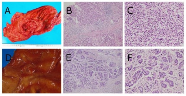
Methods: Autopsy records were searched to extract cases of pulmonary tumor embolism induced by metastasis of gastric carcinoma in the Toho University Omori Medical Center from 2000 to 2006. And then, tissue sections of extracted cases were prepared for not only light microscopic observation but morphometric analysis with the use of selected PTTM cases.

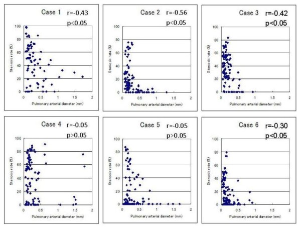
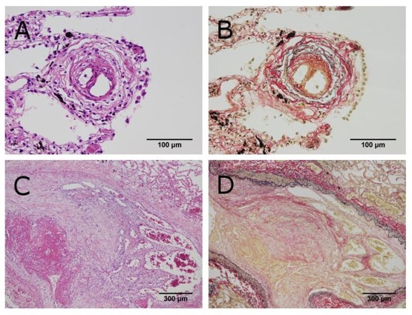
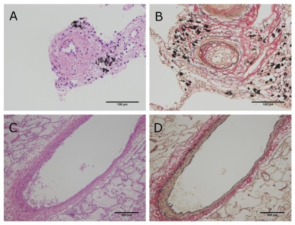
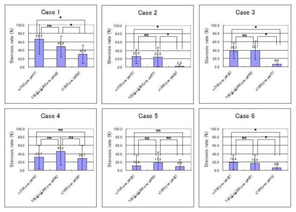
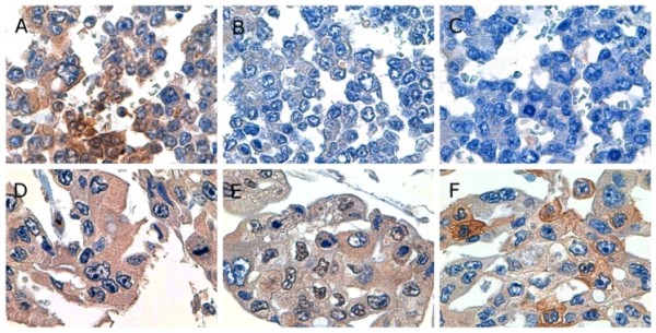
Results: Six autopsies involved PTTM and clinicopathological data of them were summarized. There was a significant negative association between pulmonary arterial diameter and stenosis rate in four cases. Although all cases showed an increase of stenosis rate to some degree, the degree of stenosis rate varied from case to case. Significant differences were found for average stenosis rate between the under 100 micrometer group or the 100 to 300 micrometer group and the 300 micrometer group in four cases. However, no significant differences were found for average stenosis rate between the under 100 micrometer group and the 100 to 300 micrometer group in all cases. Meanwhile, all cases showed positive reactivity for tissue factor (TF), five showed positive reactivity for vascular endothelial growth factor (VEGF), and three showed positive reactivity for osteopontin (OPN).

Conclusions: In the present study, we revealed that the degree of luminal narrowing of the pulmonary arteries varied from case to case, and our results suggested that pulmonary hypertension in PTTM occurs in selected cases which have a widespread pulmonary lesion with severe luminal narrowing in the smaller arteries. Furthermore, our immunohistochemical examination indicated that gastric carcinoma indicating PTTM shows a higher TF-positive rate than typical gastric carcinoma. However, it remains still obscuring whether gastric carcinoma indicating PTTM shows a higher VEGF or OPN-positive rate as determined by immunohistochemistry.


Figures

Figure 1
Figure 1
Macroscopic and histopathological findings of gastric carcinomas in case 1 and 2. Case 1 (A) The ulcerated tumor with relatively demarcated and raised margins (Borrmann's type 3 tumor) was located in the gastric body. (B and C) Poorly differentiated adenocarcinoma cells diffusely proliferated (hematoxylin-eosin (HE) double stain, × 100, × 400, respectively). Case 2 (D) The ulcerated tumor with demarcated and slightly raised margins (Borrmann's type 2 tumor) was located in the gastric body. (E and F) Poorly differentiated adenocarcinoma cells were arranged in small nests or trabecular pattern surrounded by fibrous stroma (HE double stain, × 100, × 400, respectively).
Figure 2
Figure 2
Scatter plots of pulmonary arterial diameter and stenosis rate in pulmonary arteries. There was a significant negative association between pulmonary arterial diameter and stenosis rate in four of the six cases (Case 1, 2, 3, and 6). In contrast, there was no significant association in the remaining two cases (Case 4 and 5).
Figure 3
Figure 3
Histopathological findings of pulmonary tumor thrombotic microangiopathy in case 1. (A and B) Fibrocellular intimal proliferation occludes the lumen of the pulmonary artery under 100 μm in diameter (Hematoxylin-eosin (HE) double stain and elastica van Gieson (EVG) stain, × 400). (C and D) Fibrocellular intimal proliferation associated with carcinoma cells proliferation occludes the lumen of the pulmonary artery over 300 μm in diameter (HE double stain and EVG stain, × 40).
Figure 4
Figure 4
Histopathological findings of pulmonary tumor thrombotic microangiopathy in case 2. (A and B) Fibrocellular intimal proliferation occludes the lumen of the pulmonary artery under 100 μm in diameter (Hematoxylin-eosin (HE) double stain and elastica van Gieson (EVG) stain, × 400). (C and D) Neither prominent fibrocellular intimal proliferation nor carcinoma cells were present in the pulmonary artery over 300 μm in diameter (HE double stain and EVG stain, × 40).
Figure 5
Figure 5
Average stenosis rate in each arterial group for six patients with pulmonary tumor thrombotic microangiopathy. No significant differences were found for average stenosis rate between the under 100-μm group and the 100 to 300-μm group in all cases. On the other hand, significant differences were found for average stenosis rate between the under 100-μm group or 100 to 300-μm group and the 300-μm group in four of the six cases (Case 1, 2, 3, and 6). Statistical significance was tested using the one-way analysis of variance (ANOVA) and the Bonferroni post hoc test. Values are expressed as mean ± standard deviation. Differences were considered significant at P < 0.05 (*: P < 0.05, ns: P > 0.05).
Figure 6
Figure 6
Photomicrographs showing representative immunohistochemistry. (A, B, and C) Gastric carcinoma cells in case 1 showed positive reactivity for tissue factor and negative reactivity for vascular endothelial growth factor and osteopontin (× 1,000). (D, E, and F) Gastric carcinoma cells in case 6 showed positive reactivity for tissue factor, vascular endothelial growth factor, and osteopontin (× 1,000).

Similar articles

Cited by

References

    1. von Herbay A, Illes A, Waldherr R, Otto HF. Pulmonary tumor thrombotic microangiopathy with pulmonary hypertension. Cancer. 1990;66:587–592. doi: 10.1002/1097-0142(19900801)66:3<587::AID-CNCR2820660330>3.0.CO;2-J. - DOI - PubMed
    1. Chinen K, Tokuda Y, Fujiwara M, Fujioka Y. Pulmonary tumor thrombotic microangiopathy in patients with gastric carcinoma: an analysis of 6 autopsy cases and review of the literature. Pathol Res Pract. 2010;206:682–689. doi: 10.1016/j.prp.2010.05.002. - DOI - PubMed
    1. Malani AK, Gupta C, Kutty AV, Betlej T. Pulmonary tumor thrombotic microangiopathy from metastatic gallbladder carcinoma: an unusual cause of severe pulmonary hypertension. Dig Dis Sci. 2007;52:555–557. doi: 10.1007/s10620-006-9232-z. - DOI - PubMed
    1. Sakashita N, Yokose C, Fujii K, Matsumoto M, Ohnishi K, Takeya M. Pulmonary tumor thrombotic microangiopathy resulting from metastatic signet ring cell carcinoma of the stomach. Pathol Int. 2007;57:383–387. doi: 10.1111/j.1440-1827.2007.02111.x. - DOI - PubMed
    1. Chinen K, Kazumoto T, Ohkura Y, Matsubara O, Tsuchiya E. Pulmonary tumor thrombotic microangiopathy caused by a gastric carcinoma expressing vascular endothelial growth factor and tissue factor. Pathol Int. 2005;55:27–31. doi: 10.1111/j.1440-1827.2005.01783.x. - DOI - PubMed

Publication types

MeSH terms