. 2011 Jun;85(12):5733-44.
doi: 10.1128/JVI.00385-11. Epub 2011 Mar 30.

Herpes simplex virus 1 ICP4 forms complexes with TFIID and mediator in virus-infected cells

Affiliations

Herpes simplex virus 1 ICP4 forms complexes with TFIID and mediator in virus-infected cells

Jonathan T Lester et al. J Virol. 2011 Jun.

Abstract

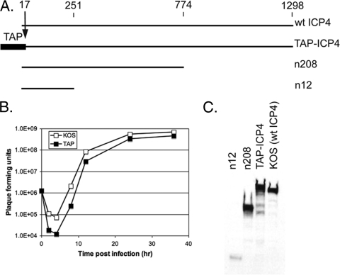
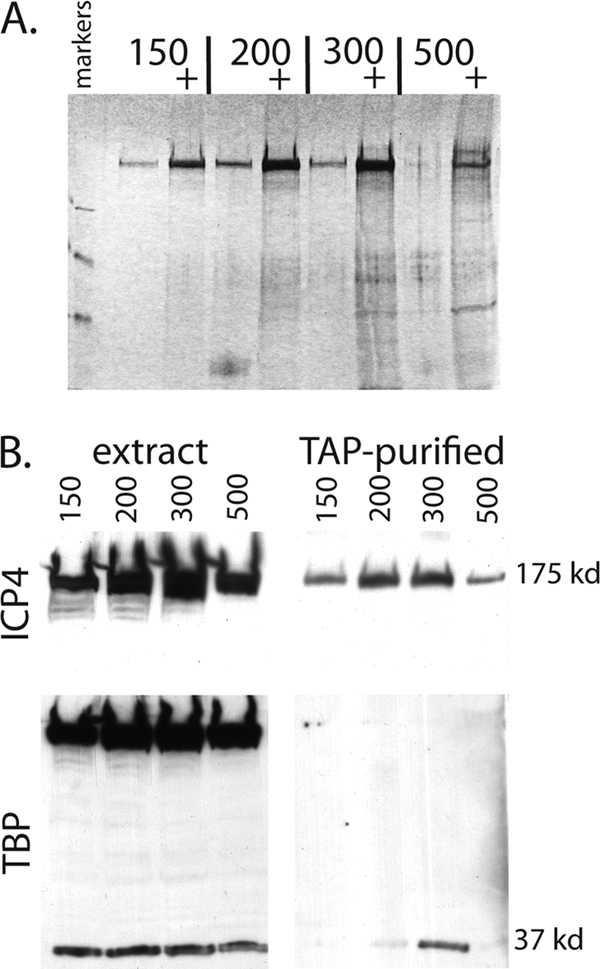
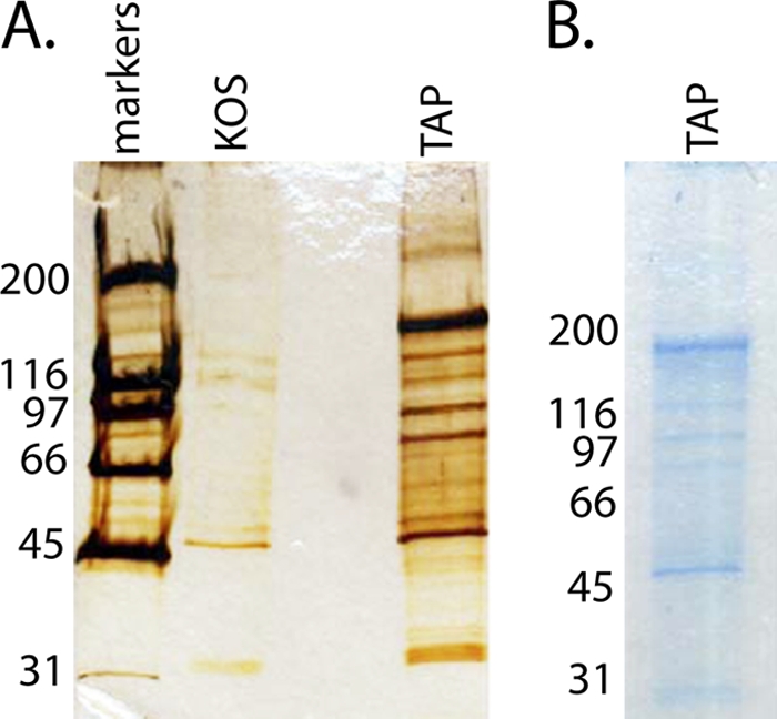
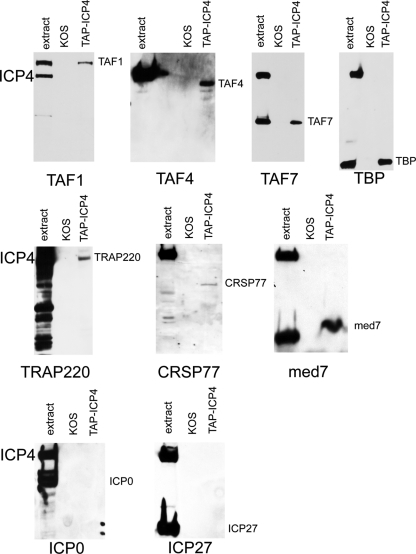
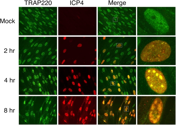
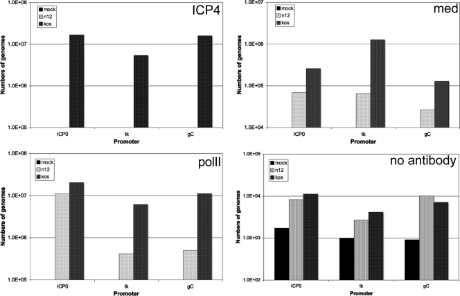
The infected cell polypeptide 4 (ICP4) of herpes simplex virus 1 (HSV-1) is a regulator of viral transcription that is required for productive infection. Since viral genes are transcribed by cellular RNA polymerase II (RNA pol II), ICP4 must interact with components of the pol II machinery to regulate viral gene expression. It has been shown previously that ICP4 interacts with TATA box-binding protein (TBP), TFIIB, and the TBP-associated factor 1 (TAF1) in vitro. In this study, ICP4-containing complexes were isolated from infected cells by tandem affinity purification (TAP). Forty-six proteins that copurified with ICP4 were identified by mass spectrometry. Additional copurifying proteins were identified by Western blot analysis. These included 11 components of TFIID and 4 components of the Mediator complex. The significance of the ICP4-Mediator interaction was further investigated using immunofluorescence and chromatin immunoprecipitation. Mediator was found to colocalize with ICP4 starting at early and continuing into late times of infection. In addition, Mediator was recruited to viral promoters in an ICP4-dependent manner. Taken together, the data suggest that ICP4 interacts with components of TFIID and Mediator in the context of viral infection, and this may explain the broad transactivation properties of ICP4.


Figures

Fig. 1.
Fig. 1.
Generation and characterization of TAP-ICP4. (A) Schematic comparing the primary sequence of the ICP4 peptides expressed by the viruses used in this study. Amino acid numbers are given at the top. (B) One-step growth curve of KOS and TAP-ICP4. Vero cells were infected at an MOI of 5 with either the TAP virus or KOS. Virus was harvested 2, 4, 8, 12, 24, and 36 hpi. Viral titers were determined via plaque assay. (C) Expression of the ICP4 peptides from cells infected with the indicated virus was assessed via Western blotting of whole-cell extracts from Vero cells infected with the indicated virus.
Fig. 2.
Fig. 2.
Effect of KCl concentration on protein extraction and complex isolation via TAP. (A) Silver-stained SDS-PAGE gel on TAP material from TAP-ICP4-infected HeLa cell nuclei extracted with the indicated concentrations of KCl. Unconcentrated and concentrated (+) samples were run for each KCl concentration. (B) Copurification of TBP with TAP-ICP4 as a function of KCl concentration used to extract infected cell nuclei. Nuclear extracts and the corresponding TAP samples from panel A were analyzed by Western blotting for ICP4 (top) and TBP (bottom).
Fig. 3.
Fig. 3.
TAP material from KOS- and TAP-ICP4-infected cells. TAP-purified samples were separated via SDS-PAGE, and total protein was visualized by silver stain (A) and colloidal blue stain (B) in preparation for MS analysis. Sizes for the proteins in the standard molecular mass ladder are given in kDa.
Fig. 4.
Fig. 4.
Western blot analysis of TAP material from KOS- and TAP-ICP4-infected cells. The extract from TAP-KOS-infected cells and the TAP material from KOS- and TAP-ICP4-infected cells was separated by SDS-PAGE and subjected to Western blot analysis. The antibody used to probe the blot is indicated at the bottom of each blot.
Fig. 5.
Fig. 5.
Localization of TRAP220 and ICP4 in HSV-infected cells. HEL cells that were either mock or KOS infected (MOI, 10 PFU/cell) for 2, 4, and 8 h were analyzed by immunofluorescence as described in Materials and Methods. Shown are the TRAP220 (green), ICP4 (red), merged, and merged single-cell fields (from white box).
Fig. 6.
Fig. 6.
ChIP analysis of the association of ICP4, Mediator (med), and pol II with HSV promoters in infected cells. The numbers of genomes precipitated with the antibodies to ICP4, Mediator (med) components TRAP220 and CRSP77, and pol II amplifying with primers specific for the ICP0, tk, and gC promoters in mock-, n12-, and KOS-infected cells is shown. A no-antibody control also is shown (lower right).

References

    1. Alwine J. C., Steinhart W. L., Hill C. W. 1974. Transcription of herpes simplex type 1 DNA in nuclei isolated from infected HEp-2 and KB cells. Virology 60:302–307 - PubMed
    1. Arnosti D. N., Merino A., Reinberg D., Schaffner W. 1993. Oct-2 facilitates functional preinitiation complex assembly and is continuously required at the promoter for multiple rounds of transcription. EMBO J. 12:157–166 - PMC - PubMed
    1. Bates P. A., DeLuca N. A. 1998. The polyserine tract of herpes simplex virus ICP4 is required for normal viral gene expression and growth in murine trigeminal ganglia. J. Virol. 72:7115–7124 - PMC - PubMed
    1. Benetti L., Munger J., Roizman B. 2003. The herpes simplex virus 1 US3 protein kinase blocks caspase-dependent double cleavage and activation of the proapoptotic protein BAD. J. Virol. 77:6567–6573 - PMC - PubMed
    1. Blazek E., Mittler G., Meisterernst M. 2005. The mediator of RNA polymerase II. Chromosoma 113:399–408 - PubMed

Publication types

MeSH terms

Substances

LinkOut - more resources