. 2011 Jun;85(11):5465-75.
doi: 10.1128/JVI.00313-11. Epub 2011 Mar 30.

Antibody-dependent cell-mediated viral inhibition emerges after simian immunodeficiency virus SIVmac251 infection of rhesus monkeys coincident with gp140-binding antibodies and is effective against neutralization-resistant viruses

Affiliations

Antibody-dependent cell-mediated viral inhibition emerges after simian immunodeficiency virus SIVmac251 infection of rhesus monkeys coincident with gp140-binding antibodies and is effective against neutralization-resistant viruses

Mohammed Asmal et al. J Virol. 2011 Jun.

Abstract

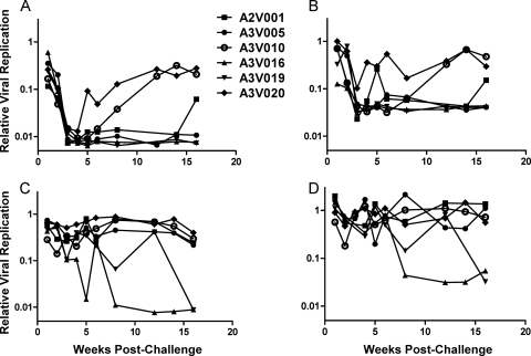
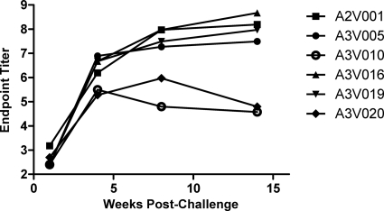
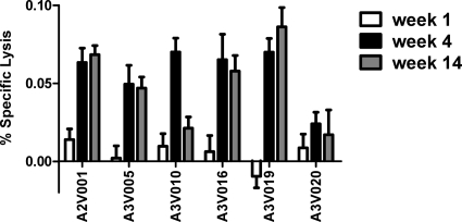
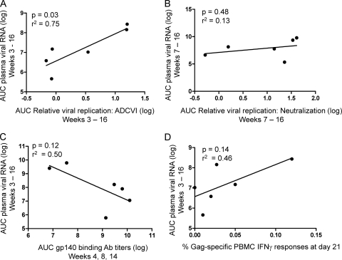
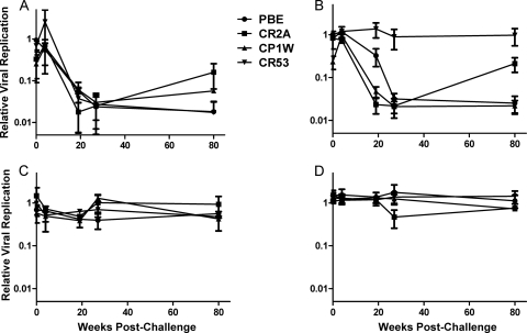
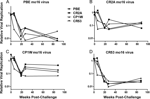
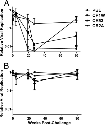
Antibody-dependent cell-mediated viral inhibition (ADCVI) is an attractive target for vaccination because it takes advantage of both the anamnestic properties of an adaptive immune response and the rapid early response characteristics of an innate immune response. Effective utilization of ADCVI in vaccine strategies will depend on an understanding of the natural history of ADCVI during acute and chronic human immunodeficiency virus type 1 (HIV-1) infection. We used the simian immunodeficiency virus (SIV)-infected rhesus monkey as a model to study the kinetics of ADCVI in early infection, the durability of ADCVI through the course of infection, and the effectiveness of ADCVI against viruses with envelope mutations that are known to confer escape from antibody neutralization. We demonstrate the development of ADCVI, capable of inhibiting viral replication 100-fold, within 3 weeks of infection, preceding the development of a comparable-titer neutralizing antibody response by weeks to months. The emergence of ADCVI was temporally associated with the emergence of gp140-binding antibodies, and in most animals, ADCVI persisted through the course of infection. Highly evolved viral envelopes from viruses isolated at late time points following infection that were resistant to plasma neutralization remained susceptible to ADCVI, suggesting that the epitope determinants of neutralization escape are not shared by antibodies that mediate ADCVI. These findings suggest that despite the ability of SIV to mutate and adapt to multiple immunologic pressures during the course of infection, SIV envelope may not escape the binding of autologous antibodies that mediate ADCVI.


Figures

Fig. 1.
Fig. 1.
Development of ADCVI and neutralizing antibodies following SIVmac251 infection. Heat-inactivated plasma obtained from between 1 and 16 weeks postinfection was diluted 1 to 250 and incubated with SIVmac251-infected human CD4+ T cells. Replicating virus in the culture supernatant was quantified after 5 days of incubation using a TZM-bl reporter assay. Results are shown for ADCVI and neutralization utilizing NK cells and CD4+ T cells isolated from two different human donors. (A) ADCVI using CD4+ T and NK cells from a single donor. Infected CD4+ T cells were incubated with plasma as well as a 2-to-1 ratio of human NK cells. Results of the TZM-bl assay were normalized to infected cells cultured with natural killer cells in the absence of plasma and plotted as relative viral replication (absolute RLU values for viral replication control, 20,000 to 50,000; RLU for no-virus control, 200 to 400). (B) ADCVI using CD4+ T and NK cells from a second donor. (C) Plasma neutralization using CD4+ T cells from the donor in panel A. Infected CD4+ T cells were incubated with plasma alone. Results of the TZM-bl assay were normalized to infected cells cultured in the absence of plasma and plotted as relative viral replication. (D) Plasma neutralization using CD4+ T cells from the donor in panel B.
Fig. 2.
Fig. 2.
Longitudinal gp140-binding antibody titers. EIA plates were coated with purified SIVmac251 gp140 and then incubated with serial dilutions of plasma from infected animals.
Fig. 3.
Fig. 3.
Specific lysis of SIVmac251-infected cells by plasma from infected animals. Human CD4+ T cells were infected with an MOI of 1.0 of SIVmac239, incubated for 3 days, and then cocultured in the presence of autologous NK cells at a ratio of 2 to 1 and plasma from the noted infected monkeys. After 4 h, cytotoxicity and total cell viability were measured utilizing the Cytotox-glo system (Promega). Infected cells in the presence of NK cells but without plasma were used to calculate percent specific lysis.
Fig. 4.
Fig. 4.
Correlation of plasma viral RNA with ADCVI, neutralizing antibody, and Gag-specific CD8+ T cell responses. Area under curve (AUC) was calculated for plasma viral RNA levels for each animal between either day 21 or day 49 and day 112 postinfection. These values were plotted against AUC calculations for relative viral replication for ADCVI (A) or neutralizing antibody responses (B) for the corresponding animals. Aggregate binding antibody (Ab) titers over the time points tested were plotted against AUC viral copy number during that time frame (C). Finally, we also plotted AUC plasma viral RNA levels against previously obtained data on the frequencies of CD8+ T cell Gag-specific IFN-γ responses following exposure to a pool of 15-mer Gag peptides (D).
Fig. 5.
Fig. 5.
ADCVI responses to SIVmac251 infection. Human CD4+ T cells were infected with an MOI of 0.01 of the same SIVmac251 stock used to challenge the animals in this study. These target cells were incubated with plasma from infected animals obtained at various times postchallenge in the presence of autologous NK cells. After 5 days of coincubation, virus was quantified by TZM-bl assay and normalized to a plasma-free control to derive a relative viral replication value. Relative viral replication is plotted as a function of time after challenge for the 4 animals in this study. Samples were assayed in duplicate in each of two different donors for effector and target cells, and the means and standard errors of the quadruplicate results are plotted. (A) ADCVI assay with 1:100 dilution plasma. (B) ADCVI assay with 1:250 dilution of plasma. (C) Neutralization assay with 1:100 dilution of plasma. (D) Neutralization assay with 1:250 dilution of plasma.
Fig. 6.
Fig. 6.
ADCVI responses to chronic viruses. SIV molecular clones expressing month 16 envelopes were used to infect human CD4+ T cells. Four molecular clones were used, constructed with envelopes representing the dominant circulating viruses isolated from each of 4 chronically infected animals. These viruses were assayed against plasma from all 4 animals at all time points postinfection, from week 0 to week 22. ADCVI results were corrected for plasma neutralizing activity. Each panel, A to D, represents the ADCVI sensitivity of the noted month 16 virus to plasma from all of the animals.
Fig. 7.
Fig. 7.
ADCVI to the heterologous virus SIVsmE660. Human CD4+ T cells were infected with SIVsmE660 and then incubated with plasma from SIVmac251-infected monkeys and human NK cells. (A) ADCVI at 1:250 dilution of plasma. (B) Neutralization at 1:250 dilution of plasma. Means and standard errors for ADCVI activity and neutralization from two different human donors are presented.

References

    1. Aasa-Chapman M. M., et al. 2004. Development of the antibody response in acute HIV-1 infection. AIDS 18:371–381 - PubMed
    1. Ahmad A., Menezes J. 1996. Antibody-dependent cellular cytotoxicity in HIV infections. FASEB J. 10:258–266 - PubMed
    1. Ahmad R., et al. 2001. Evidence for a correlation between antibody-dependent cellular cytotoxicity-mediating anti-HIV-1 antibodies and prognostic predictors of HIV infection. J. Clin. Immunol. 21:227–233 - PubMed
    1. AIDS Vaccine Evaluation Group 022 Protocol Team 2001. Cellular and humoral immune responses to a canarypox vaccine containing human immunodeficiency virus type 1 Env, Gag, and Pro in combination with rgp120. J. Infect. Dis. 183:563–570 - PubMed
    1. Alsmadi O., Tilley S. A. 1998. Antibody-dependent cellular cytotoxicity directed against cells expressing human immunodeficiency virus type 1 envelope of primary or laboratory-adapted strains by human and chimpanzee monoclonal antibodies of different epitope specificities. J. Virol. 72:286–293 - PMC - PubMed

Publication types

MeSH terms

LinkOut - more resources