. 2011 May 19;117(20):5350-61.
doi: 10.1182/blood-2011-01-333476. Epub 2011 Mar 30.

Kit-Shp2-Kit signaling acts to maintain a functional hematopoietic stem and progenitor cell pool

Affiliations

Kit-Shp2-Kit signaling acts to maintain a functional hematopoietic stem and progenitor cell pool

Helen He Zhu et al. Blood. .

Abstract

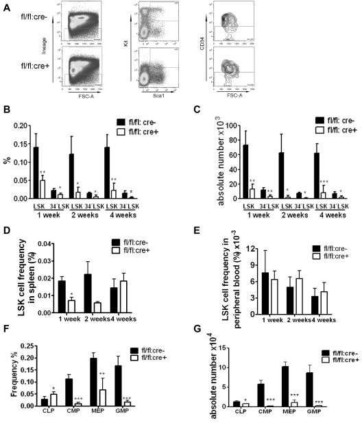
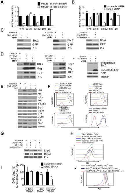
The stem cell factor (SCF)/Kit system has served as a classic model in deciphering molecular signaling events in the hematopoietic compartment, and Kit expression is a most critical marker for hematopoietic stem cells (HSCs) and progenitors. However, it remains to be elucidated how Kit expression is regulated in HSCs. Herein we report that a cytoplasmic tyrosine phosphatase Shp2, acting downstream of Kit and other RTKs, promotes Kit gene expression, constituting a Kit-Shp2-Kit signaling axis. Inducible ablation of PTPN11/Shp2 resulted in severe cytopenia in BM, spleen, and peripheral blood in mice. Shp2 removal suppressed the functional pool of HSCs/progenitors, and Shp2-deficient HSCs failed to reconstitute lethally irradiated recipients because of defects in homing, self-renewal, and survival. We show that Shp2 regulates coordinately multiple signals involving up-regulation of Kit expression via Gata2. Therefore, this study reveals a critical role of Shp2 in maintenance of a functional HSC/progenitor pool in adult mammals, at least in part through a kinase-phosphatase-kinase cascade.


Figures

Figure 1
Figure 1
Inducible deletion of Shp2 in vivo leads to decreased cellularity in peripheral blood, BM, and spleen. (A) Deletion of the floxed region was verified by PCR analysis of peripheral blood cells isolated from mice 2 weeks after final Poly-I:C injection. (B) Deletion of Shp2 in BM was verified by immunoblotting at 2 weeks after final Poly-I:C injection. Erk was used as loading control. (C) Complete blood cell counting. Peripheral blood was collected at time points after final Poly-I:C injection (n = 5-9). HCT indicates hematocrit; PLT, platelet; RBC, red blood cells; WBC, white blood cells. (D) Representative H&E staining of trabecular femur 2 weeks after final Poly-I:C injection indicates a severe hypocellular phenotype in fl/fl:cre+ BM. Top panels show images scanned by ScanScope Digital Slide Scanners with 100× magnification. Bottom panels scanned with 400× magnification. (E) BM cellularity decreased significantly in fl/fl:cre+ mice starting from 1 week after final Poly-I:C injection (n = 3-15). (F) Splenic cellularity in the mutant animal was significantly reduced. Time points were after final Poly-I:C injection (n = 3-4). ***P < .001, **P < .01,*P < .05, error bars are SD; see also supplemental Figure 1.
Figure 2
Figure 2
Loss of phenotypic HSPCs and myeloid progenitors in Shp2 knockout animals. (A) Representative FACS plots for linKit+Sca1+ CD34 (34LSK) staining 2 weeks after final Poly-I:C injection. (B) Quantification of CD34LSK cell frequency per 2 hindlimbs at indicated time points after final Poly-I:C injection (n = 3-4). (C) Quantification of CD34LSK cell number per 2 hindlimbs at indicated time points after final Poly-I:C injection (n = 3-4). (D) LSK cell frequency in the spleen at indicated time points after final Poly-I:C injection (n = 3-4). (E) LSK cell frequency in the peripheral blood was not changed in mutant animal. Time points were after final Poly-I:C injection (n = 3-4). (F) CMPs, GMPs, MEPs, and CLPs frequency in the BM at 2 weeks after final Poly-I:C injection. CMPs were gated as LinSca-1Kit+FcgRloCD34+. CLPs were gated as LinIL-7R Sca-1loKitlo. GMPs were gated as LinSca-1Kit+FcgRhiCD34+. Megakaryocyte-erythrocyte progenitors were gated as LinSca-1Kit+FcgRloCD34. (G) Absolute numbers of CMPs, GMPs, MEPs, and CLPs at 2 weeks after final Poly-I:C injection (n = 4). ***P < .001, **P < .01,*P < .05, data are presented as mean ± SD.
Figure 3
Figure 3
Shp2 ablation results in significant decrease in BM HSCs/progenitors. (A, B) In vitro CFU assays for whole BM cells or purified LSK cells. 50 000 BM (A) or 250 sorted LSK (B) cells were seeded on methylcellulose medium Methocult 3434 (StemCell Technologies Inc) with cytokines for 14 days before colony numeration. (n = 3-6). (C) Representative spleen colonies from CFU-S day 12 assay. Quantification of CFU-S12 suggests a decrease of HSCs and multipotent progenitors (n = 5-6). (D) Competitive reconstitution strategy 1: injection first/transplantation later. Donor mice were treated with poly-I:C before BM transplantation. (E) Representative FACS plots illustrate BM chimerism in 1:1 donor versus competitor reconstitution assay. (F) Percentage of CD45.2 donor derived cells using 1:1 donor to competitor ratio (n = 6-10). (G) Percentage of CD45.2 donor-derived cells within different lineages in 1:1 ratio competitive reconstitution (n = 4-5). (H) Shp2Δ/Δ BM contributes to less than 1% of LSK chimerism in 1:1 ratio competitive reconstitution (n = 4). (I) 500 sorted LSK cells were transplanted into lethally irradiated recipients in competition with 5 × 10⁵ CD45.1 cells in the injection first/transplantation later setting. Peripheral chimerism was analyzed at indicated time points. fl/fl:cre LSK cells barely gave rise to any peripheral blood cells (n = 4-5). ***P < .001, **P < .01,*P < .05, error bars are SD; see also supplemental Figure 2.
Figure 4
Figure 4
Homing defect in Shp2Δ/Δ BM cells. (A) Competitive reconstitution strategy 2: transplantation first/injection later. The recipient mice were treated with poly-I:C after BM transplantation. (B) Percentage of CD45.2 donor-derived cells using 1:1 donor to competitor ratio with poly-I:C injection after transplantation (n = 4-7). (C) Shp2Δ/Δ BM contributed significantly less LSK cells in the 1:1 ratio competitive reconstitution experiment (n = 3). (D) Percentage of CD45.2 donor-derived cells within different lineages in 1:1 ratio competitive reconstitution (n = 3). (E) Declined reconstitution efficacy of Shp2Δ/Δ BM cells in the secondary transplantation (n = 4-6). (F) Sorted LSK cells were used as donor cells in the transplantation first/injection later competitive reconstitution assay. Peripheral chimerism was analyzed at indicated time points (n = 4). (G) In vivo homing assays. Results were shown with percentage of homed BM cells 16 hours after transplantation (n = 4, 5). (H) Homing defects in Shp2Δ/Δ progenitors. 5 × 10⁶ wild-type or Shp2Δ/Δ BM cells were transplanted to lethally irradiated mice. Recipient BM was harvested 24 hours later and seeded in Methocult 3434 medium (StemCell Technologies Inc). Colonies were counted on day 8 (n = 7). We used lethally irradiated but nontransplanted recipients as negative control, and no colonies were detected from the BM. Significantly fewer colonies were formed from Shp2Δ/Δ marrow transplantation, suggesting that the homing defect resides in primitive progenitors. (I) Shp2 deletion severely reduces the chemotactic response of lin cells to SDF-1α. 106 lineage-depleted BM cells were plated in the upper chamber for migration toward 5 nm SDF-1α in the lower chamber (performed in triplicate). ***P < .001, **P < .01,*P < .05; error bars are SD.
Figure 5
Figure 5
Disturbed quiescence and increased apoptosis for LSK cells derived from Shp2Δ/Δ BM. (A) Representative FACS plots illustrate BrdU/7-aminoactinomycin D (7-AAD) staining in LSK cells. (B) Quantification of S-phase or G0/G1-phase cell percentage in LSK subpopulation indicating more actively proliferating LSK cells in Shp2Δ/Δ animals. Results were obtained from 3 independent experiments. (C) Quantification of G0-G1, S, and G2-M phase cell percentage in 34LSK subpopulation. (D) linckit+Sca1 and LSK cell but not lin population contained more Annexin V+ cells. (E, F) Elevated Annexin V+ 7-AAD and Annexin V+ 7-AAD+ percentage in Shp2Δ/Δ 34LSK cells (n = 4). ***P < .001, **P < .01,*P < .05, error bars are SD; see also supplemental Figure 3.
Figure 6
Figure 6
Kit down-regulation in SLAM marker-labeled HSCs. (A) Representative FACS plots for linSca1+CD48CD150+ staining 4 weeks post final injection indicates severe reduction of Kit expression in SLAM marker-enriched HSCs from fl/fl:cre+ BM. (B) Median fluorescence intensity (MFI) for Kit staining of linSca1+CD48CD150+ cells (n = 4). (C) FACS histogram plots illustrating Kit staining of linSca1+CD48CD150+ cells at different time points after first poly-I:C injection, indicating immediate Kit down-regulation after acute Shp2 deletion. (D) Immunoblotting of BM cells after first poly-I:C injection showed gradually increased deletion from day 7 to 16. Tubulin was used as loading control. ***P < .001, **P < .01,*P < .05, error bars are SD; see also supplemental Figure 4.
Figure 7
Figure 7
Shp2 modulates Kit expression through Gata2. (A, B) qRT-PCR of Kit and Kit regulatory transcription factors Scl, Sp1, Gata2, and Gata1 mRNA demonstrated consistent suppression of Kit and Gata2 transcription in primary Shp2Δ/Δ lineage cells and Shp2 knockdown EML cells. β-actin was used as a reference gene for normalization. (C) Shp2 knockdown repressed Kit promoter activity. Constructs with Kit regulatory elements fusion to GFP reporter were transfected into HEK293 cells. Shp2 knockdown by specific siRNA lead to GFP down-regulation in both p13kit and p70kit transfectants but not GFP vector transfectant. Erk was used as loading control. (D) Enforced expression of flag-tagged wild-type (WT) and dominant-active (DA) Shp2-enhanced, dominant-negative (DN) Shp2 suppressed Kit promoter activity, with no effect for truncated Shp2 containing the 2 SH2 domains. Erk or Tubulin was used as loading control. (E) Signaling modification in Shp2 knockdown EML cells elicited by SCF. Tubulin was used as loading control. (F) Treatment of EML cells with PI3K inhibitor LY294004, MEK inhibitor U0126, or a low dose (500 nm) of LY294004, U0126, and STAT3 inhibitor AG490 led to down-regulation of Kit expression on cell surface. (G) Shp2 knockdown in EML cells suppressed Gata2 protein expression. Tubulin was used as loading control. (H) Ectopic expression of Gata2 rescued Kit down-regulation in Shp2 knockdown EML cells. (I) Quantitative analysis of Gata2 binding to the −114 kb, +5 kb, and promoter region of Kit gene by the CHIP assay. Binding of Gata2 to Kit promoter but not −114 kb or +5 kb region was significantly reduced in Shp2 knockdown EML cells. *P < .05; error bars are SD. (J) Kit staining profile of wt linSca1+CD48CD150+ cells, Shp2Δ/Δ linSca1+CD48CD150+ cells, Shp2Δ/Δ/PtenΔ/Δ linSca1+CD48CD150+ cells. At least 3 animals were examined in each group. Representative FACS plot was shown; see also supplemental Figure 5.

Similar articles

Cited by

References

    1. Xie T, Li L. Stem cells and their niche: an inseparable relationship. Development. 2007;134(11):2001–2006. - PubMed
    1. Méndez-Ferrer S, Michurina TV, Ferraro F, et al. Mesenchymal and haematopoietic stem cells form a unique bone marrow niche. Nature. 2010;466(7308):829–834. - PMC - PubMed
    1. Chabot B, Stephenson DA, Chapman VM, Besmer P, Bernstein A. The proto-oncogene c-kit encoding a transmembrane tyrosine kinase receptor maps to the mouse W locus. Nature. 1988;335(6185):88–89. - PubMed
    1. Chui DH, Loyer BV, Russell ES. Steel (Sl) mutation in mice: identification of mutant embryos early in development. Dev Biol. 1976;49(1):300–303. - PubMed
    1. Russell ES. Hereditary anemias of the mouse: a review for geneticists. Adv Genet. 1979;20:357–459. - PubMed

Publication types

MeSH terms

Substances