

Review
doi: 10.1167/iovs.10-6997f.
Print 2011 Mar.
The international workshop on meibomian gland dysfunction: report of the diagnosis subcommittee
Affiliations
- PMID: 21450918
- PMCID: PMC3072162
- DOI: 10.1167/iovs.10-6997f
Item in Clipboard
Review
The international workshop on meibomian gland dysfunction: report of the diagnosis subcommittee
Invest Ophthalmol Vis Sci.
.
No abstract available
Figures
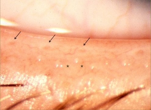
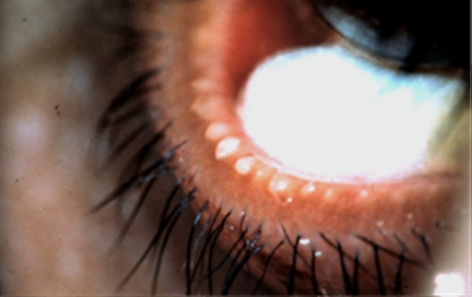
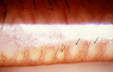
Normal lid margin, showing meibomian orifices (arrows) and clear, expressed oil (courtesy of A. Bron).
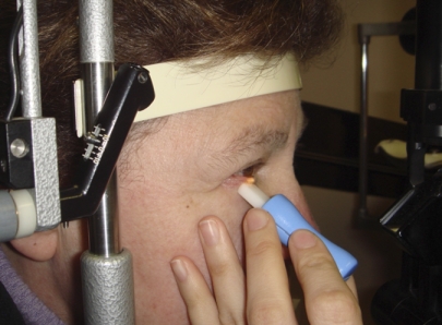
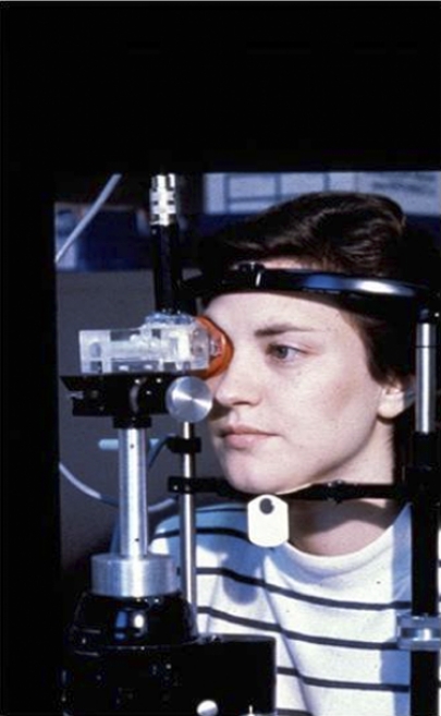
Standardized meibomian gland expression performed at the slit lamp using a diagnostic expression instrument (Korb and Blackie52). See text for further details (courtesy of D. Korb). Reprinted with permission from Korb DR, Blackie CA. Meibomian gland diagnostic expressibility: correlation with dry eye symptoms and gland location. Cornea. 2008;27(10):1142–1147.
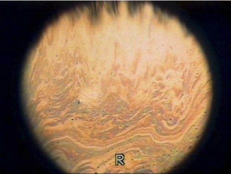
Spreading of a normal tear film lipid layer image by interferometry (courtesy of N. Yokoi).
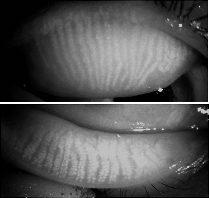
Normal meibomian glands of a 38-year-old woman, viewed by infrared meibography shows scattered gland absence or irregularity (courtesy of R. Arita).
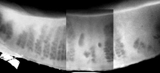
Photographic montage of the lower lid viewed by transillumination meibography. There is extensive meibomian gland dropout in a patient with meibomian gland dysfunction (courtesy of N. Yokoi).
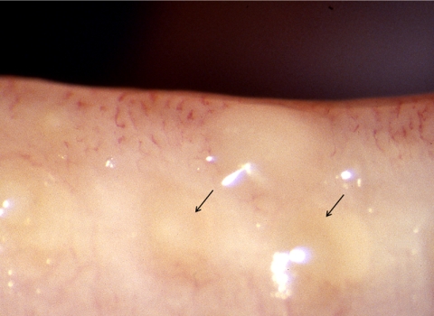
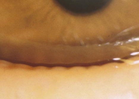
Meibomian gland dysfunction. Cloudy expressed meibum (arrows) (courtesy of A. Bron).
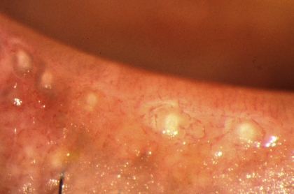
Meibomian gland dysfunction: expression of opaque meibum (courtesy of D. Korb).
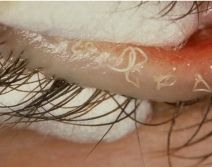
Meibomian gland dysfunction: strings of toothpaste-like opaque meibum expressed in response to forceful bimanual gland expression (courtesy of D. Korb).
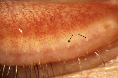
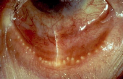
Cicatricial meibomian gland dysfunction. Lid margin hyperemia with orifice opacity with plugging (arrows); (courtesy of A. Bron).
Advanced non-cicatricial meibomian gland dysfunction: dense orifice opacification with periductal fibrosis (courtesy of A. Bron).
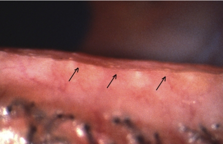
Cicatricial meibomian gland dysfunction: All meibomian orifices open onto the marginal conjunctiva, with some exposure of terminal ducts (arrows) (courtesy of A. Bron).
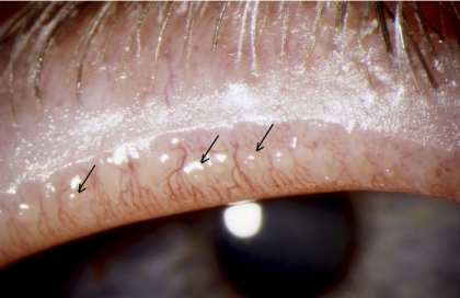
Cicatricial meibomian gland dysfunction: All meibomian orifices open onto the hyperemic marginal conjunctiva, with some exposure of terminal ducts (arrows) (courtesy of A. Bron).
Advanced cicatricial meibomian gland dysfunction: orifice retroplacement and opacity (courtesy of G. Foulks).
Dimpling or notching of the posterior lid margin due to tissue absorption in the region of the orifices (courtesy of J. Shimazaki).
Advanced meibomian gland dysfunction: epithelial ridging extending between opacified meibomian gland orifices (courtesy of A. Bron).
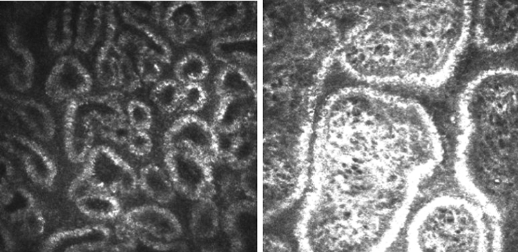
In vivo confocal microscopy of meibomian glands, showing the dilatation of acinar units in a patient with obstructive meibomian gland dysfunction (right) compared to that in a healthy control (left) (courtesy of M. Dogru).
Evaporimetry (courtesy of A. Tomlinson).
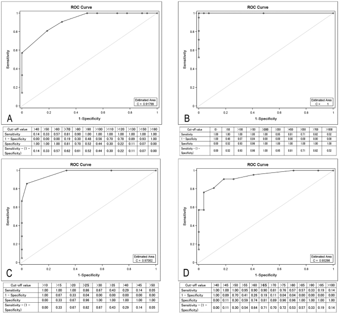
This figure was published in Ophthalmology, Vol 117, Ibrahim OM, Matsumoto Y, Dogru M et al., The efficacy, sensitivity, and specificity of in vivo laser confocal microscopy in the diagnosis of meibomian gland dysfunction. Page 670, ©Elsevier (2010). Reprinted with permission.
References
-
- Foulks G, Bron AJ. A clinical description of meibomian gland dysfunction. Ocul Surf. 2003;1:107–126 - PubMed
-
- Kozak I, Bron AJ, Kucharova K, et al. Morphologic and volumetric studies of the meibomian glands in elderly human eyelids. Cornea. 2007;26:610–614 - PubMed
-
- Arita R, Itoh K, Inoue K, Amano S. Noncontact infrared meibography to document age-related changes of the Meibomian glands in a normal population. Ophthalmology. 2008;115:911–915 - PubMed
-
- Nicolaides N, Kaitaranta JK, Rawdah TN, Macy JI, Boswell FM, 3rd, Smith RE. Meibomian gland studies: comparison of steer and human lipids. Invest Ophthalmol Vis Sci. 1981;20:522–536 - PubMed
-
- Chew CKS, Hykin PG, Jansweijer C, Dikstein S, Tiffany JM, Bron AJ. The casual level of meibomian lipids in humans. Curr Eye Res. 1993;12:255–259 - PubMed
Publication types
MeSH terms
LinkOut - more resources
Full Text Sources
Other Literature Sources
Medical
