Controlled Clinical Trial
. 2011 Apr 15;203(8):1165-73.
doi: 10.1093/infdis/jiq175.

A trimeric, V2-deleted HIV-1 envelope glycoprotein vaccine elicits potent neutralizing antibodies but limited breadth of neutralization in human volunteers

Affiliations
Controlled Clinical Trial

A trimeric, V2-deleted HIV-1 envelope glycoprotein vaccine elicits potent neutralizing antibodies but limited breadth of neutralization in human volunteers

Paul Spearman et al. J Infect Dis. .

Abstract

Background: A key missing element in the development of a successful human immunodeficiency virus (HIV) vaccine is an immunogen that can generate broadly cross-neutralizing antibodies against primary isolates of the virus.

Methods: This phase 1 clinical trial employed a DNA prime and subunit envelope protein boost in an attempt to generate cellular and humoral immune responses that might be desirable in a protective HIV vaccine. Priming was performed via intramuscular injection with gag and env DNA adsorbed to polylactide coglycolide microspheres, followed by boosting with a recombinant trimeric envelope (Env) glycoprotein delivered in MF59 adjuvant.

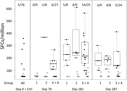
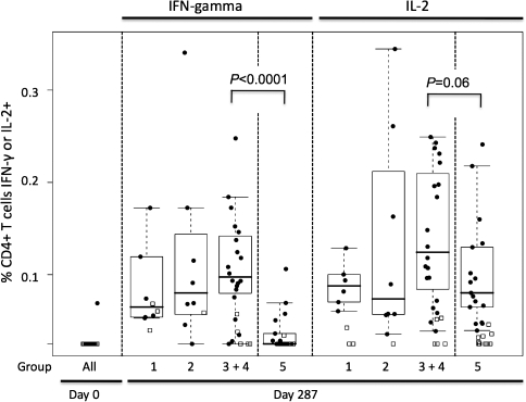
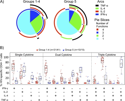
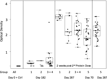
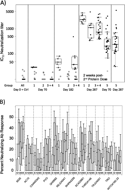
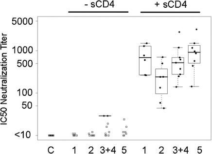
Results: The DNA prime and protein boost were generally safe and well-tolerated. Env-specific CD4(+) cellular responses were generated that were predominantly detected after Env protein boosting. Neutralizing antibody responses against the homologous SF162 viral isolate were remarkably strong and were present in the majority of vaccine recipients, including a strong response against CD4-induced epitopes on gp120. Despite the promising potency of this vaccine approach, neutralization breadth against heterologous tier 2 strains of HIV-1 was minimal.

Conclusions: Potent neutralization against neutralization-sensitive strains of HIV is achievable in humans through a DNA prime, recombinant oligomeric Env protein boost regimen. Eliciting substantial breadth of neutralization remains an elusive goal.

Clinical trials registration: NCT00073216.


Figures

Figure 1.
Figure 1.
Interferon-γ enzyme-linked immunosorbent spot assay Responses to Env Following DNA/ polylactide coglycolide (PLG) Priming and Oligomeric, V2-deleted Glycoprotein Boosting. Responses are expressed as the number of spot-forming units (SFUs) per 106 cells. Env responses are shown at 2 weeks following the completion of DNA/PLG priming (day 70) and 2 weeks after receipt of the first and second protein boosts (days 182 and 287, respectively). The numbers of positive responders in each group are indicated at the top of the figure.
Figure 2.
Figure 2.
CD4+ T cell Responses Measured by intracellular cytokine staining for interferon (IFN)–γ and interleukin (IL)–2. The percentage of the CD3+/CD4+ population expressing either cytokine following exposure to Env peptides is depicted. IFN-γ– and IL-2–specific responses are shown for each vaccination group. Note that for group 5, day 70 represents 2 weeks after receipt of the second protein dose, whereas day 182 for the DNA-primed groups represents 2 weeks after receipt of the first protein boost.
Figure 3.
Figure 3.
Analysis of Polyfunctional CD4+ T cell Responses. A, interferon (IFN)–γ, interleukin (IL)–2, tumor necrosis factor (TNF)–α, and IL-4 responses to Env peptides were assessed 2 weeks after receipt of the final protein boost by intracellular cytokine staining. Pie charts show the percentage of Env-specific cells responding with secretion of 1, 2, 3, or 4 cytokines averaged for individuals receiving DNA priming (groups 1–4; left) or for individuals receiving protein alone (group 5; right). Arcs show the percentage of cells producing each of the 4 cytokines. The color keys for the pie slices and arcs are shown. B, Specific cytokine secretion patterns are shown for those cells expressing 1 cytokine (left), 2 cytokines (middle), or 3 cytokines (right) following in vitro peptide stimulation. Red dots indicate responses from individuals primed with DNA (groups 1–4); blue dots indicate responses from individuals receiving protein alone (group 5).
Figure 4.
Figure 4.
Binding Antibody Responses Measured by enzyme-linked immunosorbent assay, Shown as Optical Density Units. The heavy bar indicates the median, with a box around the 25th and 75th percentiles, with whiskers on the plot representing the 5th and 95th percentiles. Responses to Env are shown for serum samples 2 weeks after receipt of the first and second protein boosts for groups 1–4 (days 182 and 287, respectively) and 2 weeks after receipt of the second and third protein doses for group 5 (days 70 and 287, respectively). For comparison of responses following the second protein boost in primed (groups 1–4) versus unprimed (group 5) individuals, we present these side by side, as indicated. Ctrl, results from volunteers receiving saline.
Figure 5.
Figure 5.
Neutralizing Antibody Titers Measured Using the TZM-bl Luciferase Reporter Assay. A, Neutralization of SF162, shown as the reciprocal dilution providing 50% reduction in reporter signal. Group assignments are indicated below the plot and the time points in the trial are indicated above the plot. For comparison of responses following receipt of the second protein boost in primed (groups 1–4) versus unprimed (group 5) individuals, we present these side by side, as indicated. B, Neutralization of a panel of clade B primary isolate pseudoviruses at a single dilution (1:10). Isolate designation is provided below the plot. The dashed line indicates 50% neutralization cutoff. Group designation indicated below each box plot. Ab, antibody; Ctrl, results from subjects receiving saline; IC50, half maximal inhibitory concentration.
Figure 6.
Figure 6.
Measurement of Neutralizing Antibodies Against CD4i Epitopes. Human immunodeficiency virus (HIV)–2 7312A/V434M is a molecularly-cloned HIV-2 isolate that is susceptible to neutralization by CD4i antibodies in the presence of subinhibitory concentrations of sCD4. In this assay, SF162 or HIV-2 were incubated in the absence (left) or presence (right) of 0.5 μg/mL sCD4, and neutralization of virus assayed using TZM-bl luciferase reporter cells.

Comment in

Similar articles

Cited by

References

    1. Flynn NM, Forthal DN, Harro CD, Judson FN, Mayer KH, Para MF. Placebo-controlled phase 3 trial of a recombinant glycoprotein 120 vaccine to prevent HIV-1 infection. J Infect Dis. 2005;191:654–65. - PubMed
    1. Pitisuttithum P, Gilbert P, Gurwith M, et al. Randomized, double-blind, placebo-controlled efficacy trial of a bivalent recombinant glycoprotein 120 HIV-1 vaccine among injection drug users in Bangkok, Thailand. J Infect Dis. 2006;194:1661–71. - PubMed
    1. Buchbinder SP, Mehrotra DV, Duerr A, et al. Efficacy assessment of a cell-mediated immunity HIV-1 vaccine (the Step Study): a double-blind, randomised, placebo-controlled, test-of-concept trial. Lancet. 2008;372:1881–93. - PMC - PubMed
    1. Rerks-Ngarm S, Pitisuttithum P, Nitayaphan S, et al. Vaccination with ALVAC and AIDSVAX to prevent HIV-1 infection in Thailand. N Engl J Med. 2009;361:2209–20. - PubMed
    1. Srivastava IK, Stamatatos L, Kan E, et al. Purification, characterization, and immunogenicity of a soluble trimeric envelope protein containing a partial deletion of the V2 loop derived from SF162, an R5-tropic human immunodeficiency virus type 1 isolate. J Virol. 2003;77:11244–59. - PMC - PubMed

Publication types

MeSH terms

Substances

Associated data