Comparative Study
. 2011 Mar 30;31(13):4844-51.
doi: 10.1523/JNEUROSCI.6181-10.2011.

Thyroid hormone controls cone opsin expression in the retina of adult rodents

Affiliations
Comparative Study

Thyroid hormone controls cone opsin expression in the retina of adult rodents

Anika Glaschke et al. J Neurosci. .

Abstract

Mammalian retinas display an astonishing diversity in the spatial arrangement of their spectral cone photoreceptors, probably in adaptation to different visual environments. Opsin expression patterns like the dorsoventral gradients of short-wave-sensitive (S) and middle- to long-wave-sensitive (M) cone opsin found in many species are established early in development and thought to be stable thereafter throughout life. In mouse early development, thyroid hormone (TH), through its receptor TRβ2, is an important regulator of cone spectral identity. However, the role of TH in the maintenance of the mature cone photoreceptor pattern is unclear. We here show that TH also controls adult cone opsin expression. Methimazole-induced suppression of serum TH in adult mice and rats yielded no changes in cone numbers but reversibly altered cone patterns by activating the expression of S-cone opsin and repressing the expression of M-cone opsin. Furthermore, treatment of athyroid Pax8(-/-) mice with TH restored a wild-type pattern of cone opsin expression that reverted back to the mutant S-opsin-dominated pattern after termination of treatment. No evidence for cone death or the generation of new cones from retinal progenitors was found in retinas that shifted opsin expression patterns. Together, this suggests that opsin expression in terminally differentiated mammalian cones remains subject to control by TH, a finding that is in contradiction to previous work and challenges the current view that opsin identity in mature mammalian cones is fixed by permanent gene silencing.


Figures

Figure 1.
Figure 1.
Experimental models of adult-onset hypothyroidism. w, Postnatal week.
Figure 2.
Figure 2.
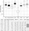
Serum iodothyronine levels of C57BL/6J mice used for cone opsin immunostaining. For description of the groups, see Figure 1. The top and bottom lines of the boxes correspond to the 75th percentile and 25th percentile, respectively. The solid lines represent median hormone levels. The dashed lines indicate the arithmetic mean. Minimum and maximum observations are indicated as filled circles. Numbers of samples are indicated above each whisker box. Data were compared using Kruskal–Wallis one-way ANOVA on ranks and Bonferroni's t test for post hoc pairwise multiple comparison. ***Significantly different from control, recovery, and control recovery groups with values of p < 0.001 at α = 0.05.
Figure 3.
Figure 3.
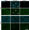
Cone photoreceptors in adult C57BL mice switch opsin on changes in serum TH concentrations. S- and M-opsin expression in matching fields of dorsal and ventral retina (75 and 50% eccentricity, respectively). Systemic hypothyroidism induced terminally differentiated M-cones to activate S-opsin in cones in dorsal retina and repress M-opsin (arrowheads). As a result, all M-cones also expressed S-opsin. Restoration of normal serum TH levels recovered the normal cone pattern. Scale bar, 50 μm.
Figure 4.
Figure 4.
Serum iodothyronine levels (free and total T3 and T4) of Brown Norway rats used for cone opsin immunostaining. Data are shown as mean ± SEM (n = 5) and compared with one-way ANOVA after passing the Kolmogorov–Smirnov test for normality. ***p < 0.001 at α = 0.05. n.d., Not detectable.
Figure 5.
Figure 5.
Cone photoreceptors in adult Brown Norway rats switch opsin on changes in serum TH concentration. S- and M-opsin expression in matching fields of dorsal and ventral retina. Adult rat cones expressed only one of the two cone opsins (Szél et al., 1994), S or M (examples indicated by circles). On induction of hypothyroidism, M-cones turned on S-opsin to coexpress both pigments in a single cell, but after recovery, cones expressed either M-opsin or S-opsin exclusively. Note that, in hypothyroid retinas, M-opsin was repressed, whereas S-opsin was activated. Scale bar, 50 μm.
Figure 6.
Figure 6.
Thyroid hormone changes mRNA expression in the adult mouse retina. RT-qPCR confirmed upregulation of iodothyronine deiodinase 2 (Dio2) in wild-type mice that were treated with MMI/perchlorate. M-opsin (Opn1mw) and deiodinase 3 (Dio3) were downregulated compared with controls. In MMI-treated mice, S-opsin (Opn1sw) transcript levels were not significantly different from controls in homogenates of entire retinas, although immunohistochemistry clearly shows upregulation in dorsal retina (Fig. 3). No differences between hypothyroid and control groups were found in the expression levels of Rho (rhodopsin, rod opsin), Thrb2 (thyroid hormone receptor β2), Nrl (neural retina leucine zipper), and Nr2e3 (nuclear receptor subfamily 2, group E, member 3). Relative gene expression was normalized to Hprt expression as determined by REST 2009, version 2.0.13. The boxes represent interquartile range, or middle 50% of observations. The dotted line represents median gene expression. The whiskers represent minimum and maximum observations. P(H1), Probability of alternate hypothesis that difference between sample and control groups is attributable only to chance; TRG, target; REF, reference.
Figure 7.
Figure 7.
Thyroid hormone reversibly changes S-cone opsin patterns in Pax8−/− mice. A, TH replacement to hypothyroid Pax8−/− mice by subcutaneous T4 injections restored the wild-type pattern of S-cone opsin expression when administered for 12 weeks. B, Pax8−/− mutant retinas exhibited the wild-type pattern of cones after 4 weeks of T4 supplementation but reverted to the mutant S-cone opsin pattern within 8 weeks after T4 supplementation was terminated. The illustrated regions are located dorsal (75% eccentricity) to the level of the optic disc. Scale bar, 30 μm. w, Postnatal week.
Figure 8.
Figure 8.
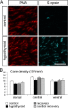
Induction of hypothyroidism activates S-opsin but does not increase total cone number. A, Flat-mounted mouse retina labeled with S-opsin antibody (blue) and peanut agglutinin (PNA) (red) to label the total population of cones. Region illustrated is from the dorsal retina. Scale bar, 30 μm. B, There was no significant difference in the number of PNA-positive cones between control, hypothyroid, recovery, and control recovery groups. Images were collected with a Zeiss Axio Imager.Z1 microscope at 40×, and total cones were quantified from 100 × 100 μm dorsal, central, and ventral regions in retinas of each experimental group. Data are expressed as mean ± SEM (number of animals n = 6, 7, 5, 5 for control, hypothyroid, recovery, control recovery group, respectively). Regions were compared with one-way ANOVA after validation of normality of data (Kolmogorov–Smirnov) and homogeneity of variances (Bartlett box F test).
Figure 9.
Figure 9.
Apoptosis and cell proliferation in the adult mouse retina. Activated caspase-3 and Ki67 immunoreactivity were detected in the retina of newborn mice serving as a positive control but absent in all retinal layers of adult wild-type and MMI-treated C57BL/6 mice. OS, Outer segment layer; ONL, outer nuclear layer; INL, inner nuclear layer; GCL, ganglion cell layer; OPL, outer plexiform layer; NBL, neuroblast layer.

References

    1. Ahnelt PK, Kolb H. The mammalian photoreceptor mosaic-adaptive design. Prog Retin Eye Res. 2000;19:711–777. - PubMed
    1. Applebury ML, Antoch MP, Baxter LC, Chun LL, Falk JD, Farhangfar F, Kage K, Krzystolik MG, Lyass LA, Robbins JT. The murine cone photoreceptor: a single cone type expresses both S and M opsins with retinal spatial patterning. Neuron. 2000;27:513–523. - PubMed
    1. Applebury ML, Farhangfar F, Glösmann M, Hashimoto K, Kage K, Robbins JT, Shibusawa N, Wondisford FE, Zhang H. Transient expression of thyroid hormone nuclear receptor TRbeta2 sets S opsin patterning during cone photoreceptor genesis. Dev Dyn. 2007;236:1203–1212. - PubMed
    1. Bianco AC, Larsen PR. Cellular and structural biology of the deiodinases. Thyroid. 2005;15:777–786. - PubMed
    1. Cheng CL, Flamarique IN. Chromatic organization of cone photoreceptors in the retina of rainbow trout: single cones irreversibly switch from UV (SWS1) to blue (SWS2) light sensitive opsin during natural development. J Exp Biol. 2007;210:4123–4135. - PubMed

Publication types

MeSH terms

LinkOut - more resources