Comparative Study
. 2011 Mar 30;31(13):4944-54.
doi: 10.1523/JNEUROSCI.0154-11.2011.

The Wnt5/planar cell polarity pathway regulates axonal development of the Drosophila mushroom body neuron

Affiliations
Comparative Study

The Wnt5/planar cell polarity pathway regulates axonal development of the Drosophila mushroom body neuron

Kazumichi Shimizu et al. J Neurosci. .

Abstract

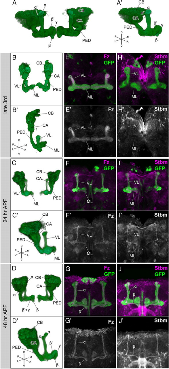
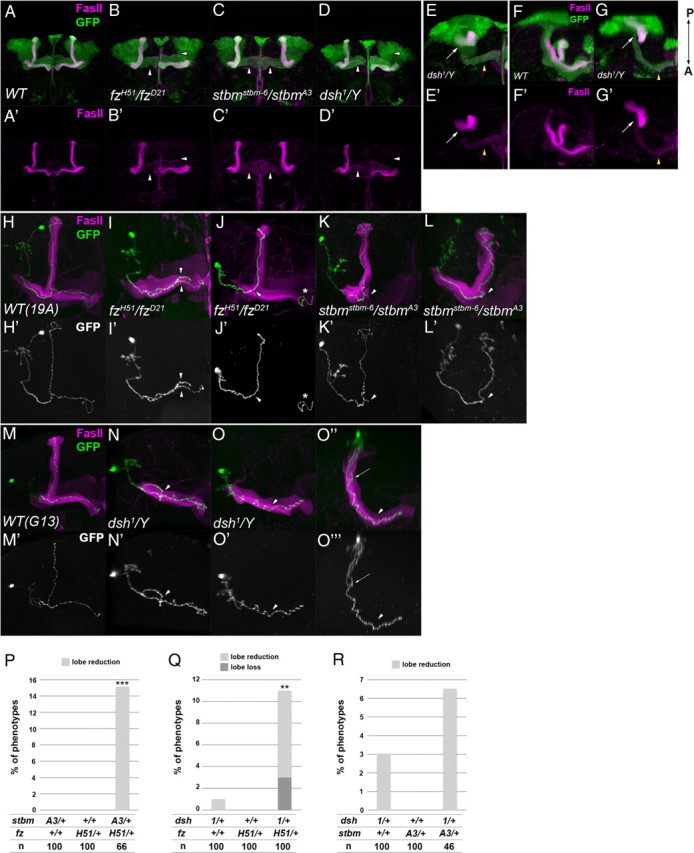
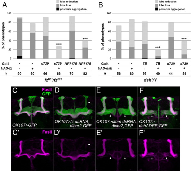
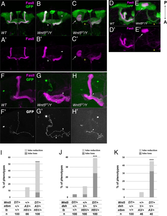
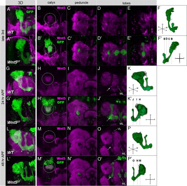
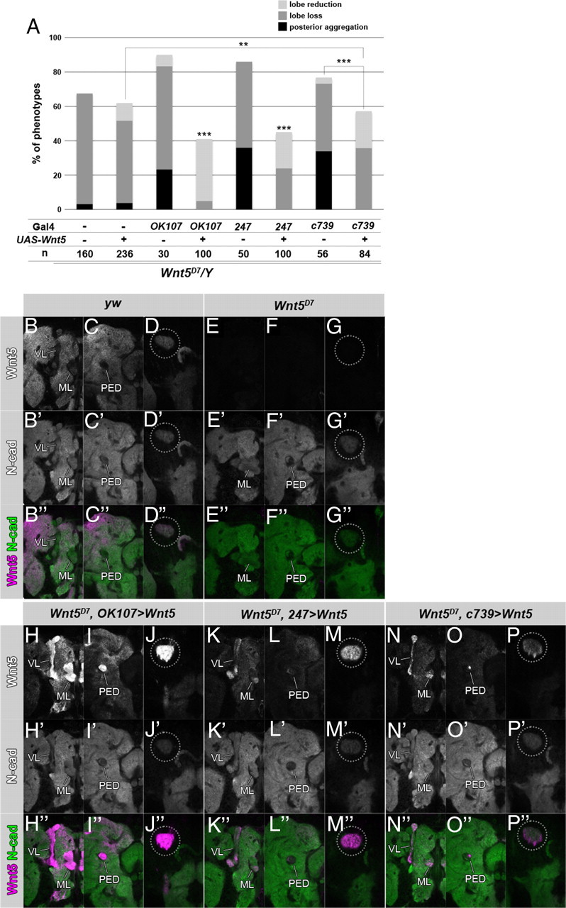
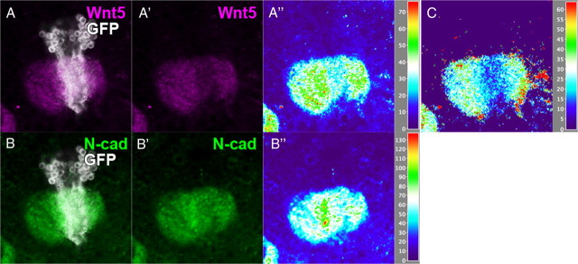
Axonal development is a fundamental process for circuit formation in the nervous system and is dependent on many cellular events, including axon initiation, elongation, guidance, and branching. The molecular mechanisms underlying these events have been well studied, especially for axon guidance. In the presence of a guidance cue, the polarization of a growth cone precedes the turning response, which is one example of the diverse forms of cell polarity. Planar cell polarity (PCP) is another example of cell polarity. Although some PCP genes are required for axonal tract formation in vertebrates, it remains elusive whether these genes participate in a common PCP pathway concertedly. Here, we show that essential PCP signaling components, encoded by frizzled (fz), strabismus (stbm), flamingo (fmi), and dishevelled (dsh), are cooperatively required for axonal targeting and branching of the Drosophila mushroom body (MB) neurons. A detailed analysis of these mutants revealed that these components were required for the correct targeting and bifurcation of axons. In addition, we suggest that Wnt5 functions as a ligand in the PCP pathway in this process. Wnt5 mutants showed similar phenotypes to PCP mutants at the single-cell level and genetically interacted with PCP genes. Wnt5 was broadly expressed in the developing brain. We propose that Wnt5 and the PCP pathway concertedly regulate axonal development of the MB.


Figures

Figure 1.
Figure 1.
The expression patterns of the PCP components during the development of the MB. A–D, Structure of the MB in the adult (A, A′), at the late third larval instar (B, B′), 24 h APF (C, C′), and 48 h APF (D, D′). Anterior perspectives of whole MBs (A–D) and oblique perspectives of right MBs (A′–D′) are indicated. E–J′, Expression patterns of Fz (E–G, magenta; E′–G′, white) and Stbm (H–J, magenta; H′–J′, white) at the third larval instar (E, E′, H, H′), 24 h APF (F, F′, I, I′), and 48 h APF (G, G′, J, J′). All of the PCP components examined were expressed in the core of the MB during the entire period of MB development. The structures of the MBs were visualized by the expression of GFP (green) driven by OK107-Gal4. The abbreviations are as follows: CB, Cell bodies; CX, calyx; PED, peduncle; VL, vertical lobe; ML, medial lobe. Because the lobes at the late third larval instar and 24 h APF do not have clearly discrete lobes that are composed of distinct subtypes of KCs, the lobes are simply indicated as the vertical lobes (VL) and the medial lobes (ML) in B–C′, E–F′, and H–I′.
Figure 2.
Figure 2.
The phenotypes of the PCP gene mutants. A–G′, The structure of the MB in wild type (A, A′, F, F′), fzH51/fzD21 (B, B′), stbmstbm-6/stbmA3 (C, C′), and dsh1/Y (D, D′, E, E′, G, G′) mutants. The vertical or medial lobes were lost or reduced in the PCP mutants (B–D′, arrowheads). Anterior (A–E′) and dorsal views with the posterior side up and the anterior side down (F–G′) are indicated. In dsh1 mutants, a loss or reduction of the lobes (D, D′, arrowheads) or an aggregation of axons beside the calyx (E, E′, G, G′, arrow) was observed. In the case of axon aggregation, only a small portion of the γ neurons projected toward the anterior side (E, E′, G, G′, yellow arrowhead). The MB was visualized by the expression of GFP driven by OK107-Gal4 (green) and the γ and α/β lobes were visualized by anti-FasII antibody (magenta). H–O‴, The axonal projections of α/β KCs marked by GFP (H–O, O″, green; H′–O′, O‴, white) in wild type (FRT 19A control clone in H, H′; FRT G13 control clone in M, M′), fzH51/fzD21 mutants (I–J′), stbmstbm-6/stbmA3 mutants (K–L′), and dsh1 mutants (N–O‴). The axon bifurcated into both the vertical and the medial lobes in the wild type (H, H′, M, M′). The projection of sister branches in the same direction (I, I′, arrowheads), the stalling of the branched axon (K, K′, arrowhead), and the reversed projection of the branched axon into the peduncle (N, N′, arrowhead) are shown as examples of guidance defects. The absence of branches (J, J′, L, L′, O–O‴, arrowhead) is shown as an example of branching defects. In O–O‴, an ectopic branch at the peduncle projected in the posterior direction (arrow). O″ and O‴ are the dorsal views of O and O′, respectively. The asterisks in J and J′ indicate an axon of a clone induced in the contralateral MB. P–R, Genetic interaction between the PCP genes. Double heterozygotes of the stbmA3 and fzH51 (P), dsh1 and fzH51 (Q), or dsh1 and stbmA3 (R) showed a higher percentage of defects in the MB than single heterozygotes of fzH51, stbmA3, and dsh1 (***p < 0.001, **p < 0.01, Fisher's exact test).
Figure 3.
Figure 3.
The PCP genes function in the developing MB. A, B, Quantification of MB defects in fzH51/fzD21 (A) and dsh1 mutants (B) with the indicated transgenes. Specific expression of exogenous fz or dsh in the MB by c739-Gal4 or NP7175-Gal4 and c739-Gal4 or 7B-Gal4 significantly rescued the defects of fzH51/fzD21 and dsh1 mutants, respectively (***p < 0.001, Fisher's exact test). Each MB was assessed by the criteria described in Materials and Methods. C–F′, The phenotypes of the MB in which specific RNAi against fz (D, D′) and stbm (E, E′) or specific overexpression of a dominant-negative form of Dsh (dshΔDEP) (F, F′) was used. The cells expressing fz dsRNA, stbm dsRNA, or dshΔDEP were marked by the expression of GFP (green) under the control of OK107-Gal4, and the γ and α/β lobes were visualized by anti-FasII (magenta). fz RNAi, stbm RNAi, and the expression of dshΔDEP resulted in the loss or reduction of the lobes (arrowheads) in 28.3% (n = 60), 42.3% (n = 52), and 22.5% (n = 40) of cases, respectively.
Figure 4.
Figure 4.
The mutant phenotype of Wnt5D7 and the genetic interaction between Wnt5D7 and mutants of the PCP genes. A–E′, The structure of the MB of wild type (A, A′, D, D′) and Wnt5D7 mutants (B, B′, C, C′, E, E′). The MB was visualized by the expression of GFP driven by OK107-Gal4 (green), and the γ and α/β lobes were visualized by an anti-FasII antibody (magenta). Anterior (A–C′) and dorsal views with the posterior side up and the anterior side down (D–E′) are indicated. In Wnt5D7 mutants, a loss or reduction of the lobes (B, B′, arrowheads) or an aggregation of axons beside the calyx (C, C′, E, E′, arrows) was observed. In the case of axon aggregation, only a small portion of the γ neurons projected toward the anterior side (C, C′, E, E′, yellow arrowheads). F–H′, The axonal projection of α/β KCs marked by GFP (F–H, green; F′–H′, white) in wild type (F, F′) and Wnt5D7 mutants (G–H′). The axon bifurcated into both the vertical and the medial lobes in the wild type (F, F′). Projection of sister branches in the same direction (G, G′, arrowheads) and the absence of the branch (H, H′, arrowhead) are shown as examples of guidance defects and branching defects, respectively. I–K, Genetic interaction between Wnt5 and the PCP genes. The percentage of the observed phenotypes increased in the triple heterozygotes of Wnt5D7, stbmA3, and fzH51 (I), Wnt5D7, dsh1, and fzH51 (J), and Wnt5D7, dsh1, and stbmA3 (K), but single heterozygotes of Wnt5D7 or double heterozygotes of any two of fzH51, stbmA3, and dsh1 showed little or no defect in the MB (***p < 0.001, Fisher's exact test).
Figure 5.
Figure 5.
The expression pattern of Wnt5 in the brain. The Wnt5 protein (magenta) was broadly expressed in the brain at the late third larval instar (A, B–E′), 24 h APF (G, H–J′), and 48 h APF (L, M–O′). The signal of Wnt5 was abolished in the brains of Wnt5 mutants at the late third larval instar (A′), 24 h APF (G′), and 48 h APF (L′). Three-dimensional reconstructed images (A, A′, G, G′, L, L′) and sections at the level of the calyx (B, B′, H, H′, M, M′), the peduncle (C, C′, I, I′, N, N′), and the lobes (D–E′, J, J′, O, O′) are indicated. The structure of the MB was visualized by the expression of GFP (green) driven by OK107-Gal4. The level of each section is schematically illustrated in lateral views of the MB at each stage (F′, K′, P′). Wnt5 was localized in the calyx (B, B′, H, H′, M, M′, dotted circles) at all stages. At 24 h APF, Wnt5 was also detected at the branch point of the lobes (J, J′, arrow) and at the tip of the medial lobe (J, J′, ML). At 48 h APF, Wnt5 was accumulated at the branch point of the lobes (O, O′, arrow) and at the tips of both the vertical and the medial lobes (O, O′, VL and ML).
Figure 6.
Figure 6.
MB specific rescue of the Wnt5 mutants. A, Specific restoration of Wnt5 expression in the MB using the indicated Gal4 lines rescued the Wnt5 mutant phenotypes (***p < 0.001, **p < 0.01, Fisher's exact test). B–P″, Wnt5 was restored in the calyx by the expression of Wnt5 in the MBs of the Wnt5D7 mutants. Wnt5 is indicated in white (B–P) or magenta (B″–P″), and N-cadherin is indicated in white (B′–P′) or green (B″–P″). OK107-Gal4 (H–J″), 247-Gal4 (K–M″), or c739-Gal4 (N–P″) was used. The expression patterns of Wnt5 in yw (B–D″) and Wnt5D7 (E–G″) are shown as positive and negative staining controls, respectively. Sections of the hemispheres of 48 h pupal brains at the level of the lobes (B–B″, E–E″, H–H″, K–K″, N–N″), the peduncle (C–C″, F–F″, I–I″, L–L″, O–O″), and the calyx (D–D″, G–G″, J–J″, M–M″, P–P″) are indicated. Wnt5 was restored in the calyx to a level higher than that of the wild type (compare the encircled areas in D, D″, and J, J″, M, M″, P, P″). Wnt5 was detected not only in the whole MB (H, H″, K, K″, N, N″, VL, ML; I, I″, L, L″, O, O″, PED) but also in the surrounding neuropile regions (H, H″, I, I″, K, K″, L, L″, N, N″, O, O″).
Figure 7.
Figure 7.
Wnt5 is expressed in the KCs. Expression of Wnt5 (A, A′, magenta) is slightly reduced in a clone of the Wnt5 mutant (A, B, white) induced in the MB, but the signal intensity of N-cadherin (B, B′, green) is not changed within the clone. The signal intensities of Wnt5 (A″) and N-cadherin (B″) were visualized by the height of the peaks in 2.5-dimensional images. C, The signal intensity of Wnt5 was calibrated using the signal intensity of N-cadherin to eliminate the influence from potential differences in dendritic densities. Wnt5 was clearly decreased in the Wnt5 mutant clone.

Similar articles

Cited by

References

    1. Axelrod JD. Unipolar membrane association of Dishevelled mediates Frizzled planar cell polarity signaling. Genes Dev. 2001;15:1182–1187. - PMC - PubMed
    1. Axelrod JD. Progress and challenges in understanding planar cell polarity signaling. Semin Cell Dev Biol. 2009;20:964–971. - PubMed
    1. Axelrod JD, Miller JR, Shulman JM, Moon RT, Perrimon N. Differential recruitment of Dishevelled provides signaling specificity in the planar cell polarity and Wingless signaling pathways. Genes Dev. 1998;12:2610–2622. - PMC - PubMed
    1. Bastock R, Strutt D. The planar polarity pathway promotes coordinated cell migration during Drosophila oogenesis. Development. 2007;134:3055–3064. - PMC - PubMed
    1. Bastock R, Strutt H, Strutt D. Strabismus is asymmetrically localised and binds to Prickle and Dishevelled during Drosophila planar polarity patterning. Development. 2003;130:3007–3014. - PubMed

Publication types

LinkOut - more resources