Comparative Study
. 2011 Mar 30;31(13):4991-9.
doi: 10.1523/JNEUROSCI.5216-10.2011.

The role of the medial prefrontal cortex in innate fear regulation in infants, juveniles, and adolescents

Affiliations
Comparative Study

The role of the medial prefrontal cortex in innate fear regulation in infants, juveniles, and adolescents

Thomas Chan et al. J Neurosci. .

Abstract

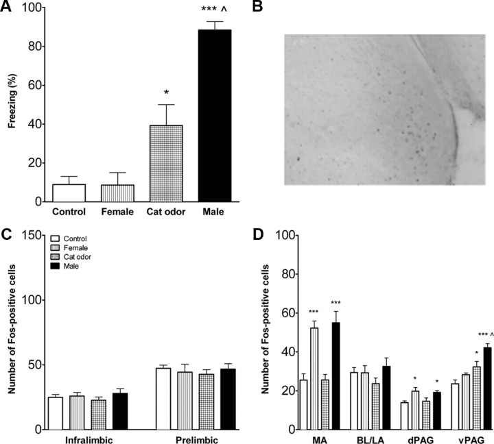
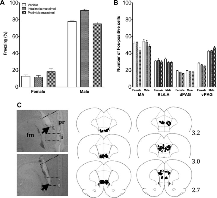
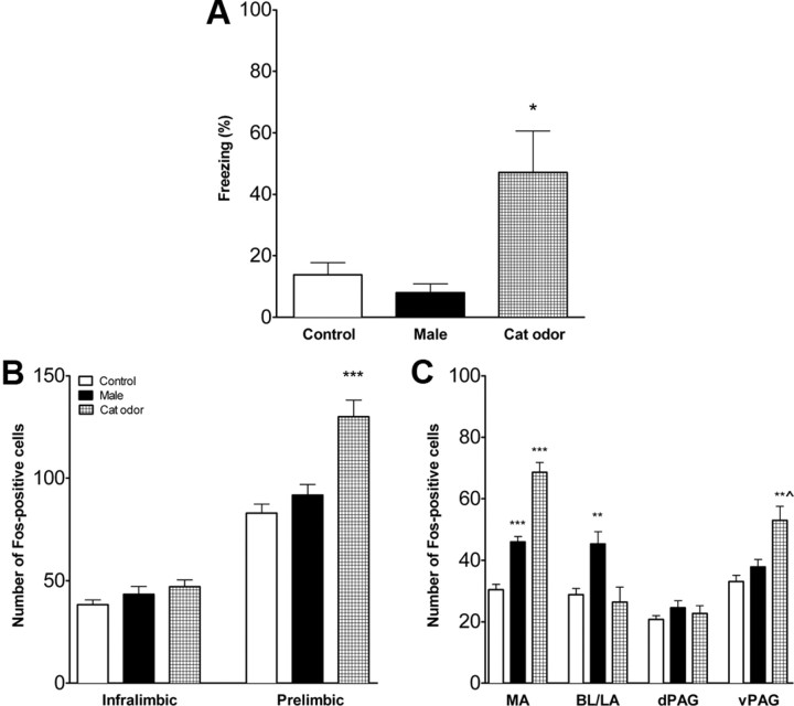
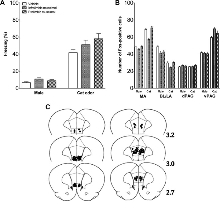
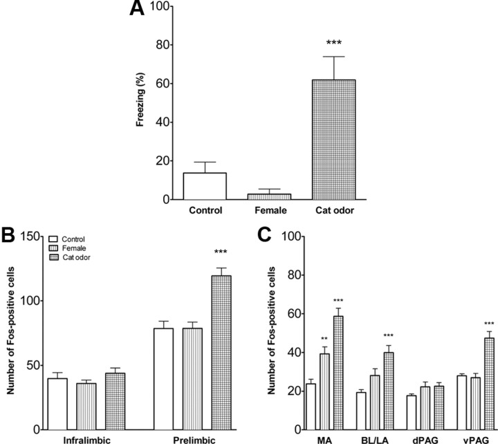
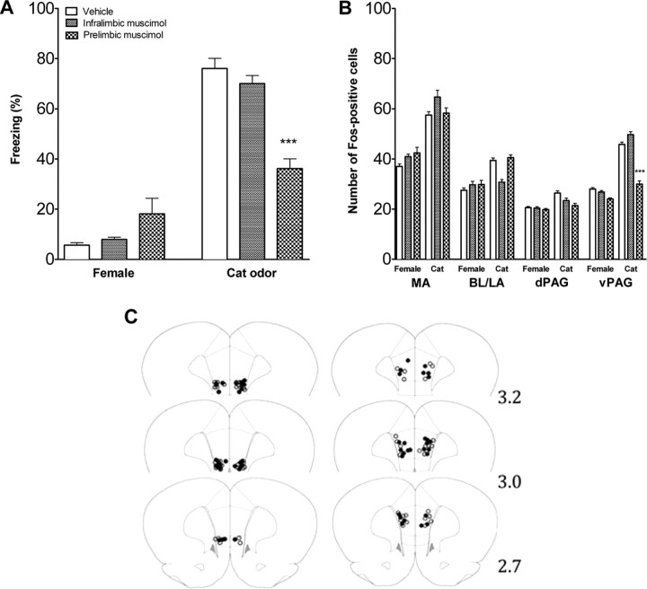
In adult animals, the medial prefrontal cortex (mPFC) plays a significant role in regulating emotions and projects to the amygdala and periaqueductal gray (PAG) to modulate emotional responses. However, little is known about the development of this neural circuit and its relevance to unlearned fear in pre-adulthood. To address these issues, we examined the mPFC of 14-d-old (infants), 26-d-old (juveniles), and 38- to 42-d-old (adolescents) rats to represent different developmental and social milestones. The expression patterns of the neuronal marker FOS were used to assess neurological activity. Muscimol, a GABA agonist, was used to inactivate the prelimbic and infralimbic mPFC subdivisions (400 ng in 200 nl). Animals were exposed to either a threatening or nonthreatening stimulus that was ecologically relevant and age specific. Freezing was measured as an indicator of innate fear behavior. The data indicated that the mPFC is neither active nor responsive to innate fear in infant rats. In juveniles, the prelimbic mPFC became responsive in processing aversive sensory stimulation but did not regulate freezing behavior. Finally, during adolescence, inactivation of the prelimbic mPFC significantly attenuated freezing and decreased FOS expression in the ventral PAG. Surprisingly, across all ages, there were no significant differences in FOS levels in the medial and basolateral/lateral amygdala when either mPFC subdivision was inactivated. Together, unlearned fear has a unique developmental course with different brain areas involved in unlearned fear in the immature animal than the adult. In particular, the mPFC neural circuitry is different in young animals and progressively develops more capacities as the animal matures.


Figures

Figure 1.
Figure 1.
Fear response and FOS expression in infant rats with intact mPFC. A, Percentage time freezing (mean ± SEM) during exposure to an adult female rat, cat fur odor, or an adult male rat. Controls were exposed to an empty cage. Male-exposed rats froze significantly more than cat-odor-exposed, female-exposed, or control rats (p < 0.001 for all). Pups exposed to the cat odor froze more than pups exposed to either the control or female (p < 0.05 for each). B, Representative micrograph to show FOS staining in the medial amygdala of a male exposed 14-d-old pup. C, FOS expression in the mPFC subdivisions in infant rats with intact mPFC. There were no differences in the number of FOS-positive cells (mean ± SEM) in infralimbic and prelimbic mPFC. D, Activation of the amygdala and PAG in infant rats with intact mPFC. Exposure to the nonthreatening stimulus (adult female) or the threatening stimulus (adult male), but not the cat odor, increased the number of FOS-positive cells in the medial amygdala and dPAG. Exposure to the adult male and to a lesser extent the cat odor increased FOS in the ventrolateral PAG. MA, Medial amygdala; BL/LA, basolateral/lateral amygdala. n = 6–9 for all conditions. **p < 0.01, ***p < 0.001 compared with controls; ∧p < 0.05 compared with control or female exposed. Note the y-axis scale differs for Figures 1, 3, and 5 for the prefrontal cortex FOS counts.
Figure 2.
Figure 2.
Fear response and brain activation in infant rats infused with muscimol or vehicle in mPFC. A, Percentage time freezing in infant rats infused with muscimol or vehicle in mPFC and exposed to a nonthreatening female or threatening male rat. Mean ± SEM. B, Number of FOS-positive cells in the amygdala and PAG of infant rats injected with vehicle or muscimol into infralimbic and prelimbic mPFC and exposed to a nonthreatening female or threatening male rat. Mean ± SE. n = 6–9 in each condition. C, Cannula placement in mPFC of infant rats. Photographs of cresyl-violet-stained sections with placement in prelimbic (pr) and infralimbic (i) mPFC. fm, Forceps minor corpus callosum; arrow, cannula tip. Drawing of placement in infralimbic (middle) and prelimbic (right) mPFC. Open circles represent the location of cannula tips in rats infused with vehicle; filled circles represent the location of cannula tips in rats infused with muscimol. Numbers indicate the distance in millimeters from bregma.
Figure 3.
Figure 3.
Fear response and FOS expression in juvenile rats with intact mPFC. A, Percentage time freezing (mean ± SE) during exposure to an adult male rat or cat fur odor. Controls were exposed to an empty cage. Cat-odor-exposed rats froze significantly more than male-exposed or control rats. B, Activation of the mPFC in juvenile rats with intact mPFC. Number of FOS-positive cells (mean ± SE) was significantly increased in prelimbic mPFC of rats exposed to the threatening stimulus (cat odor). C, Activation of the amygdala and PAG in juvenile rats with intact mPFC. Exposure to the threatening stimulus (cat odor) increased the number of FOS-positive cells (mean ± SE) in the medial amygdala and ventrolateral PAG. Exposure to the nonthreatening stimulus (adult male) increased FOS in the medial and basolateral/lateral amygdala. MA, Medial amygdala; BL/LA, basolateral/lateral amygdala. *p < 0.05, **p < 0.01, ***p < 0.001 compared with control; ∧p < 0.01 vs male exposure. n = 6–7 in each condition.
Figure 4.
Figure 4.
Fear response and brain activation in juvenile rats infused with muscimol or vehicle in mPFC. A, Percentage time freezing in juvenile rats infused with vehicle or muscimol in mPFC and exposed to a nonthreatening male or threatening cat odor. Mean ± SE. B, Number of FOS-positive cells in the amygdala and PAG of juvenile rats injected with vehicle or muscimol into infralimbic and prelimbic mPFC and exposed to a nonthreatening male or threatening cat odor. Mean ± SE. n = 6–7 in each condition. C, Cannula placement in infralimbic (left) and prelimbic (right) mPFC of juvenile rats. Open circles represent the location of cannula tips in rats infused with vehicle, and filled circles represent the location of cannula tips in rats infused with muscimol. Numbers indicate the distance in millimeters from bregma.
Figure 5.
Figure 5.
Fear response and brain activation in adolescent rats with intact mPFC. A, Percentage time freezing in adolescent rats exposed to an adult female rat or cat fur odor. Controls were exposed to empty cage. Cat-odor-exposed rats froze significantly more than female-exposed or control rats. n = 6–7 in each condition. B, Activation of the mPFC in adolescent rats with intact mPFC. Number of FOS-positive cells was significantly increased in prelimbic mPFC of rats exposed to the threatening stimulus (cat odor). n = 6 in each condition. C, Activation of the amygdala and PAG in adolescent rats with intact mPFC. Exposure to the threatening stimulus (cat odor) increased the number of FOS-positive cells in the medial and basolateral/lateral amygdala and vPAG, and exposure to the nonthreatening stimulus (adult female) increased FOS in the medial amygdala. MA, Medial amygdala; BL/LA, basolateral/lateral amygdala. **p < 0.01, ***p < 0.001. n = 7 in each condition.
Figure 6.
Figure 6.
Fear response and brain activation in adolescent rats infused with muscimol or vehicle in mPFC. A, Percentage time freezing in adolescent rats infused with vehicle or muscimol in the infralimbic and prelimbic mPFC and exposed to a nonthreatening female or threatening cat odor (mean ± SEM). There were no significant differences for rats infused in the infralimbic mPFC, but when infused into the prelimbic mPFC, freezing to the cat was significantly reduced. B, Number of FOS-positive cells in the amygdala and PAG of adolescent rats injected with vehicle or muscimol into infralimbic or prelimbic mPFC and exposed to a nonthreatening female or threatening cat odor. Mean ± SEM. FOS expression was selectively reduced in the vPAG only by prelimbic infusion. n = 6–8 in each condition. C, Cannula placement in infralimbic (left) and prelimbic (right) mPFC of adolescent rats. Open circles represent the location of cannula tips in rats infused with vehicle, and filled circles represent the location of cannula tips in rats infused with muscimol. Numbers indicate the distance in millimeters from bregma. ***p < 0.001.

Similar articles

Cited by

References

    1. An X, Bandler R, Ongür D, Price JL. Prefrontal cortical projections to longitudinal columns in the midbrain periaqueductal gray in macaque monkeys. J Comp Neurol. 1998;401:455–479. - PubMed
    1. Apfelbach R, Blanchard CD, Blanchard RJ, Hayes RA, McGregor IS. The effects of predator odors in mammalian prey species: a review of field and laboratory studies. Neurosci Biobehav Rev. 2005;29:1123–1144. - PubMed
    1. Beach FA, Jaynes J. Studies of maternal retrieving in rats. I. Recognition of young. J Mammal. 1956;37:177–180.
    1. Benes FM, Taylor JB, Cunningham MC. Convergence and plasticity of monoaminergic systems in the medial prefrontal cortex during the postnatal period: Implications for the development of psychopathology. Cereb Cortex. 2000;10:1014–1027. - PubMed
    1. Boice R. Burrows of wild and albino rats: effects of domestication, outdoor raising, age, experience, and maternal state. J Comp Physiol Psychol. 1977;91:649–661. - PubMed

Publication types

LinkOut - more resources