. 2011 Jun;35(6):904-12.
doi: 10.1097/PAS.0b013e318212ae22.

The anterior gradient homolog 3 (AGR3) gene is associated with differentiation and survival in ovarian cancer

Affiliations

The anterior gradient homolog 3 (AGR3) gene is associated with differentiation and survival in ovarian cancer

Erin R King et al. Am J Surg Pathol. 2011 Jun.

Abstract

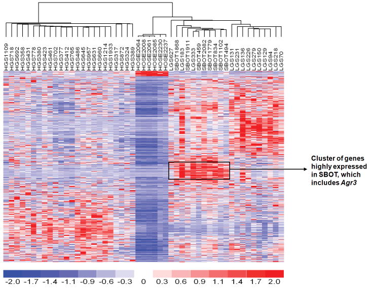
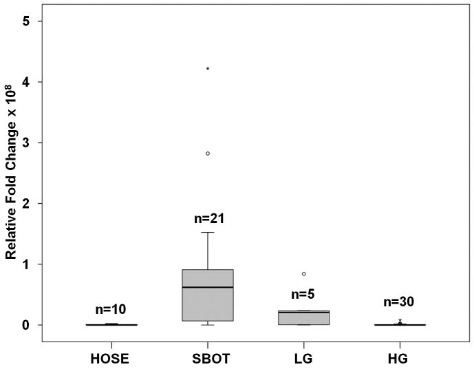
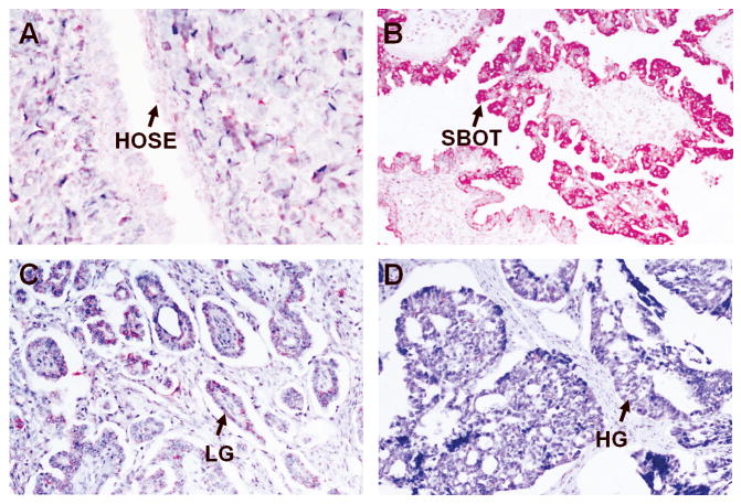
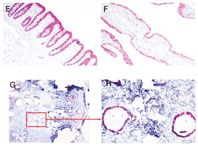
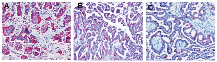
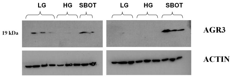
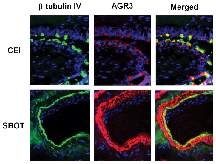
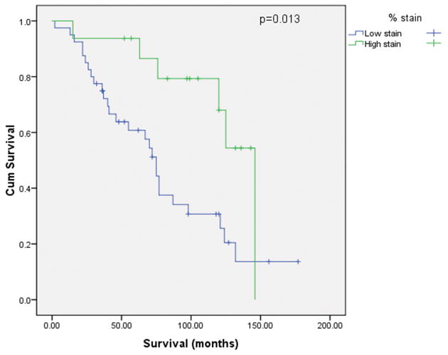
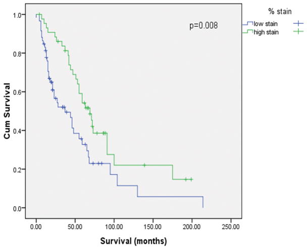
Low-grade (LG) serous ovarian carcinoma is believed to arise from serous borderline ovarian tumors; yet the progression from serous borderline tumors to LG serous ovarian carcinoma remains poorly understood. The purpose of this study was to identify differentially expressed genes between the 2 groups. Expression profiles were generated from 6 human ovarian surface epithelia, 8 serous borderline ovarian tumors (SBOTs), 13 LG serous ovarian carcinomas, and 24 high-grade (HG) serous ovarian carcinomas. The anterior gradient homolog 3 (AGR3) gene was found to be highly upregulated in serous borderline ovarian tumors. This finding was validated by real-time quantitative reverse-transcription polymerase chain reaction, Western blotting, and immunohistochemistry. Anti-AGR3 immunohistochemistry was performed on an additional 56 LG and 103 HG tissues, and the results were correlated with clinical data. Expression profiling determined that 1254 genes were differentially expressed (P<0.005) among SBOT, LG, and HG tumors. SBOTs exhibited robust positive staining for AGR3, with a lower percentage of tumor cells stained in LG and HG. Immunofluorescence staining indicated that AGR3 expression was limited to ciliated cells. Tumor samples with a high percentage (>10%) of AGR3 positively stained tumor cells were associated with improved longer median survival in both the LG (P=0.013) and HG (P=0.008) serous ovarian carcinoma groups. The progression of SBOT to LG serous ovarian carcinoma may involve the dedifferentiation of ciliated cells. AGR3 could serve as a prognostic marker for survival in patients with LG and HG serous ovarian carcinomas.


Figures

Figure 1
Figure 1
Differentially expressed genes between 6 HOSE, 8 SBOT, 13 LG, and 24 HG tumors were identified by t-test with 50 permutations to estimate false discovery rate (FDR) using dChip2009. A list of genes with median 2.2% FDR was identified. Unsupervised analysis using this set of genes was able to separate the SBOT/LG from HG.
Figure 2
Figure 2
Real-time RT-PCR validating overexpression of AGR3 in SBOT compared to LG, HG, and HOSE tumors. AGR3 was significantly up-regulated in SBOT and LG (p <0.001) but not in HG samples.
Figure 3
Figure 3
Representative stains of various tissue sections for AGR3 expression. All figures were obtained at 100× unless otherwise noted. A. Normal ovarian surface epithelium (400×); B. Serous borderline ovarian tumor; C. Low grade OSC; D. High grade OSC; E. Intestinal epithelium; F. Fallopian tube; G/H. Ovarian endosalpingiosis.
Figure 3
Figure 3
Representative stains of various tissue sections for AGR3 expression. All figures were obtained at 100× unless otherwise noted. A. Normal ovarian surface epithelium (400×); B. Serous borderline ovarian tumor; C. Low grade OSC; D. High grade OSC; E. Intestinal epithelium; F. Fallopian tube; G/H. Ovarian endosalpingiosis.
Figure 4
Figure 4
Examples of low-grade serous ovarian carcinomas with decreasing percentage of AGR3 positively stained tumor cells (from A to C). All images at 200×.
Figure 5
Figure 5
Western blot of AGR3 protein expression in low-grade serous ovarian carcinoma, high-grade serous ovarian carcinoma, and serous borderline ovarian tumor.
Figure 6
Figure 6
Immunofluorescent stain of β-tubulin IV and AGR3 in cliliated epithelial inclusion cyst versus serous borderline ovarian tumor.
Figure 7
Figure 7
Kaplan-Meier survival curve according to high- and low-AGR3 staining in serous ovarian carcinoma paraffin sections. A. Low-grade; B. High-grade.
Figure 7
Figure 7
Kaplan-Meier survival curve according to high- and low-AGR3 staining in serous ovarian carcinoma paraffin sections. A. Low-grade; B. High-grade.

References

    1. Adam PJ, Boyd R, Tyson KL, et al. Comprehensive proteomic analysis of breast cancer cell membranes reveals unique proteins with potential roles in clinical cancer. J Biol Chem. 2003;278:6482–6489. - PubMed
    1. Bonome T, Lee JY, Park DC, et al. Expression profiling of serous low malignant potential, low-grade, and high-grade tumors of the ovary. Cancer Res. 2005;65:10602–10612. - PubMed
    1. Castleman VH, Romio L, Chodhari R, et al. Mutations in radial spoke head protein genes RSPH9 and RSPH4A cause primary ciliary dyskinesia with central-microtubular-pair abnormalities. Am J Hum Genet. 2009;84:197–209. - PMC - PubMed
    1. Fletcher GC, Patel S, Tyson K, et al. hAG-2 and hAG-3, human homologues of genes involved in differentiation, are associated with oestrogen receptor-positive breast tumours and interact with metastasis gene C4.4a and dystroglycan. Br J Cancer. 2003;88:579–585. - PMC - PubMed
    1. Fliegauf M, Olbrich H, Horvath J, et al. Mislocalization of DNAH5 and DNAH9 in respiratory cells from patients with primary ciliary dyskinesia. Am J Respir Crit Care Med. 2005;171:1343–1349. - PMC - PubMed

Publication types

MeSH terms