. 2011 Jun 1;519(8):1546-61.
doi: 10.1002/cne.22584.

A specific antibody to neuropeptide AF1 (KNEFIRFamide) recognizes a small subset of neurons in Ascaris suum: differences from Caenorhabditis elegans

Affiliations

A specific antibody to neuropeptide AF1 (KNEFIRFamide) recognizes a small subset of neurons in Ascaris suum: differences from Caenorhabditis elegans

Paisarn Sithigorngul et al. J Comp Neurol. .

Abstract

A monoclonal antibody, AF1-003, highly specific to the Ascaris suum neuropeptide AF1 (KNEFIRFamide), was generated. This antibody binds strongly to AF1 and extremely weakly to other peptides with C-terminal FIRFamide: AF5 (SGKPTFIRFamide), AF6 (FIRFamide), and AF7 (AGPRFIRFamide). It does not recognize 35 other AF (A. suum FMRFamide-like) peptides at the highest concentration tested, nor does it recognize FMRFamide. When crude peptide extracts of A. suum are fractionated by two-step HPLC, the only fractions recognized by AF1-003 are those comigrating with synthetic AF1. By immunocytochemistry, antibody AF1-003 recognizes a small subset of the 298 neurons of A. suum: these include the paired URX and RIP neurons, two pairs of lateral ganglion neurons in the head, and the unpaired PQR and PDA or -B tail neurons that send processes to the head along the dorsal and ventral nerve cords, respectively. AF1 immunoreactivity is also seen in three pairs of pharyngeal neurons. Mass spectroscopy (MS) shows the presence of AF1 in the head, pharynx, and dorsal and ventral nerve cords. In A. suum, the neurons that contain AF1 show little overlap with neurons that express green fluorescent protein constructs targeting the flp-8 gene, which encodes AF1 in Caenorhabditis elegans (Kim and Li [2004] J. Comp. Neurol. 475:540-550); the URX neurons express AF1 in both species, but, in C. elegans, flp-8 expression was not detected in RIP, PQR, and PDA or -B or in the pharynx. Other, less specific monoclonal antibodies recognize AF1, as well as other peptides to differing degrees; these antibodies are useful reagents for determination of neuronal morphology.


Figures

Figure 1
Figure 1
A: Diagrams of head and tail ganglia. At left is shown an intact 30-cm female worm in a locomotory posture; the extent of the pharynx is shown by the shaded area near the tip of the head; insets show the neuronal cell bodies of the head (top; modified from Gold-schmidt, 1908) and tail ganglia (bottom) after the worm has been cut longitudinally and flattened after muscle, pharynx, intestine, and gonad have been removed; the lips have been cut away. The dotted line in the tail shows the rectum. B: Diagrams of cell bodies and neurites of paired URX neurons. C: Paired RIP neurons. The diagrams are radial projections and show the major neurites of the featured neurons (varicosities and fine branches are not indicated). The lighter background image is of the head structures as in A. NR, nerve ring; VC, ventral nerve cord; DC, dorsal nerve cord; VG, ventral ganglion; DG, dorsal ganglion; RVG, retrovesicular ganglion; LL, lateral line; LLL, left lateral line; RLL, right lateral line; AC, amphidial commissures; DeC, deirid commissures. Adjacent to the NR, the ganglia between the DC and the lateral lines are the subdorsal ganglia (both anterior and posterior to the NR), and those between the VC and the lateral lines are the subventral ganglia (anterior to the NR).
Figure 2
Figure 2
Dot-ELISA of antibody AF1-003 against synthetic endogenous A. suum peptides. Aliquots of 5 and 1 pmole of each peptide were spotted with BSA on nitrocellulose paper and cross-linked with glutaraldehyde. After exposure to antibody AF1-003, bound antibody was detected by indirect immunoperoxidase staining with a second antibody. Only AF1 shows detectable immunoreactivity.
Figure 3
Figure 3
Chromatogram of head extract (A; N = 100) and pharynx extract (B; N = 100) separated by gradient reversed-phase HPLC on a C18 column and assayed for immunoreactivity by dot-ELISA. The top trace records the OD at 214 nm. The graphs below represent the immunoreactivity of each fraction after dot-ELISA with monoclonal antibodies AF1-003, AF1-243, and AF1-62. Dot intensity is estimated by comparison with dot-ELISA of a standard twofold dilution series of AF1 and is recorded on a linear scale. The elution times of synthetic AF1, AF5, AF6, and AF7, determined in separate experiments, are shown.
Figure 4
Figure 4
Whole-mount preparations of A. suum treated with monoclonal antibody AF1-003. A: Head. B: Dorsal nerve cord. C: Tail. D: Pharynx. Anterior is to the top. DC, dorsal nerve cord; LC, lateral nerve cord in the lateral line; NR, nerve ring; VC, ventral nerve cord; VG, ventral ganglion; R, rectum. In A–C, numbers indicate individual neurons: 1 = URX; 2 = RIP; 3, 4 = lateral ganglion neurons; 5 = PDA or -B process; 6 = PQR process. In the preparation shown in A, the PDA or -B process in the ventral nerve cord ends in a prominent anterior varicosity. In D, three pairs of cells (labeled 2, 3, and 5) are stained. Scale bar = 100 µm.
Figure 5
Figure 5
Whole-mount preparation of A. suum treated with monoclonal antibody AF1-243. A: Head. B: Higher magnification of right lateral ganglion in a different preparation. C: Tail. D: Pharynx. Abbreviations and cell identities as in Figure 4. 8 = ADL; 12 = RMEV; 13 = RMED; 15 = AVK; 16 = RIS; 17 = RIR; 19 = PHA. In A, the other numbers are given to neurons for which the identification is not definitive. In C, the commissure from PQR (6), which supplies the dorsal nerve cord process extending to the head, is shown. In the pharynx, four pairs of neurons (labeled 2–5) are stained. Scale bar = 100 µm for A,C,D; 50 µm for B.
Figure 6
Figure 6
MALDI-TOF mass spectrum of dissected pharynx. A peak with m/z (mass to charge ratio) of 952.5, the calculated m/z of protonated AF1, is present.
Figure 7
Figure 7
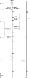
Diagram of the position of neurons (solid circles) in left and right lateral lines. The two vertical lines represent the ventral nerve cord, and the horizontal lines represent the position of dorsoventral commissures as they exit the ventral cord. The positions of some of the morphologically equivalent neurons in the left and the right lateral lines are different. Roman numerals indicate the five repeating units found in the motor nervous system. As indicated by the brackets, there is preparation-to-preparation variation in the location of some of these cell bodies, especially SDQ and the right AVM neuron. The circles at the anterior and posterior ends represent the head and tail ganglia. Vu, vulva.
Figure 8
Figure 8
Whole-mount preparations of A. suum treated with AF1-62 antibody. A: Head. B: Pharynx (focused on subventral nerves). C: Tail. D: Pharynx (focused on pharyngeal dorsal nerve). Abbreviations as in Figure 4. SDG, subdorsal ganglion; SVG, subventral ganglion. In the pharynx, there are five pairs of stained neurons (2–6 in C) and four unpaired neurons (7 and 8 in C, 9 and 10 in D) Scale bar = 100 µm.

Similar articles

Cited by

References

    1. Berman ES, Fortson SL, Checchi KD, Wu L, Felton JS, Wu KJ, Kulp KS. Preparation of single cells for imaging/profiling mass spectrometry. J Am Soc Mass Spectrom. 2008;19:1230–1236. - PubMed
    1. Brownlee DJA, Fairweather I. Exploring the neurotransmitter labyrinth in nematodes. Trends Neurosci. 1999;22:16–24. - PubMed
    1. Brownlee DJA, Walker RJ. Actions of nematode FMRFa-miderelated peptides on the pharyngeal muscle of the parasitic nematode, Ascaris suum . Ann NY Acad Sci. 1999;897:228–238. - PubMed
    1. Brownlee DJA, Fairweather I, Johnston CF, Smart D, Shaw C, Halton DW. Immunocytochemical demonstration of neuropeptides in the central nervous system of the round-worm, Ascaris suum (Nematoda, Ascaroidea) Parasitology. 1993;106:305–316. - PubMed
    1. Cowden C, Stretton AOW. AF2, an Ascaris neuropeptide: isolation, sequence, and bioactivity. Peptides. 1993;14:423–430. - PubMed

Publication types