Review
. 2011 Jun 1;519(8):1413-34.
doi: 10.1002/cne.22580.

The Edinger-Westphal nucleus: a historical, structural, and functional perspective on a dichotomous terminology

Affiliations
Review

The Edinger-Westphal nucleus: a historical, structural, and functional perspective on a dichotomous terminology

Tamás Kozicz et al. J Comp Neurol. .

Abstract

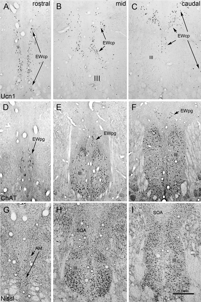
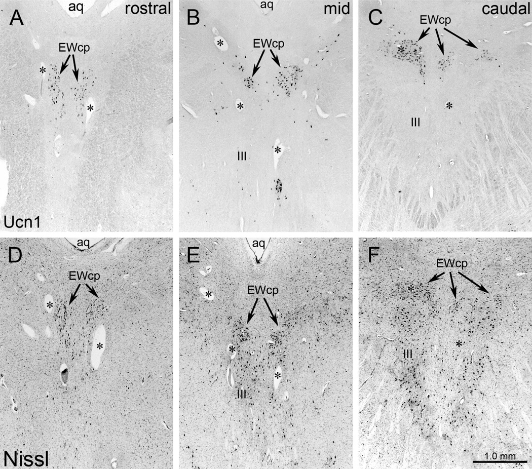
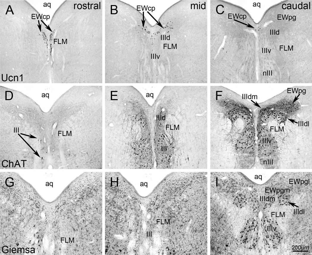
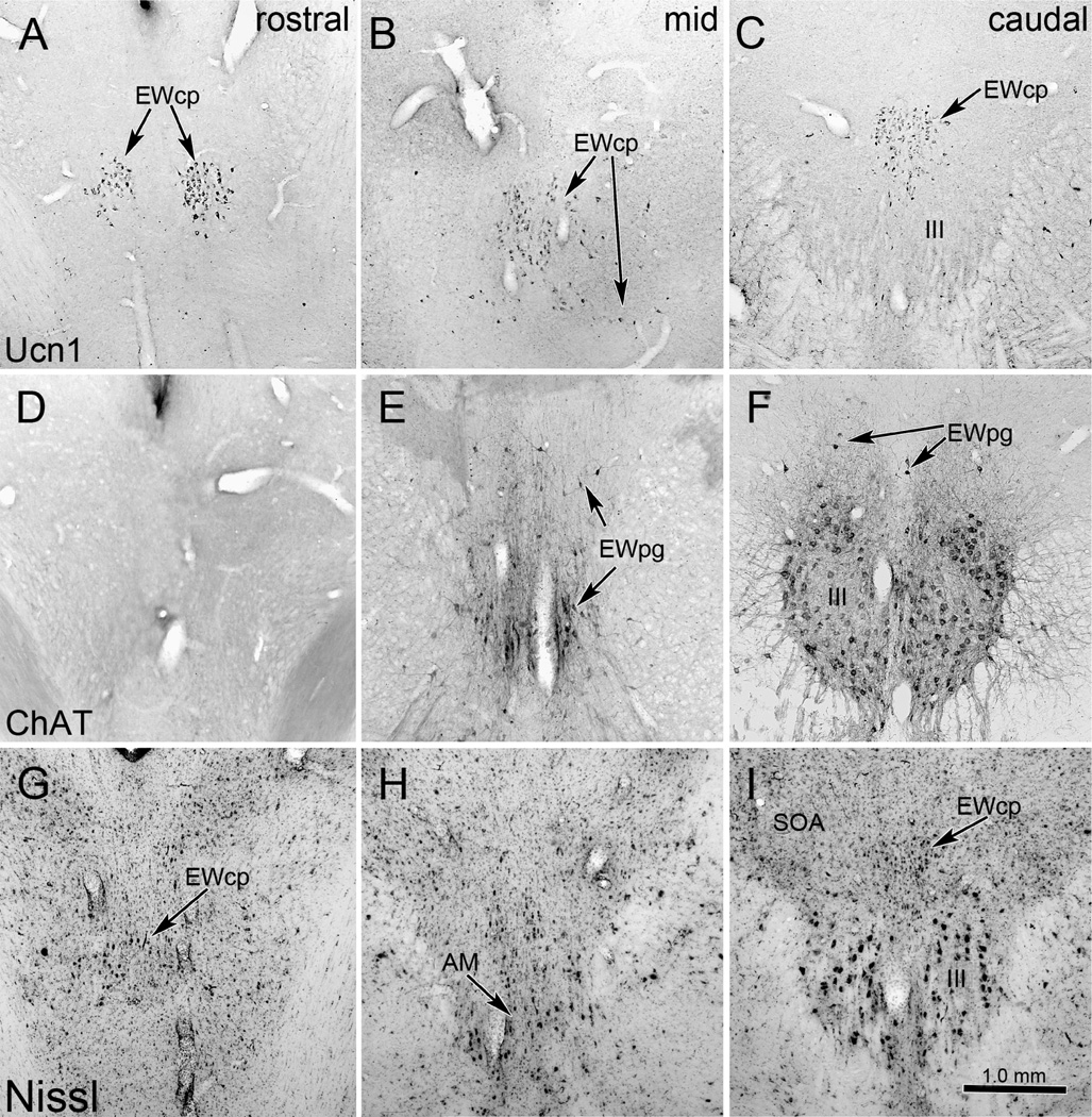
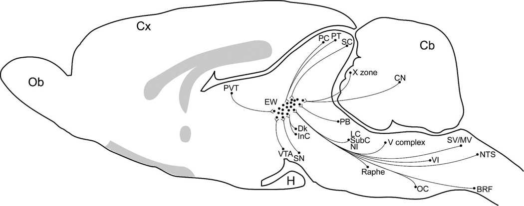
The eponymous term nucleus of Edinger-Westphal (EW) has come to be used to describe two juxtaposed and somewhat intermingled cell groups of the midbrain that differ dramatically in their connectivity and neurochemistry. On one hand, the classically defined EW is the part of the oculomotor complex that is the source of the parasympathetic preganglionic motoneuron input to the ciliary ganglion (CG), through which it controls pupil constriction and lens accommodation. On the other hand, EW is applied to a population of centrally projecting neurons involved in sympathetic, consumptive, and stress-related functions. This terminology problem arose because the name EW has historically been applied to the most prominent cell collection above or between the somatic oculomotor nuclei (III), an assumption based on the known location of the preganglionic motoneurons in monkeys. However, in many mammals, the nucleus designated as EW is not made up of cholinergic, preganglionic motoneurons supplying the CG and instead contains neurons using peptides, such as urocortin 1, with diverse central projections. As a result, the literature has become increasingly confusing. To resolve this problem, we suggest that the term EW be supplemented with terminology based on connectivity. Specifically, we recommend that 1) the cholinergic, preganglionic neurons supplying the CG be termed the Edinger-Westphal preganglionic (EWpg) population and 2) the centrally projecting, peptidergic neurons be termed the Edinger-Westphal centrally projecting (EWcp) population. The history of this nomenclature problem and the rationale for our solutions are discussed in this review.


Figures

Figure 1
Figure 1
Digital photomicrographs of the macaque monkey perioculomotor area (pIII). Sections were stained using antibody to urocortin 1 (Ucn1) (A–C), antibody to choline acetyl transferase (ChAT) (D–F) and with cresyl violet (Nissl) (G–I), and are shown in a rostral to caudal sequence. The mid section is not midway between the others, but is instead located near the front of the oculomotor nucleus (III) because the interactions of the different populations are particularly complex at this point. Rostral to III, the Ucn1 cells are oriented dorsoventrally, with many of them occupying the anteromedian nucleus (AM). Note that EWpg motoneurons form a discrete nucleus in the monkey that is dorsal to somatic motoneurons in III (i.e, the former EW). The EWcpneurons do not form an obvious nucleus, but instead extend from between the two nuclei of III into the supraoculomotor area (SOA). A few are scattered amongst the myelinated bundles lateral to III. Scale bar: 1mm.
Figure 2
Figure 2
Photomicrographs of transverse sections through pIII in human immunostained for Ucn1 (A–C). Note that at caudal levels the Ucn1-positive neurons of EWcp form two separate cell groups dorsal to III, which merge at more rostral levels to form a large compact group. This is delineated in Nissl-stained sections as well (D–F). Portions of this population were formerly termed EW. At mid levels, sometimes an additional small ventral part of the Ucn1-positive cell group is apparent, which merges with the dorsal group rostraly to III (B). For clarity, corresponding blood vessels are indicated by asterisks in the matching picture pairs. Scale bar: 1 mm.
Figure 3
Figure 3
Digital images of oculomotor (III) region in the pigeon brain. In the upper row (A–C), Ucn1-positive neurons form the two medial columns of the EWcp which is located below the central aqueduct (aq). More caudally, the major group of the Ucn1-labelled neurons moves dorsally to form a winglike structure above III. D–F present ChAT-positive neurons that constitute the subdivisions of the III and EWpg. The latter is located dorsolateral to the former. G–I present sections stained with Giemsa, showing the relationship of III and EWpg. The latter is composed of medial (EWpgm) and the lateral (EWpgl) subdivisions. This is the nucleus formerly termed EW. Other abbreviations: FLM: fasciculus longitudinalis medialis; nIII: oculomotor nerve. Scale bar: 200µm.
Figure 4
Figure 4
Photomicrographs of the feline oculomotor (III) region. Note that the ChAT-positive motoneurons of EWpg do not form a discrete nucleus in the cat (D–F), and are instead scattered dorsal, rostral and ventral to somatic motoneurons in III. The Ucn1-positive EWcp neurons do form a discrete nucleus dorsal to III that extends as a paired column at the most rostral levels (AC). As shown in the Nissl sections (G–I), this nucleus was formerly termed EW. However a few neurons are scattered in the supraoculomotor area (SOA) and amongst the myelinated bundles lateral to III. Scale bar: 1mm.
Figure 5
Figure 5
Photographs of transverse sections through the oculomotor (III) region in rat, with combined immunofluorescence for Ucn1 (green, A–C) and ChAT (red, D–F). At caudal and mid levels, the Ucn1-positive neurons of EWcp form a rather compact nucleus visible in the Nissl sections (G–I). This nucleus was formerly called EW, with its neurons more scattered at rostral levels. ChAT-positive neurons within III represent extraocular motoneurons, and scattered ChAT-positive neurons dorsal to III may represent preganglionic neurons (EWpg). These overlap with EWcp rostral to III (A,D.G). For clarity, corresponding blood vessels are labelled by asterisks in the picture pairs. Scale bar:200 µm. Other abbreviations: aq; cerebral aqueduct.
Figure 6
Figure 6
Line drawings showing the organization of EWpg and EWcp in several selected species: avian, rat, cat, monkey and human. Representative rostral (left column), middle (middle column) and caudal (right column) sections are shown. The EWcp is indicated by light gray shading and EWpg is indicated by dark gray shading. Scattered cells located outside the nuclear boundaries are indicated by appropriately shaded circles.
Figure 7
Figure 7
Three dimensional (3-D) representations of human, macaque, cat, rat and pigeon oculomotor complex, to illustrate the 3-D organization of EWpg and EWcp. The 3-D models are cut at selected points to illustrate how frontal sections through this level would look. In cases where the population is scattered, and so not contained in a discrete nucleus, dots are used.
Figure 8
Figure 8
Schematic diagram of a midsagittal section through the rat brain to summarize the known afferents to the classical EW. Species differences in the location of Ewcp and EWpg were not considered. Abbreviations: BRF, bulbar reticular formation; Cb, cerebellum; CN, cerebellar nuclei; Cx, cerebral cortex; Dk, Darkeschewitsch nucleus; H, hypophysis; InC, nucleus interstitial of Cajal; LC, locus coeruleus; LPB, lateral parabrachial nucleus; MV, medial vestbibular nucleus; NI, nucleus incertus; Ob, Olfactory bulb; OC, olivary complex; PB, parabrachial nuclei; PC, nucleus of the posterior commissure; PTm, medial pretectal nucleus; Raphe, raphe nuclei; SC, superior colliculus; SubC, subcoeruleus; SV, superior vestibular nucleus; V complex, trigeminal nuclei complex; VTA, ventral tegmental area; VI, abducens nucleus; X zone, the X zone of the cerebellar cortex. The shaded gray areas represent the corpus callosum, fornix, anterior commissure and the optical chiasm and tract.
Figure 9
Figure 9
Schematic diagram of a midsagittal section through the rat brain to summarize the known sources of inputs supplying the EWpg and EWcp (together EW). Possible species differences are not indicated. Abbreviations: Cb, cerebellum; CG, ciliary ganglion; Cx, cerebral cortex; H, hypophysis; IO, inferior olivary nucleus; LPB, lateral parabrachial nucleus; LRN, lateral reticular nucleus; LS, lateral septal nucleus; MeON, medial olivary nucleus; MON, main olivary nucleus; Ob, Olfactory bulb; RVRN, rostroventral reticular nucleus; V complex, trigeminal complex; VII, facial nucleus. The shaded gray areas represent the corpus callosum, fornix, anterior commissure and the optical chiasm and tract.

References

    1. Adler A. Zur Lokalisation des Konvergenzentrums und der Kerne der glatten Augenmuskeln. Ztschr ges Neurol Psychiat. 1933;145:185–207.
    1. Akert K, Glicksman MA, Lang W, Grob P, Huber A. The Edinger-Westphal nucleus in the monkey. A retrograde tracer study. Brain Res. 1980;184:491–498. - PubMed
    1. Bach L. Über das Verhalten der motorischen Kerngebiete nach Läsion der peripheren Nerven und uber die physiologische Bedeutung der Edinger-Westfalschen Kerne. Zbl Nervenheilk. 1906;29:140.
    1. Bachtell RK, Tsivkovskaia NO, Ryabinin AE. Strain differences in urocortin expression in the Edinger-Westphal nucleus and its relation to alcohol-induced hypothermia. Neuroscience. 2002a;113:421–434. - PubMed
    1. Bachtell RK, Tsivkovskaia NO, Ryabinin AE. Alcohol-induced c-Fos expression in the Edinger-Westphal nucleus: pharmacological and signal transduction mechanisms. J Pharmacol Exp Ther. 2002b;302:516–524. - PubMed

Publication types