. 2011 Jul 1;519(10):1829-46.
doi: 10.1002/cne.22596.

Fezf1 and Fezf2 are required for olfactory development and sensory neuron identity

Affiliations

Fezf1 and Fezf2 are required for olfactory development and sensory neuron identity

Matthew J Eckler et al. J Comp Neurol. .

Abstract

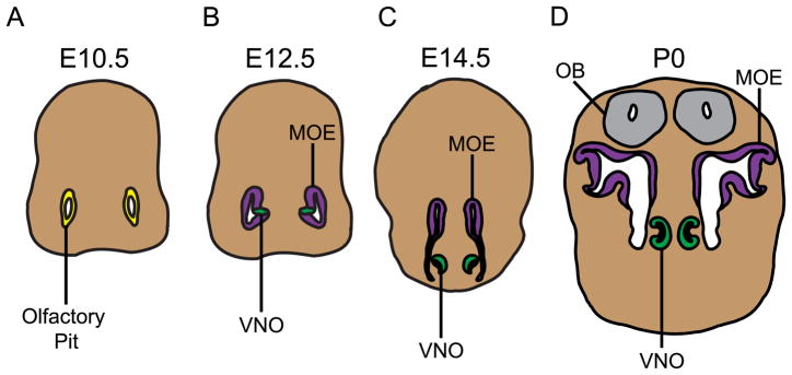
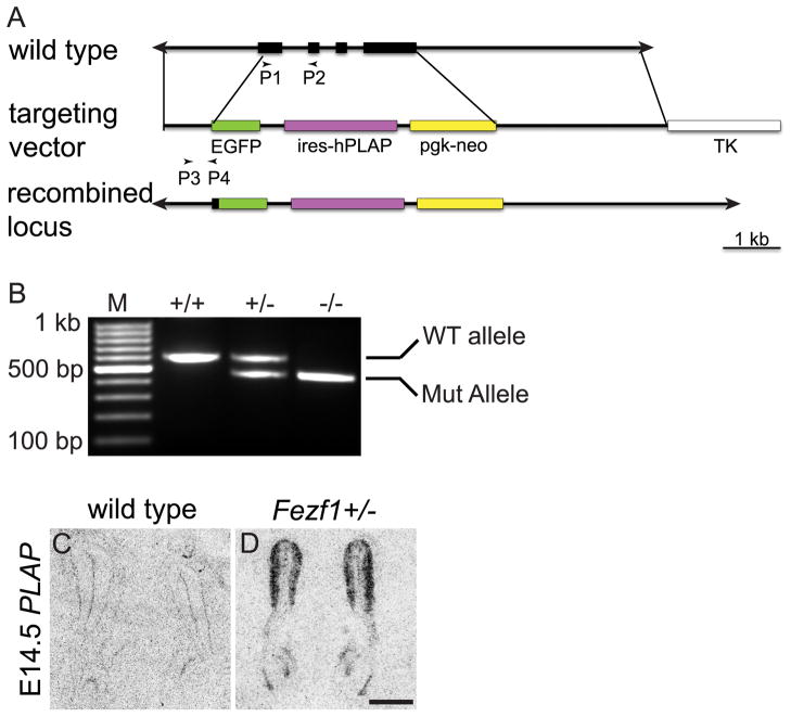
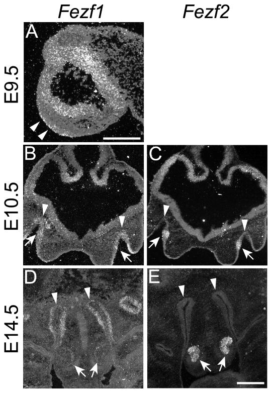
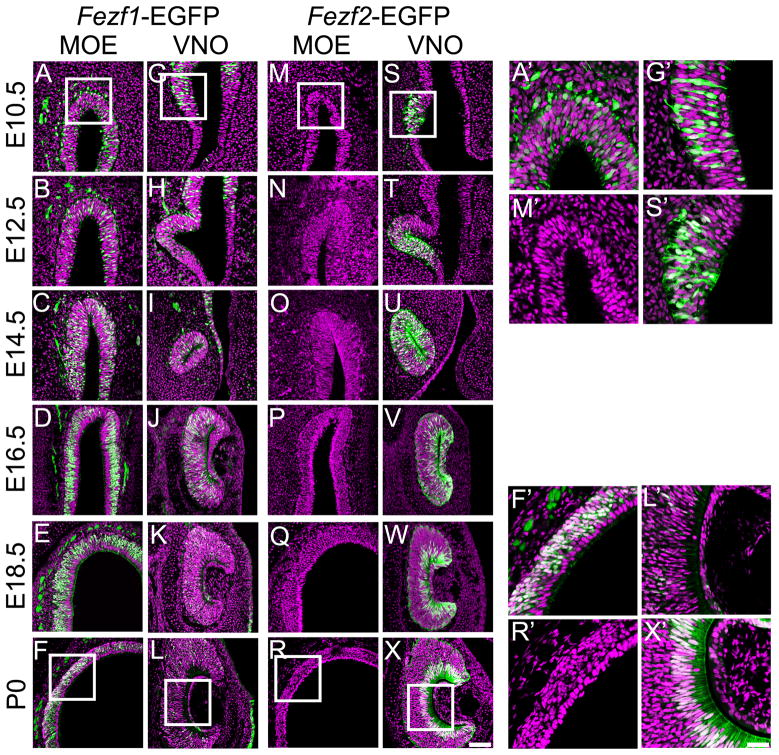
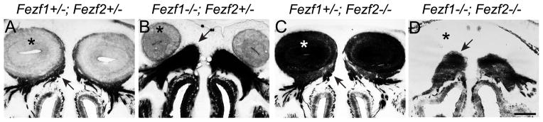
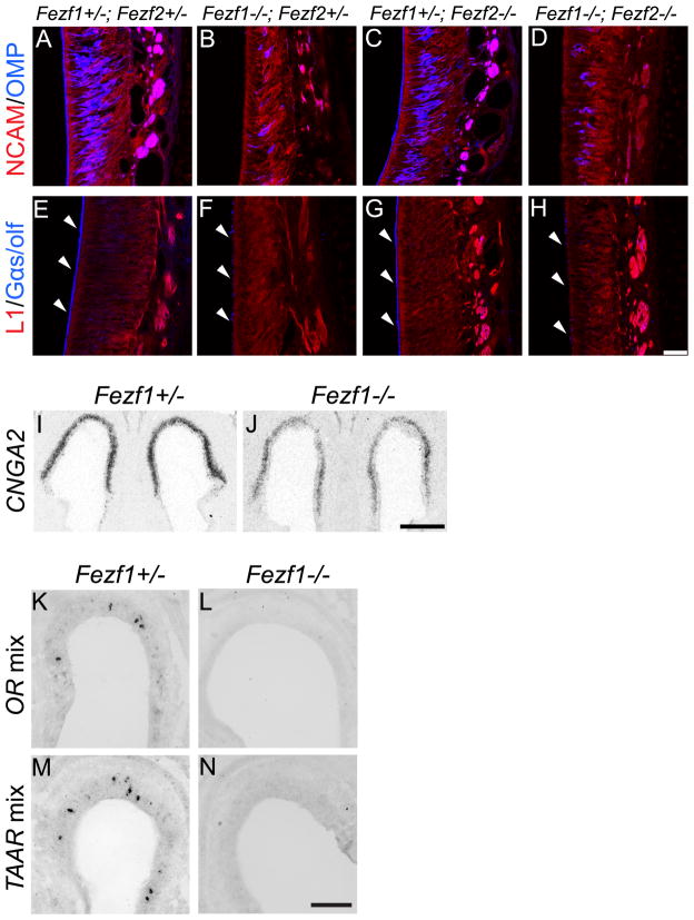
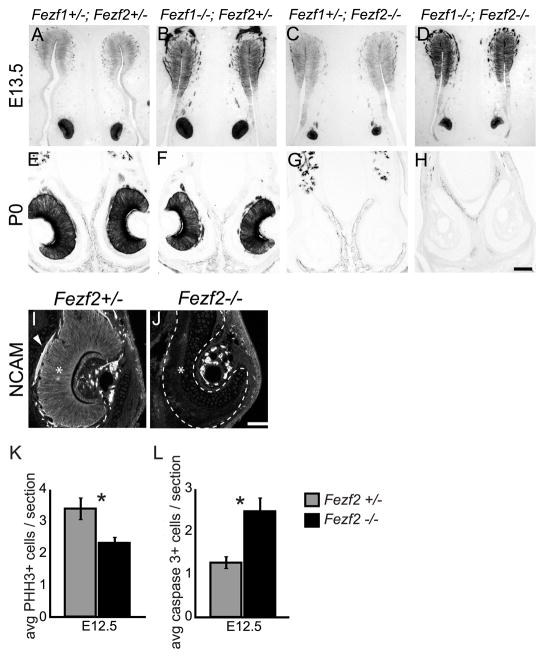
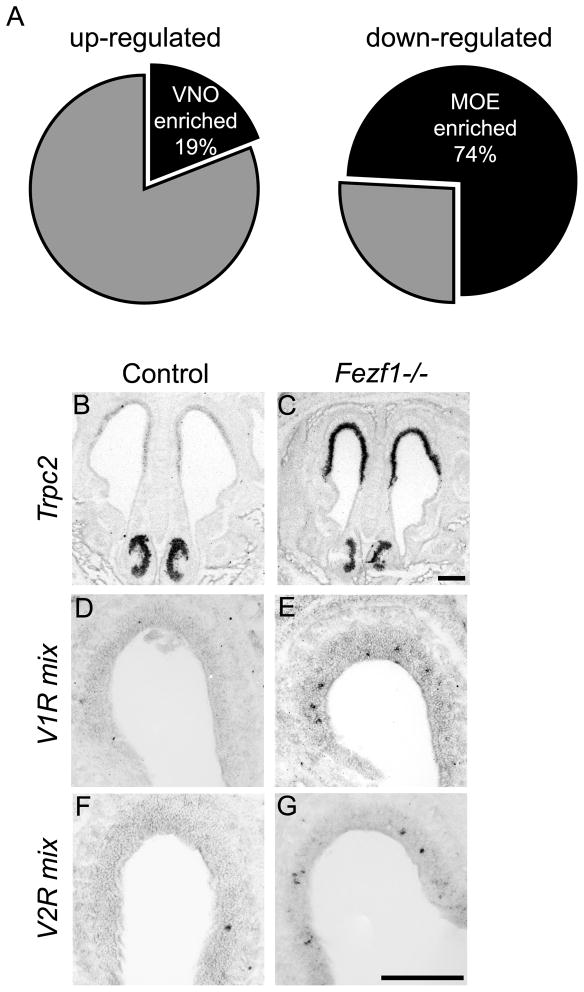
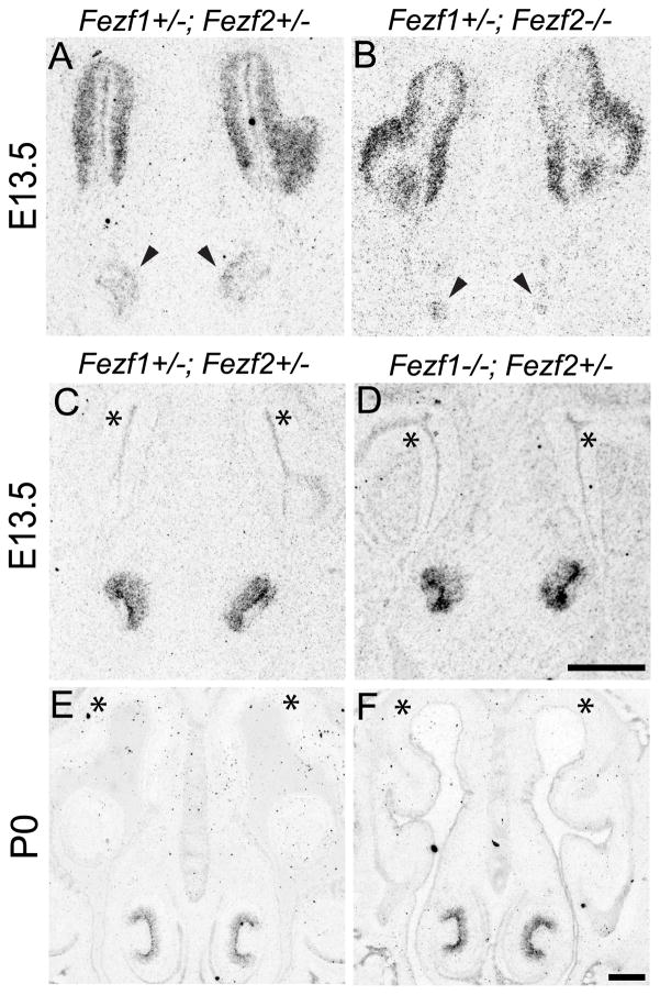
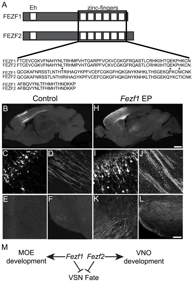
The murine olfactory system consists of main and accessory systems that perform distinct and overlapping functions. The main olfactory epithelium (MOE) is primarily involved in the detection of volatile odorants, while neurons in the vomeronasal organ (VNO), part of the accessory olfactory system, are important for pheromone detection. During development, the MOE and VNO both originate from the olfactory pit; however, the mechanisms regulating development of these anatomically distinct organs from a common olfactory primordium are unknown. Here we report that two closely related zinc-finger transcription factors, FEZF1 and FEZF2, regulate the identity of MOE sensory neurons and are essential for the survival of VNO neurons respectively. Fezf1 is predominantly expressed in the MOE while Fezf2 expression is restricted to the VNO. In Fezf1-deficient mice, olfactory neurons fail to mature and also express markers of functional VNO neurons. In Fezf2-deficient mice, VNO neurons degenerate prior to birth. These results identify Fezf1 and Fezf2 as important regulators of olfactory system development and sensory neuron identity.


Figures

Figure 1
Figure 1
A diagram depicting the developmental origins of the main olfactory epithelium (MOE) and vomeronasal organ (VNO) from the olfactory pit. All schematics shown depict a coronal orientation. (A) At E10.5 the olfactory pit (shown in yellow) is morphologically discernible but does not show any distinction between the regions that will give rise to the MOE and VNO. (B) By E12.5 the pit has undergone an initial invagination, distinguishing the dorsal region that will give rise to the MOE (purple) from the ventromedial region that will become the VNO (green). (C) By E14.5 the VNO has separated from the pit and become anatomically distinct. (D) This spatial and molecular distinction is clear at birth (P0), with both the MOE and VNO projecting axons to innervate the olfactory bulb (OB) (grey). Diagrams are not drawn to scale.
Figure 2
Figure 2
Fezf1 knockout strategy. The endogenous Fezf1 locus was replaced with a cassette containing EGFP-IRES-hPLAP (A) and transmission of the targeted allele was confirmed by PCR using primers P1 and P2 (for wild type allele), P3 and P4 (for mutant allele) (B). Appropriate expression of the targeted Fezf1 allele was confirmed by radioactive in situ hybridization to detect PLAP expression in Fezf1+/ (D), but not in wild type mice (C). The scale bar in D is 100 μm.
Figure 3
Figure 3
Expression of Fezf1 and Fezf2 transcripts during olfactory system development. Radioactive in situ hybridization was used to detect Fezf1 (A, B, and D) or Fezf2 (C, and E) expression. At E9.5 Fezf1 was detected in the olfactory placode (arrowheads in A). At E10.5 it is detected throughout the newly formed olfactory pits (arrowheads in B). Fezf1 remains strongly expressed in the MOE (arrowheads in D) and weakly in the VNO (arrows in D) at E14.5 and later stages (D, and data not shown). Alternatively, Fezf2 is specifically expressed in the VNO throughout olfactory system development (arrows in C and E). A, sagittal section; B–E, coronal sections. The scale bar in A is 25 μm and in E is 100 μm.
Figure 4
Figure 4
Expression patterns of Fezf1 and Fezf2 during development of the olfactory system detected by EGFP reporter expression. GFP immunohistochemistry was used to visualize cells expressing either Fezf1-EGFP (A–L, A′, F′, G′, L′) or Fezf2-EGFP (M–X, M′, R′, S′, X′). Nuclei were visualized using SYTOX orange. At E10.5 and E12.5, Fezf1-EGFP is expressed throughout the future MOE and VNO (A, B, G, H, A′, and G′). After E14.5 Fezf1-EGFP expression starts to decrease in the VNO and becomes restricted to progenitor and neuronal cell layers of the MOE (D–F, J–L, F′, and L′). This transition is complete by E18.5 and persists postnatally (E, F, K, L, F′, and L′). In contrast to the Fezf1-EGFP expression pattern, Fezf2-EGFP expression is restricted to the VNO throughout olfactory system development (M–X, M′, S′, R′, and X′). At E10.5, when the VNO is first starting to differentiate from the olfactory pit, Fezf2-EGFP is strongly expressed throughout cells in the ventromedial anlage that will give rise to the VNO (S, S′). Beginning at E14.5, Fezf2-EGFP expression undergoes a dynamic transition and becomes restricted to the sustentacular cell layer of the VNO (O–R; U–X, R′, X′). This expression pattern of Fezf2-EGFP is maintained postnatally (R, X, R′, X′). For all images, EGFP expression is shown in green and SYTOX orange staining is in purple. Images in A′, F′, G′, L′, M′, R′, S′ and X′ represent the enlarged regions shown in the white boxes in panels A, F, G, L, M, R, S and X. The scale bar in X is 75 μm and in X′ is 30 μm.
Figure 5
Figure 5
Fezf1 is required for OSN axons to reach the olfactory bulb. (A–D) PLAP staining was used to visualize Fezf1 and Fezf2 expressing cells and their projections. In Fezf1+/; Fezf2+/− (A) and Fezf1+/ ; Fezf2/ (C)mice the axons of OSNs (arrows) extended from the MOE and innervated the OB (asterisk). However in Fezf1/; Fezf2+/− (B) and Fezf1−/−; Fezf2−/− (D) mice OSN axons (arrowheads) were unable to penetrate the cribriform plate and innervate the OB. Note also the OB is not present in the Fezf1−/−; Fezf2−/− mice. The very dark PLAP staining in the OB of Fezf1+/; Fezf2−/− mice is due to PLAP-labeled axons from deep-layer neurons of the cerebral cortex which projected ectopically into the olfactory bulb. The scale bar in D is 200 μm.
Figure 6
Figure 6
The OSNs in the MOE of Fezf1−/− mice fail to mature. Immunohistochemistry shows a reduction in olfactory marker protein (OMP) expression in the MOE of Fezf1−/− (B) and Fezf1−/−; Fezf2−/− (D) compared with control (A) and Fezf2−/− mice (C). Similarly, Gαs/olf expression is markedly reduced in the dendrites (white arrowheads in E–H) of Fezf1−/− (F) and Fezf1−/−; Fezf2−/− (H) when compared with control (E) and Fezf2−/− mice (G). Neural cell adhesion molecule (NCAM) and L1 cell adhesion molecule (L1) expression both appeared unaffected in Fezf1−/− (B and F), Fezf2−/− (C and G) and Fezf1−/−; Fezf2−/− (D and H) animals. Reduction in CNGA2 expression was visualized by radioactive in situ hybridization for the control and Fezf1 mutant MOE (I and J). Non-radioactive in situ hybridization using probes corresponding to a mixture of olfactory receptors (ORs) (K and L) or trace amine associated receptors (TAARs) (M and N) showed loss of expression in Fezf1−/− animals. Scale bars are H: 75 μm; J: 200 μm; and N: 100 μm.
Figure 7
Figure 7
VNO degeneration in Fezf2−/− mice. (A–H) PLAP staining was used to visualize Fezf1 and Fezf2 expressing cells of the olfactory system. At E13.5 the VNO properly separated from the olfactory pit in Fezf1−/−; Fezf2+/, Fezf1+/;Fezf2−/−, and Fezf1−/−; Fezf2−/− mice (A–D). However in Fezf2−/−, and Fezf1−/−; Fezf2−/− animals the VNO is smaller (compare C, D with A, B). This defect is exacerbated by P0, with a complete absence of PLAP-positive cells in Fezf1+/; Fezf2−/−, and Fezf1−/−; Fezf2−/− animals (compare G, H to E, F). The loss of neurons in the Fezf2 mutant VNO was confirmed by NCAM staining at P0 (I and J). NCAM+ cells (*) and their axons (arrowhead) are clearly visible in the Fezf2+/ but not in the Fezf2−/− VNO. Quantification of phosphohistone H3 (PHH3+) and cleaved-caspase 3+ cells indicates a significant decrease in proliferation (average of 3.38 vs. 2.31 per VNO section, p = 0.0269) (K) and increase in apoptosis (average of 1.28 vs. 2.48 per VNO section, p = 0.0003) (L). For Fezf2+/, n = 80 sections and for Fezf2−/−, n = 52 sections. The scale bar in H is 100 μm and in J is 75 μm.
Figure 8
Figure 8
Fezf1−/− MOE acquires a VNO-like transcriptional identity. Microarray analysis of E18.5 Fezf1−/− cells showed that 74% of down-regulated transcripts and 19% of up-regulated transcripts correspond to MOE- and VNO-enriched transcripts, respectively (A). In situ hybridization confirming mis-expression of VNO-enriched transcripts in the MOE of Fezf1−/− mice (B–G). Transient receptor potential cation channel 2 (Trpc2) (B, C), some vomeronasal receptor class 1 genes (V1Rs) (D, E), and some vomeronasal receptor class 2 genes (V2Rs) (F, G) were mis-expressed in the MOE of Fezf1 mutant animals. Trpc2 expression was visualized by radioactive in situ hybridization. Expression of V1Rs and V2Rs was visualized by digoxigenin in situ hybridization. Scale bars in C and G are 100 μm.
Figure 9
Figure 9
Fezf1 and Fezf2 are not mutually repressive. Radioactive in situ hybridization is used to visualize Fezf1 (A, B) or Fezf2 (C–F) transcripts. At E13.5 expression of Fezf1 is not increased in Fezf2−/− mice (A, B). Similarly, loss of Fezf1 has no effect on expression of Fezf2 at E13.5 or P0 (C–F). The VNO is marked by arrowheads (A, B). The MOE is indicated by asterisks (C–F). Scale bars in D and F are 100 μm.
Figure 10
Figure 10
Fezf1 and Fezf2 can function interchangeably during cell fate specification in vivo. (A) A schematic presentation of identifiable FEZF1 and FEZF2 protein domains showing their N-terminal engrailed homology domain (Eh) and C-terminal C2H2 zinc-finger motifs. An alignment of the zinc-finger motifs shows only three amino acid differences (*), corresponding to 97% identity between the two proteins. (B–L) Misexpression of Fezf1 in corticocortical projection neurons of the cerebral cortex is sufficient to redirect their axons subcortically, similar to previously reported results for Fezf2 misexpression (Chen et al., 2008). When Fezf1 and EGFP were electroporated into layer 2/3 neurons in utero (H–L) their axons are redirected to the striatum (J), thalamus (K) and pons (L). No GFP+ axons were detected in these brain regions when only EGFP was misexpressed (B–F). Our model predicts that Fezf1 promotes MOE development and Fezf2 promotes VNO development by preventing progenitors and sensory neurons of the MOE and progenitors and supporting cells of the VNO from assuming a VNO sensory neuron identity (M). The diagram in A is not drawn to scale. The scale bar in H is 1 mm and in L is 75 μm.

References

    1. Baker H, Grillo M, Margolis FL. Biochemical and immunocytochemical characterization of olfactory marker protein in the rodent central nervous system. J Comp Neurol. 1989;285(2):246–261. - PubMed
    1. Balmer CW, LaMantia AS. Noses and neurons: induction, morphogenesis, and neuronal differentiation in the peripheral olfactory pathway. Dev Dyn. 2005;234(3):464–481. - PubMed
    1. Beites CL, Kawauchi S, Crocker CE, Calof AL. Identification and molecular regulation of neural stem cells in the olfactory epithelium. Exp Cell Res. 2005;306(2):309–316. - PubMed
    1. Cau E, Casarosa S, Guillemot F. Mash1 and Ngn1 control distinct steps of determination and differentiation in the olfactory sensory neuron lineage. Development. 2002;129(8):1871–1880. - PubMed
    1. Cau E, Gradwohl G, Casarosa S, Kageyama R, Guillemot F. Hes genes regulate sequential stages of neurogenesis in the olfactory epithelium. Development. 2000;127(11):2323–2332. - PubMed

Publication types

MeSH terms