

Development of a physical 3D anthropomorphic breast phantom
- PMID: 21452726
- PMCID: PMC4108620
- DOI: 10.1118/1.3533896
Development of a physical 3D anthropomorphic breast phantom
Abstract
Purpose: Develop a technique to fabricate a 3D anthropomorphic breast phantom with known ground truth for image quality assessment of 2D and 3D breast x-ray imaging systems.
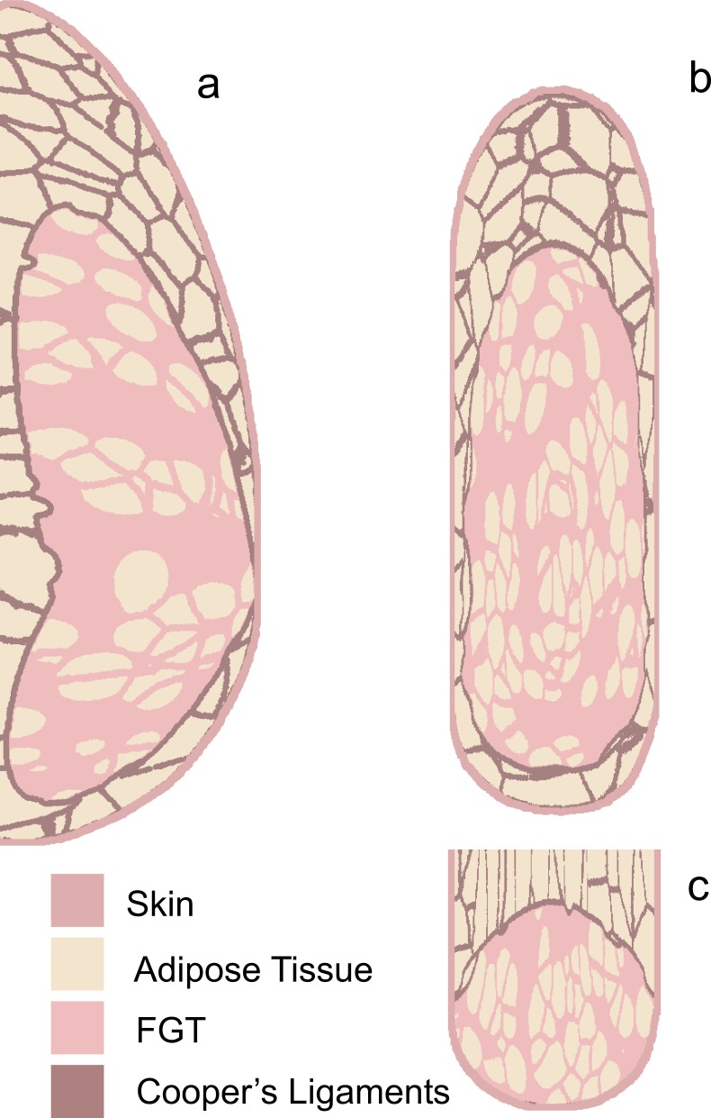
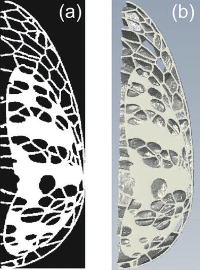
Methods: The phantom design is based on an existing computer model that can generate breast voxel phantoms of varying composition, size, and shape. The physical phantom is produced in two steps. First, the portion of the voxel phantom consisting of the glandular tissue, skin, and Cooper's ligaments is separated into sections. These sections are then fabricated by high-resolution rapid prototyping using a single material with 50% glandular equivalence. The remaining adipose compartments are then filled using an epoxy-based resin (EBR) with 100% adipose equivalence. The phantom sections are stacked to form the physical anthropomorphic phantom.
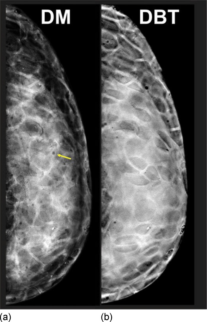
Results: The authors fabricated a prototype phantom corresponding to a 450 ml breast with 45% dense tissue, deformed to a 5 cm compressed thickness. Both the rapid prototype (RP) and EBR phantom materials are radiographically uniform. The coefficient of variation (CoV) of the relative attenuation between RP and EBR phantom samples was <1% and the CoV of the signal intensity within RP and EBR phantom samples was <1.5% on average. Digital mammography and reconstructed digital breast tomosynthesis images of the authors' phantom were reviewed by two radiologists; they reported that the images are similar in appearance to clinical images, noting there are still artifacts from air bubbles in the EBR.
Conclusions: The authors have developed a technique to produce 3D anthropomorphic breast phantoms with known ground truth, yielding highly realistic x-ray images. Such phantoms may serve both qualitative and quantitative performance assessments for 2D and 3D breast x-ray imaging systems.
Figures


References
-
- Caldwell C. B., Fishell E. K., Jong R. A., Weiser W. J., and Yaffe M. J., “Evaluation of mammographic image quality: Pilot study comparing five methods,” AJR, Am. J. Roentgenol. 159, 295–301 (1992). - PubMed
-
- Zhang C., Bakic P. R., and Maidment A. D. A., “Development of an anthropomorphic breast software phantom based on region growing algorithm,” in Proceedings of the Medical Imaging Conference 2008: Visualization, Image-guided Procedures, and Modeling (volume 6918), 69180V, edited by Miga M. I. and Cleary K. R. (SPIE, San Diego, CA, 2008).
Publication types
MeSH terms
Grants and funding
LinkOut - more resources
Full Text Sources
