. 2011 May 24;5(5):4037-45.
doi: 10.1021/nn200629g. Epub 2011 Apr 5.

Magnetic virus-like nanoparticles in N. benthamiana plants: a new paradigm for environmental and agronomic biotechnological research

Affiliations

Magnetic virus-like nanoparticles in N. benthamiana plants: a new paradigm for environmental and agronomic biotechnological research

Xinlei Huang et al. ACS Nano. .

Abstract

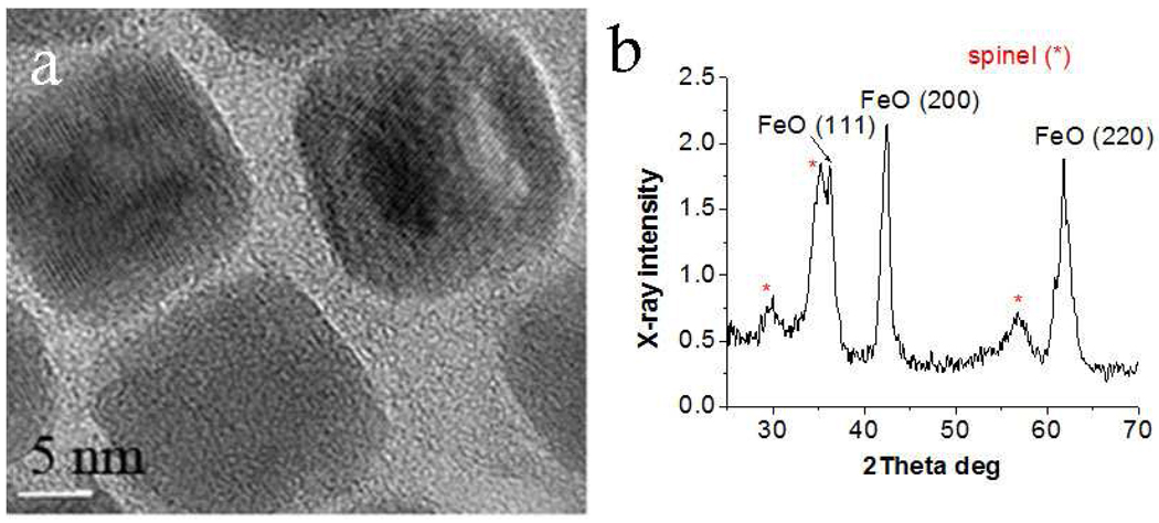
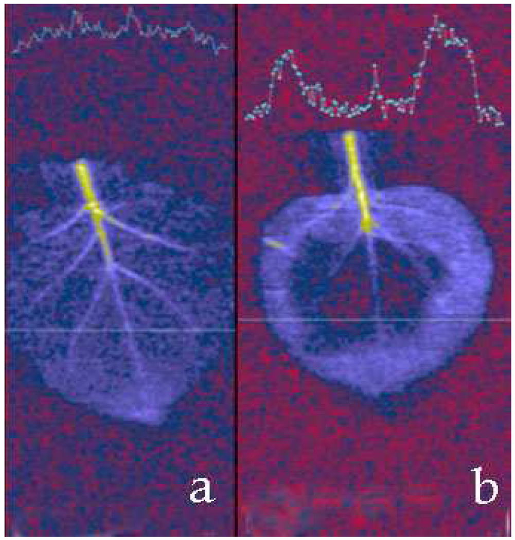
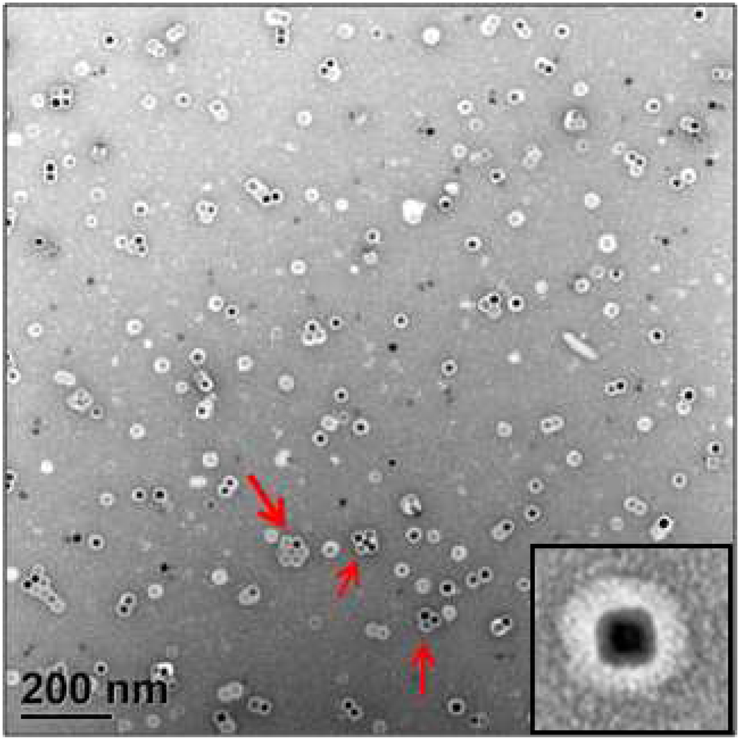
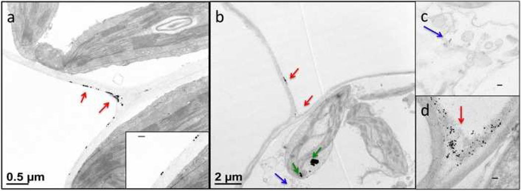
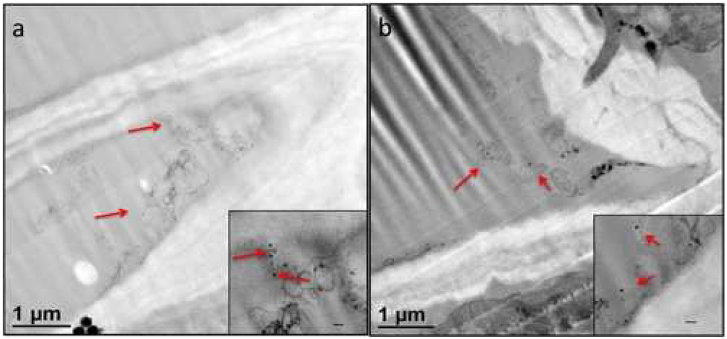
This article demonstrates the encapsulation of cubic iron oxide nanoparticles (NPs) by Brome mosaic virus capsid shells and the formation, for the first time, of virus-based nanoparticles (VNPs) with cubic cores. Cubic iron oxide NPs functionalized with phospholipids containing poly(ethylene glycol) tails and terminal carboxyl groups exhibited exceptional relaxivity in magnetic resonance imaging experiments, which opens the way for in vivo MRI studies of systemic virus movement in plants. Preliminary data on cell-to-cell and long-distance transit behavior of cubic iron oxide NPs and VNPs in Nicotiana benthamiana leaves indicate that VNPs have specific transit properties, i.e., penetration into tissue and long-distance transfer through the vasculature in N. benthamiana plants, even at low temperature (6 °C), while NPs devoid of virus protein coats exhibit limited transport by comparison. These particles potentially open new opportunities for high-contrast functional imaging in plants and for the delivery of therapeutic antimicrobial cores into plants.


Figures

Figure 1
Figure 1
HRTEM image (a) and XRD profile (b) of the as-prepared 18.6 nm iron oxide NPs.
Figure 2
Figure 2
(a) Longitudinal T1 and (b) T2 map images of the 18.6 nm cubic iron oxide NPs coated with HOOC-PEG-PL and dispersed in Milli-Q water. Scale bar in (a) is linear (unit: milliseconds), while in (b) it is in logarithmic units. Sample concentrations are (1): 0 mM Fe (pure Milli-Q water), (2): 0.93 mM Fe, (3): 0. 37 mM Fe, (4): 0.14 mM Fe, (5): 0.06 mM Fe, (6): 0.04 mM Fe, and (7): 0.01 mM Fe.
Figure 3
Figure 3
T2 -weighted image (sample 2 and 3 spots are circled because the intensities are as low as the background signal due to the high T2 effect of the NPs in water). The sample concentrations are the same as those of the caption to Figure 2.
Figure 4
Figure 4
(a) T2-weighted MR image (repetition time 3 s, echo time 37 ms) of N. benthamiana leafs before (a) and after (b) infiltration with HOOC-PEG-PL-coated 18.6 nm cubic NPs. A cross section of the MRI map is shown on the top part of each image illustrating contrast enhancement induced through NPs infiltration.
Figure 5
Figure 5
Negatively stained TEM image of VNPs formed by self-assembling BMV proteins around the 18.6 nm cubic NPs coated with HOOC-PEG-PL. The dark spots are iron oxide NPs. Light rings around NPs are BMV shells including the HOOC-PEG-PL shells. The red arrows illustrate possibly pre-assembled VNP clusters. Inset shows a higher magnification image of a single VNP.
Figure 6
Figure 6
Stained TEM images of histological preparations of leaf sections. (a) Iron oxide NPs attached to the exterior of the cell wall in the apoplast; red arrows indicate the iron oxide NPs while the enlarged inset shows cubic particles sticking to the cell wall. (b) Magnetic VNPs after entering the plant leaf also gathered at the cell junctions of the apoplast (red arrows) but some particles were present within plant cells (blue arrow). The dark spots inside the chloroplast are indicated by green arrows and are impurities in the staining solution which may have been confused for NPs, were it not for their specific shape. (c) Image of VNPs in the cytoplasm.. (d) Image of VNPs gathered at the cell junction area of the apoplast. All the scale bars in the insets are 100 nm.
Figure 7
Figure 7
Magnetic VNPs in (a) phloem and (b) xylem after 25 °C incubation for 24 hr. Insets show enlarged images, scale bars are 100 nm. Red arrows indicate VNPs.
Scheme 1
Scheme 1
Sample infiltration pattern of magnetic NPs (blue zones) and VNPs (green zones) in N. benthamiana plant leaves. Blue and green zones 1–4 were infiltrated with 0.5 mg/mL, 0.25 mg/mL, 0.1 mg/mL, and 0.05 mg/mL of NPs or VNPs. A single zone near the base of the leaf was infiltrated with the infiltration buffer (10 mM MgCl2 and 10 mM MES, pH 5.9) as control.

References

    1. Douglas T, Young M. Host-Guest Encapsulation of Materials by Assembled Virus Protein Cages. Nature. 1998;393:152–155.
    1. Smith LM, Pontes O, Searle I, Yelina N, Yousafzai FK, Herr AJ, Pikaard CS, Baulcombe DC. An Snf2 Protein Associated with Nuclear Rna Silencing and the Spread of a Silencing Signal between Cells in Arabidopsis. Plant Cell. 2007;19:1507–1521. - PMC - PubMed
    1. Stephanidis B, Adichtchev S, Gouet P, McPherson A, Mermet A. Elastic Properties of Viruses. Biophys. J. 2007;93:1354–1359. - PMC - PubMed
    1. Dragnea B, Chen C, Kwak E-S, Stein B, Kao CC. Gold Nanoparticles as Spectroscopic Enhancers for in Vitro Studies on Single Viruses. J. Am. Chem. Soc. 2003;125:6374–6375. - PubMed
    1. Douglas T, Strable E, Willits D, Aitouchen A, Libera M, Young M. Protein Engineering of a Viral Cage for Constrained Nanomaterials Synthesis. Adv. Mater. 2002;14:415–418.

Publication types