Review
. 2011 Jun;11(3):155-80.
doi: 10.2174/156652311795684722.

Gene therapy and targeted toxins for glioma

Affiliations
Review

Gene therapy and targeted toxins for glioma

Maria G Castro et al. Curr Gene Ther. 2011 Jun.

Abstract

The most common primary brain tumor in adults is glioblastoma. These tumors are highly invasive and aggressive with a mean survival time of 15-18 months from diagnosis to death. Current treatment modalities are unable to significantly prolong survival in patients diagnosed with glioblastoma. As such, glioma is an attractive target for developing novel therapeutic approaches utilizing gene therapy. This review will examine the available preclinical models for glioma including xenographs, syngeneic and genetic models. Several promising therapeutic targets are currently being pursued in pre-clinical investigations. These targets will be reviewed by mechanism of action, i.e., conditional cytotoxic, targeted toxins, oncolytic viruses, tumor suppressors/oncogenes, and immune stimulatory approaches. Preclinical gene therapy paradigms aim to determine which strategies will provide rapid tumor regression and long-term protection from recurrence. While a wide range of potential targets are being investigated preclinically, only the most efficacious are further transitioned into clinical trial paradigms. Clinical trials reported to date are summarized including results from conditionally cytotoxic, targeted toxins, oncolytic viruses and oncogene targeting approaches. Clinical trial results have not been as robust as preclinical models predicted; this could be due to the limitations of the GBM models employed. Once this is addressed, and we develop effective gene therapies in models that better replicate the clinical scenario, gene therapy will provide a powerful approach to treat and manage brain tumors.


Figures

Figure 1
Figure 1. Comparative assessment of in vivo efficacy and toxicity of adenovrial vectors encoding cytotoxic transgenes after injection into normal brain
(A) Lewis rats were implanted with CNS1 cells in the brain and then treated 4 days later with an intratumoral injection of either saline, or adenoviral vectors encoding TNF-α (Ad-TNF-α), TRAIL (Ad-TRAIL), FasL (Ad-FasL) or thymidine kinase (Ad-TK). Rats treated with Ad-TK received GCV. Kaplan Meier survival curves are shown *p<0.05 vs saline, ^p<0.05 vs Ad-FasL (Mantel log-rank test). Representative microphotographs show the appearance of the tumor at the time of treatment (day 4), as assessed by vimentin staining. (B) To assess neuropathology, each vector was injected into the naïve Lewis rat brain. 7 days later neuropathological analysis of the brain was assessed by Nissl staining and immunocytochemistry using antibodies against tyrosine hydroxylase (TH).
Figure 2
Figure 2. Immunological mechanism underlying the efficacy of combined gene therapy using Ad-Flt3L and Ad-TK (+GCV)
Transduction of tumor cells with Ad-TK (+GCV treatment) leads to tumor cell death and the release of endogenous brain tumor antigens and TLR ligands. Concomittant delivery of Ad-Flt3L into the brain tumor causes the infiltration of dendritic cells (DCs) into the tumor mass. The release of TLR ligands, including the TLR2 agonist HMGB1, results in TLR activation on on tumor infiltrating DCs. DC’s phagocytose tumor cells remnants which lead to their activation and maturation. Mature, loaded DCs migrate to the draining cervical lymph nodes where they present brain tumor antigens to naïve T cells, thereby causing the proliferation of anti-GBM specific T cells. The expansion of anti-GBM specific T cells kills residual brain tumor cells leading to tumor regression and immunological memory.
Figure 3
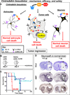
Figure 3. Intratumoral delivery of Cintredekin besudotox improves survival in nude mice bearing orthotopic U251 brain tumors but leads to severe neurotoxicity
A. Illustration depicting the mechanism of action of the protein formulation Cintredekin besudotox mediated cytotoxicity in glioma cells. B. Nude mice were implanted with human U251 glioma cells in the striatum and 5 days later, animals received an intratumoral injections of either Cintredekin besudotox at a high dose (1 µg) or a low dose (0.2 µg) or saline as control. B. Kaplan Meier survival curves of nude mice bearing intracranial U251 and treated as indicated in A. C. Three days post-delivery of Cintredekin besudotox, mice were euthanized due to severe neurological deficits and neuropathological analysis was assessed by Nissl staining and immunocytochemistry using antibodies against tyrosine hydroxylase (TH) and myelin basic protein (MBP). Note the severe local neurotoxicity of both doses of Cintredekin besudotox when injected into the normal brain parenchyma (arrows).
Figure 4
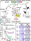
Figure 4. Intratumoral delivery of an adenoviral vector encoding mhIL-13-PE and mIL-4 leads to long-term survival with an absence of neurotoxicity
A. Illustration depicting the mechanism of action of the adenovirus encoded IL13-PE in brain tumor cells. B. Nude mice were implanted with human U251 glioma cells in the striatum and 5 days later, animals received an intratumoral injection of either a control vector Ad.mhIL-4.TRE.mhIL-13 (without PE) or the therapeutic vector Ad.mhIL-4.TRE.mhIL-13-PE. To “turn on” transgene expression, animals also received a simultaneous injection of an adenoviral vector encoding the TetOn expression cassette (Ad.TetON). Control animals received saline or an empty Ad vector (Ad.0). Animals were fed Dox-chow to switch on transgene expression. B. Kaplan Meier survival curves of nude mice bearing intracranial U251 and treated as indicated in A. *p<0.05 vs saline, ^p<0.05 vs control Ad.mhIL-4.TRE.mhIL-13. Mantel log rank test. C. Naïve wild type Balb/c mice were intracranially injected with saline, control vector Ad.mhIL-4.TRE.mhIL-13 or therapeutic vector Ad.mhIL-4.TRE.mhIL-13-PE and Ad.TetON. Animals were fed Dox-chow to activate transgene expression. Seven days post-vector delivery, neuropathological analysis of the brain was assessed by Nissl staining and immunocytochemistry using antibodies against MBP.

Similar articles

Cited by

References

    1. Wen PY, Kesari S. Malignant gliomas in adults. N Engl J Med. 2008;359:492–507. - PubMed
    1. Stupp R, Hegi ME, van den Bent MJ, et al. Changing paradigms-an update on the multidisciplinary management of malignant glioma. Oncologist. 2006;11:165–180. - PubMed
    1. Stupp R, Hottinger AF, van den Bent MJ, Dietrich PY, Brandes AA. Frequently asked questions in the medical management of high-grade glioma: a short guide with practical answers. Ann Oncol. 2008;19 Suppl 7:vii209–vii216. - PubMed
    1. Stupp R, Roila F. Malignant glioma: ESMO clinical recommendations for diagnosis, treatment and follow-up. Ann Oncol. 2008;19 Suppl 2:ii83–ii85. - PubMed
    1. Grossman SA, Ye X, Piantadosi S, et al. Current survival statistics for patients with newly diagnosed glioblastoma treated with radiation and temozolomide on research studies in the United States. ASCO Annual Meeting. 2009 - PMC - PubMed

Publication types