. 2011 Jun;133(2):221-38.
doi: 10.1111/j.1365-2567.2011.03429.x. Epub 2011 Apr 1.

The role of interleukin-12 on modulating myeloid-derived suppressor cells, increasing overall survival and reducing metastasis

Affiliations

The role of interleukin-12 on modulating myeloid-derived suppressor cells, increasing overall survival and reducing metastasis

Catherine E Steding et al. Immunology. 2011 Jun.

Abstract

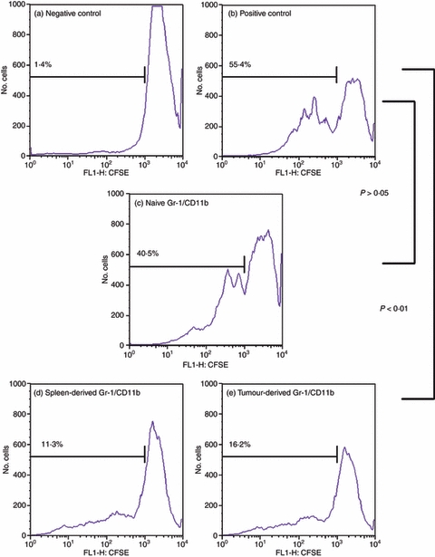
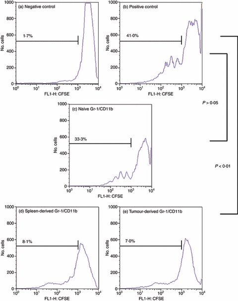
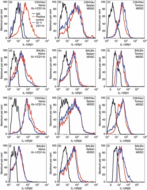
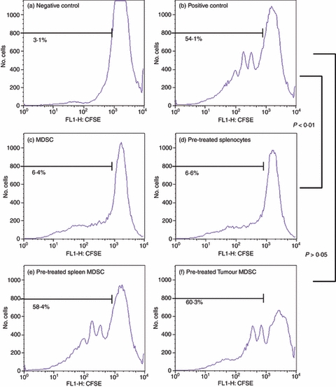
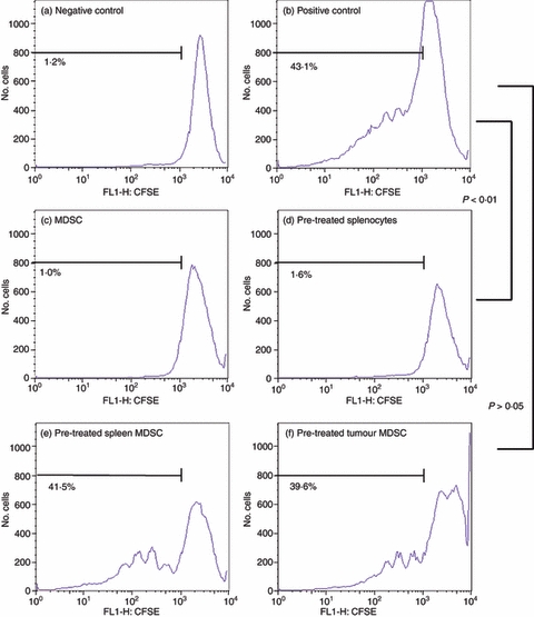
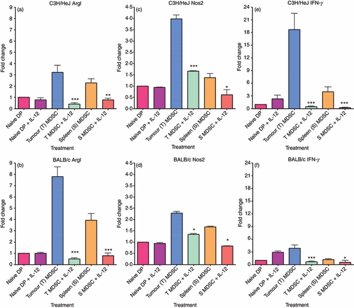
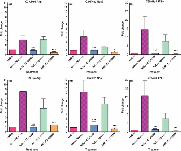
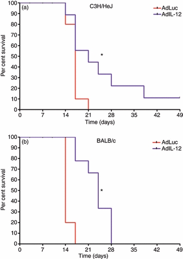
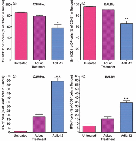
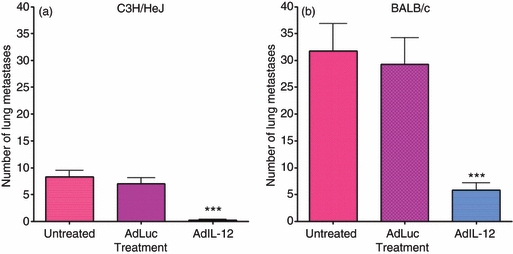
Myeloid-derived suppressor cells (MDSC) are important to the tumour microenvironment as they actively suppress the immune system and promote tumour progression and metastasis. These cells block T-cell activation in the tumour microenvironment, preventing anti-tumour immune activity. The ability of a treatment to alter the suppressive function of these cells and promote an immune response is essential to enhancing overall therapeutic efficacy. Interleukin-12 (IL-12) has the potential not only to promote anti-tumour immune responses but also to block the activity of cells capable of immune suppression. This paper identifies a novel role for IL-12 as a modulator of MDSC activity, with implications for IL-12 as a therapeutic agent. Treatment with IL-12 was found to alter the suppressive function of MDSC by fundamentally altering the cells. Interleukin-12-treated MDSC exhibited up-regulation of surface markers indicative of mature cells as well as decreases in nitric oxide synthase and interferon-γ mRNA both in vitro and in vivo. Treatment with IL-12 was also found to have significant therapeutic benefit by decreasing the percentage of MDSC in the tumour microenvironment and increasing the percentage of active CD8(+) T cells. Treatment with IL-12 resulted in an increase in overall survival accompanied by a reduction in metastasis. The findings in this paper identify IL-12 as a modulator of immune suppression with significant potential as a therapeutic agent for metastatic breast cancer.


Figures

Figure 1
Figure 1
Gr-1/CD11b double-positive cells derived from tumour-bearing C3H/HeJ animals are functional myeloid-derived suppressor cells (MDSC). Whole splenocytes from C3H/HeJ animals were stained with 10 μm CFSE and co-cultured with sorted Gr-1/CD11b double-positive cells at a 1 : 1 ratio for a total of 2 × 10⁵ cells per well. Cells were treated with 5 μg/ml anti-CD3 and anti-CD28 antibodies in 96-well plates for 4 days. The cells were harvested and stained with anti-Gr-1 and anti-CD4 antibodies to isolate only CD4+ T cells for analysis. Cells were gated for CD4+ T cells only and analysed for dilution of CFSE via flow cytometry. A negative control of unstimulated splenocytes (a) and a positive control of stimulated splenocytes only (b) were compared with splenocytes activated in the presence of Gr-1/CD11b double-positive cells from the spleen of naive animals (c) and Gr-1/CD11b double-positive cells from the spleen (d) and tumour (e) of tumour-bearing animals. Percentages indicate per cent activation.
Figure 2
Figure 2
Gr-1/CD11b double-positive cells derived from tumour-bearing BALB/c animals are functional myeloid-derived suppressor cells (MDSC). BALB/c cells were prepared as described previously (Fig. 1). Cells were gated for CD4+ T cells only and analysed for dilution of CFSE via flow cytometry. A negative control of unstimulated splenocytes (a) and a positive control of stimulated splenocytes only (b) were compared with splenocytes activated in the presence of Gr-1/CD11b double-positive cells from the spleen of naive animals (c) and Gr-1/CD11b double-positive cells from the spleen (d) and tumour (e) of tumour-bearing animals. Percentages indicate per cent activation.
Figure 3
Figure 3
Interleukin-12 receptor β1 (IL-12Rβ1) and IL-12Rβ2 expression on the surface of Gr-1/CD11b double-positive cells. Gr-1/CD11b double-positive cells, natural killer (NK) cells, and naive CD4+ T cells were blocked with 1× PBS containing 1% BSA or 2 μg/ml IgG and stained with 2 μg/ml fluorochrome-conjugated anti-IL-12Rβ1 antibody or 2 μg/ml anti-IL12Rβ2 primary antibody followed by 2 μg/ml phycoerythrin-conjugated secondary antibody and analysed via flow cytometry. IL-12Rβ1 (a–f) and IL-12Rβ2 (g–l) were analysed. Naive Gr-1/CD11b double-positive cells from C3H/HeJ (a, g) and BALB/c (d, j) animals were compared with myeloid-derived suppressor cells from spleen (C3H/HeJ b, h; BALB/c e, k) and tumour (C3H/HeJ c, i; BALB/c f, l). Graphs are representative of three independent stains.
Figure 4
Figure 4
Interleukin-12 (IL-12) alters the suppressive activity of C3H/HeJ myeloid-derived suppressor cells (MDSC). C3H/HeJ naive whole splenocytes were stained with 10 μm CFSE and co-cultured with C3H/HeJ MDSC sorted from digested tumours and spleens of C3L5 tumour-bearing animals. MDSC were pre-treated with or without 10 ng/ml IL-12 for 24 hr. Cells were then co-cultured in 96-well plates at a 1 : 1 ratio of whole splenocytes to MDSC for a total of 2 × 10⁵ cells per well stimulated with anti-CD3 and anti-CD28 antibodies for 4 days. After the 4-day incubation, cells were stained with fluorochrome-conjugated anti-CD4 and anti-Gr-1 antibodies to isolate only the CD4+ T cells for analysis. Using gating and flow cytometric analysis, CD4+ T cells from untreated cells were used as a negative control (a) while a positive control consisted of CD4+ T cells treated with anti-CD-3 and anti-CD-28 antibodies (b). Untreated MDSC co-cultured with CD4+ T cells and stimulated with anti-CD3 and anti-CD28 antibodies (c) were also included as a control for suppression. These controls were compared with whole splenocytes pre-treated with IL-12 followed by co-culture with MDSC (d) IL-12 pre-treated spleen-derived MDSC (e) and IL-12 pre-treated tumour-derived MDSC (f) both co-cultured with CD4+ T cells and stimulated with anti-CD3 and anti-CD28 antibodies. Percentages indicate per cent activation.
Figure 5
Figure 5
Interleukin-12 (IL-12) alters the suppressive activity of BALB/c myeloid-derived suppressor cells (MDSC). BALB/c cells were obtained and stained as described previously (Fig. 3). After the 4-day incubation, cells were stained with fluorochrome-conjugated anti-CD4 and anti-Gr-1 antibodies to isolate only the CD4+ T cells for analysis. Using gating and flow cytometric analysis, CD4+ T cells from untreated cells were used as a negative control (a) while a positive control consisted of CD4+ T cells treated with anti-CD-3 and anti-CD-28 antibodies (b). Untreated MDSC co-cultured with CD4+ T cells and stimulated with anti-CD3 and anti-CD28 antibodies (c) were also included as a control for suppression. These controls were compared with whole splenocytes pre-treated with IL-12 followed by co-culture with MDSC. (d) IL-12 pre-treated spleen-derived MDSC (e) and IL-12 pre-treated tumour-derived MDSC (f) both co-cultured with CD4+ T cells and stimulated with anti-CD3 and anti-CD28 antibodies. Percentages indicate per cent activation.
Figure 6
Figure 6
In vitro treatment with interleukin-12 (IL-12) reduces ArgI, Nos2 and interferon-γ (IFN-γ). Gr-1/CD11b double-positive cells were stained using fluorochrome-conjugated antibodies and sorted from the spleens of naive animals as well as the spleens and tumours of tumour-bearing animals. Sorted myeloid-derived suppressor cells (MDSC) were treated for 24 hr with 10 ng/ml recombinant mouse IL-12. RNA was extracted from sorted cell populations, converted to cDNA and analysed for mRNA expression via Real Time PCR. The expression levels for ArgI, Nos2 and IFN-γ were normalized to expression from naive double-positive cells and graphed as a fold change. The expression of ArgI in C3H/HeJ (a) and BALB/c (b), Nos2 in C3H/HeJ (c) and BALB/c (d) and expression of IFN-γ in C3H/HeJ (e) and BALB/c (f) are shown. Statistically significant reductions in mRNA expression were observed following treatment with IL-12 (*P < 0·05; **P < 0·01; ***P < 0·001).
Figure 7
Figure 7
In vivo treatment with interleukin-12 (IL-12) reduces ArgI, Nos2 and interferon-γ (IFN-γ). C3H/HeJ and BALB/c tumour-bearing animals were treated with intramuscular injections of 1 × 10⁹ adenovirus particles of either AdLuc or AdIL-12. Twenty-four hours after inoculation with virus, spleens and tumours were harvested and digested to obtain single-cell suspensions. Gr-1/CD11b double-positive cells were stained using fluorochrome-conjugated antibodies and sorted from the spleens and tumours of AdLuc- or AdIL-12-treated animals. RNA was extracted from sorted cell populations, converted to cDNA and analysed for mRNA expression via Real Time PCR. The expression levels for ArgI, Nos2 and IFN-γ were normalized to expression from naive double-positive cells and plotted on graphs as a fold change. The expression of ArgI in C3H/HeJ (a) and BALB/c (b), Nos2 in C3H/HeJ (c) and BALB/c (d) and expression of IFN-γ in C3H/HeJ (e) and BALB/c (f) are shown. Statistically significant reductions in mRNA expression were observed following treatment with IL-12 (*P < 0·05; ***P < 0·001).
Figure 8
Figure 8
In vivo treatment with interleukin-12 (IL-12) increases overall survival. C3H/HeJ and BALB/c animals were inoculated with 2 × 10⁵ C3L5 and 1 × 10⁵ 4T1 cells, respectively. Once tumours reached an average size of 65 mm3, intramuscular injections of 1 × 10⁹ adenovirus particles of AdLuc or AdIL-12 were performed. Tumours were measured twice weekly. C3H/HeJ (a) and BALB/c (b) animals were analysed for overall survival with tumour volumes of 500 mm3 as the end-point (*P < 0·05).
Figure 9
Figure 9
In vivo treatment with interleukin-12 (IL-12) increases active CD8+ T-cell infiltration into the tumour microenvironment. C3H/HeJ and BALB/c animals were inoculated with 2 × 10⁵ C3L5 and 1 × 10⁵ 4T1 cells, respectively. Once tumours reached an average size of 65 mm3, intramuscular injections of 1 × 10⁹ adenovirus particles of AdLuc or AdIL-12 were performed. Once tumours reached a volume of 500 mm3, tissues were harvested and single-cell suspensions obtained. Gr-1/CD11b double-positive cells were stained as a set of CD45+ cells using fluorochrome-conjugated anti-Gr-1, anti-CD11b and anti-CD45 antibodies. CD8+ T cells were stained using fluorochrome-conjugated anti-CD8 and anti-interferon-γ (IFN-γ) antibodies and analysed via flow cytometry. The percentage of Gr-1/CD11b double-positive cells in C3H/HeJ (a) and BALB/c (b) tumours as a percentage of CD45+ cells was determined. The percentage of CD8+ T cells that also express IFN-γ in C3H/HeJ (c) and BALB/c (d) tumours are also shown. Statistically significant increases in active IFN-γ+ CD8+ T cells were observed following treatment with AdIL-12 (*P < 0·05; **P < 0·01; ***P < 0·001).
Figure 10
Figure 10
In vivo treatment with interleukin-12 (IL-12) decreases metastasis. C3H/HeJ and BALB/c animals were inoculated with 2 × 10⁵ C3L5 and 1 × 10⁵ 4T1 cells, respectively. Once tumours reached an average size of 65 mm3, intramuscular injections of 1 × 10⁹ adenovirus particles of AdLuc or AdIL-12 were performed. Once tumours reached a volume of 500 mm3, lungs were harvested for analysis of metastasis. Lungs were fixed in Bouin's fixative and metastases were counted. Analysis of metastases from C3H/HeJ (a) and BALB/c (b) are also shown. Statistically significant reductions in metastasis were observed following treatment with AdIL-12 (***P < 0·001).

Similar articles

Cited by

References

    1. Gabrilovich DI, Bronte V, Chen SH, Colombo MP, Ochoa A, Ostrand-Rosenberg S, Schreiber H. The terminology issue for myeloid-derived suppressor cells. Cancer Res. 2007;67:425. author reply 6. - PMC - PubMed
    1. Nagaraj S, Gabrilovich DI. Tumor escape mechanism governed by myeloid-derived suppressor cells. Cancer Res. 2008;68:2561–3. - PubMed
    1. Kusmartsev S, Gabrilovich DI. Role of immature myeloid cells in mechanisms of immune evasion in cancer. Cancer Immunol Immunother. 2006;55:237–45. - PMC - PubMed
    1. Serafini P, Meckel K, Kelso M, et al. Phosphodiesterase-5 inhibition augments endogenous antitumor immunity by reducing myeloid-derived suppressor cell function. J Exp Med. 2006;203:2691–702. - PMC - PubMed
    1. Filipazzi P, Valenti R, Huber V, et al. Identification of a new subset of myeloid suppressor cells in peripheral blood of melanoma patients with modulation by a granulocyte–macrophage colony-stimulation factor-based antitumor vaccine. J Clin Oncol. 2007;25:2546–53. - PubMed

MeSH terms