. 2011 Mar 31:6:7.
doi: 10.1186/1747-1028-6-7.

The loop-less tmCdc34 E2 mutant defective in polyubiquitination in vitro and in vivo supports yeast growth in a manner dependent on Ubp14 and Cka2

Affiliations

The loop-less tmCdc34 E2 mutant defective in polyubiquitination in vitro and in vivo supports yeast growth in a manner dependent on Ubp14 and Cka2

Agnieszka Lass et al. Cell Div. .

Erratum in

Abstract

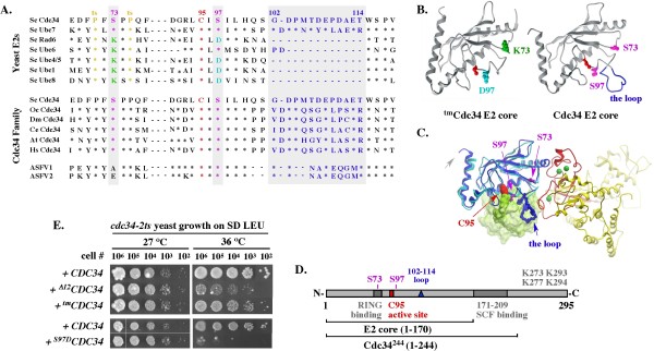
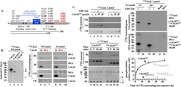
Background: The S73/S97/loop motif is a hallmark of the Cdc34 family of E2 ubiquitin-conjugating enzymes that together with the SCF E3 ubiquitin ligases promote degradation of proteins involved in cell cycle and growth regulation. The inability of the loop-less Δ12Cdc34 mutant to support growth was linked to its inability to catalyze polyubiquitination. However, the loop-less triple mutant (tm) Cdc34, which not only lacks the loop but also contains the S73K and S97D substitutions typical of the K73/D97/no loop motif present in other E2s, supports growth. Whether tmCdc34 supports growth despite defective polyubiquitination, or the S73K and S97D substitutions, directly or indirectly, correct the defect caused by the loop absence, are unknown.

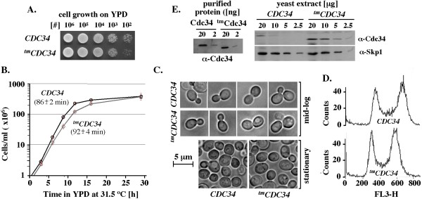
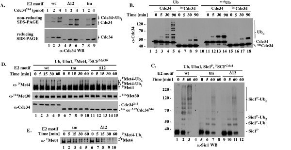
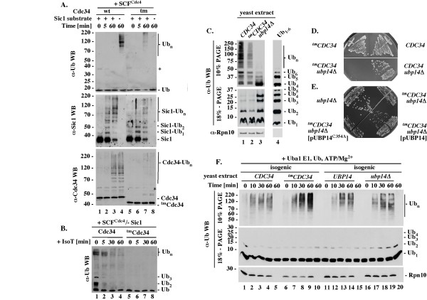
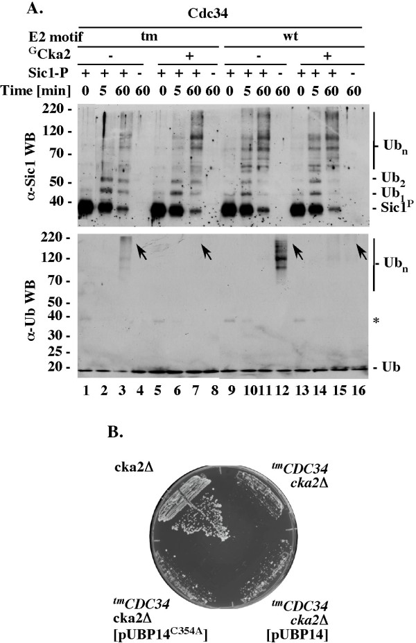
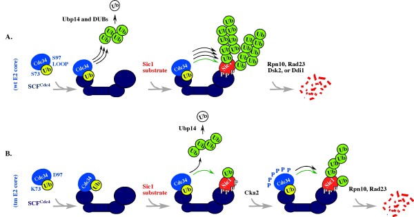
Results: tmCdc34 supports yeast viability with normal cell size and cell cycle profile despite producing fewer polyubiquitin conjugates in vivo and in vitro. The in vitro defect in Sic1 substrate polyubiquitination is similar to the defect observed in reactions with Δ12Cdc34 that cannot support growth. The synthesis of free polyubiquitin by tmCdc34 is activated only modestly and in a manner dependent on substrate recruitment to SCFCdc4. Phosphorylation of C-terminal serines in tmCdc34 by Cka2 kinase prevents the synthesis of free polyubiquitin chains, likely by promoting their attachment to substrate. Nevertheless, tmCDC34 yeast are sensitive to loss of the Ubp14 C-terminal ubiquitin hydrolase and DUBs other than Ubp14 inefficiently disassemble polyubiquitin chains produced in tmCDC34 yeast extracts, suggesting that the free chains, either synthesized de novo or recycled from substrates, have an altered structure.

Conclusions: The catalytic motif replacement compromises polyubiquitination activity of Cdc34 and alters its regulation in vitro and in vivo, but either motif can support Cdc34 function in yeast viability. Robust polyubiquitination mediated by the S73/S97/loop motif is thus not necessary for Cdc34 role in yeast viability, at least under typical laboratory conditions.


Figures

Figure 1
Figure 1
Disruption, but not replacement, of the S73/S97/loop motif conserved among the Cdc34/Ubc7 E2 family is lethal for yeast. (A). Partial sequence alignment. Asterisks represent residues identical to Cdc34 and dashes represent gaps. Sc - S. cerevisiae; Oc - O. cuniculus; Dm - D. melanogaster; Ce - C. elegans; At - A. thaliana; Hs - H. sapiens; ASFV1 - African swine fever virus (GI:9628248); ASFV2 -African swine fever virus (GI:450743). (B). Structural models of the E2 core (a.a. 1-170) of Cdc34 and tmCdc34. Residues corresponding to K73 and D97 in tmCdc34, and S73 and S97 in Cdc34, are color coded as in A and shown in the context of structures of a scRad6 and scUbc7 fragment (Methods); navy blue: the acidic loop formed by scUbc7 residues corresponding to amino acids 103-114 in Cdc34; red: the residue corresponding to the catalytic site C95 of Cdc34 and tmCdc34. (C). Model of ubiquitin-charged Cdc34 (a.a. 1-170) bound to the RING domain of Rbx1. See Methods. (D). Scheme of Cdc34 domains of interest. (E). Rescue of cdc34-2ts yeast growth with Cdc34 E2 core mutant constructs. Cultures of cdc34-2ts strain carrying the indicated constructs under the GAL10 promoter on a 2μ YEp51 plasmid were grown overnight at 27°C in SD-Leu, adjusted to a density of 1 × 10⁸ cells/ml, serially diluted, spotted onto SD-Leu plates and incubated at permissive (27°C) or non-permissive (37°C) temperature for 4 days. Note that dextrose allows only low expression of the GAL10 controlled constructs.
Figure 2
Figure 2
Isogenic CDC34 (BL2) and tmCDC34 (RC85) yeast strains have similar growth properties, cell size and cell cycle profiles under typical laboratory conditions. (A). Growth on plates. Overnight cultures were adjusted to a density of 1 × 10⁸ cells/ml, serially diluted, spotted onto YPD and grown at 30°C. (B). Growth in liquid culture. Each strain was inoculated in three biological replicates into pre-warmed 50 ml of YPD at a starting density of 5 × 10⁵ cells/ml and grown with vigorous shaking at 31.5°C. The doubling time for CDC34 (BL2) is 86 +/- 2 min and tmCDC34 (RC85) is 92 +/- 4 minutes during the exponential phase of growth (0-11.75 hrs). (C). Light microscopic images of cells. Yeast cultures were grown as in B, with images collected at 1.5 × 10⁷ cells/ml (mid-log phase) and 2 × 10⁸ cells/ml (stationary phase). (D). Cell cycle distributions. Yeast cells were analyzed by flow cytometry for their DNA content using propidium iodide staining (Methods). (E). Quantitative western blot analysis of the steady-state levels of Cdc34 and tmCdc34 proteins. Yeast extracts were prepared as described in Methods. The indicated amounts of total proteins were separated by SDS-PAGE and analyzed by α-Cdc34 WB. Control α-Skp1 WB was performed to verify equal loading of analyzed samples. Known amounts of purified Cdc34 and tmCdc34 were used to verify that the α-Cdc34 antibodies have similar affinity for each protein construct.
Figure 3
Figure 3
Purified tmCdc34 forms ubiquitin thiolester and monoubiquitinates substrates, but is defective in substrate polyubiquitination. All assays were performed at 25°C in 10-20 μL and included 1 pmol of Uba1 E1 and 1.3 nmol of ubiquitin or its derivative. (A). Formation of ubiquitin-thiolester. The indicated Cdc34244 proteins with wt, tm or Δ12 E2 core (1-4 pmol) were incubated for 5 minutes with Uba1 and ubiquitin followed by SDS-PAGE under reducing (+ βME) or non-reducing (- βME) conditions and α-Cdc34 WB. (B). Autoubiquitination of full length Cdc34 and tmCdc34. Full length Cdc34 or tmCdc34 (5 pmol) was incubated for the times indicated with Uba1 and ubiquitin or methylated ubiquitin followed by α-Cdc34 WB. (C). FSCFCdc4-dependent polyubiquitination of Sic1. The indicated Cdc34244 proteins (5 pmol) were incubated for the times indicated with Uba1, the Sic1/Clb5/GstCdc28 substrate complex (2 pmol) and FSCFCdc4 (2 pmol) followed by 10% SDS-PAGE and α-Sic1 WB. Reactions shown in lanes 4, 8 and 12 do not have Sic1. (D). GstSCFMet30-dependent polyubiquitination of Met4. Tests were performed as described in C except that with the GstSCFMet30 E3 and FMet4 substrate. (E). GstSCFMet30-dependent monoubiquitination of FMet4. Tests as in D, but analyzed with shorter (1 instead of 5 minutes) western blot exposure time, which is necessary to visualize unubiquitinated and monoubiquitinated FMet4 as separate species due to similarities in their molecular weights.
Figure 4
Figure 4
The synthesis of free polyubiquitin chains in vitro and in vivo. (A). Regulation of free polyubiquitin chain synthesis by substrate recruitment to SCFCdc4. Standard ubiquitination reactions with Cdc34 or tmCdc34 (5 pmol) were analyzed by western blots with α-Ub, α-Sic1 or α-Cdc34 antibodies. (B). IsoT sensitivity. Reactions as in A lanes 4 and 8 were analyzed for IsoT sensitivity (Methods). (C). Levels of ubiquitin and ubiquitin conjugates in vivo. Boiled cell extracts (Methods) were analyzed by western blot with α-Ub (Covance) or α-Rpn10 antibodies. Lane 4: 200 ng of free polyubiquitin chains Ub1-6 purchased from Enzo. (D). Growth of tmCDC34 but not CDC34 yeast is sensitive to loss of UBP14. Haploids with the indicated genotypes were selected on haploid selection media with G418 and nourseothricin at 30°C for three days. (E). Overexpression of Ubp14 but not Ubp15C354A supports growth of tmCDC34 upb14Δ yeast. Heterozygous diploids (RC171 and RC172) were transformed with the indicated plasmids that overexpress Ubp14 (pUBP14) or Ubp14C354A (pUBP14-C354A), patched onto sporulation media and incubated at 26°C for five days. Haploids with the indicated genotypes were selected as in D. (F). Accumulation of ubiquitin conjugates in yeast extracts enriched with Uba1, ubiquitin, ATP and MgCl2. Extracts with active ubiquitin-proteasome system (10 μg of total proteins; see Methods) were enriched with Uba1 (10 pmol), ubiquitin (1.3 nmol) ATP (2 mM) and MgCl2 (2 mM), incubated at 25°C for the times indicated and analyzed by 10% or 18% SDS-PAGE followed by α-Ub (Sigma) or α-Rpn10 WB.
Figure 5
Figure 5
Yeast GstCka1 or GstCka2 kinases phosphorylate only the most C-terminal serines in Cdc34 and tmCdc34. (A). Scheme of Cdc34. Red: the six most C-terminal serines. Residue 244 represents the C-terminal end in Cdc34244 that includes the E2 catalytic core (a.a. 1 to 170) with the active site cysteine C95, the E3 RING domain-interacting fragment, and the 39 a.a. C-terminal fragment (a.a. 171-209) also implicated in binding to SCF. (B). In a physiological range of protein concentrations, yeast GstCka1 phosphorylates only the six most C-terminal serines within Cdc34. 1 hour assays included 10 μM ATP with 0.1 μ Ci of [γ-32P]ATP (4500 Ci/mmol), 1 pmol of the indicated Cdc34 constructs and 0.5 or 2.5 pmol of GstCka1. (C). GstCka2 phosphorylates the S207 and S216 most C-terminal serines in Cdc34244 and tmCdc34244. Assays were performed for 1 hour (or as indicated), with 5 pmols (or as indicated) of the indicated constructs, 1pmol of GstCka1 kinase and 10 μM ATP with 5 μCi [32P] ATP (4500Ci/mmol), leading to ~50-fold higher sensitivity of [32P] detection than in B. Graph: quantitation of the [32P] signal of the proteins at different autoradiogram exposure times. CB: Coomassie blue.
Figure 6
Figure 6
GstCka2 inhibits synthesis of free polyubiquitin chains by Cdc34 constructs. (A). Effects of GstCka2 on Sic1 ubiquitination and synthesis of free polyubiquitin chains by Cdc34 proteins. 5 pmol of Cdc34 or tmCdc34 were phosphorylated at 25°C for 1 hour, as in 5B. The assays were then supplemented with 1 pmol E1, 660 pmol of ubiquitin, 2 pmol of FlagSCFCdc4 and 2 pmol of Sic1/Cln5/GstCdc28, where indicated, and incubated at 25°C for the times indicated, followed by α-Sic1 and α-Ub WBs. (B). Ectopic expression of Ubp14 or Ubp15C354A does not rescue growth of tmCDC34 cka2Δ yeast. The heterozygous diploid RC174 was transformed with the indicated plasmids that overexpress Ubp14 (pUBP14) or catalytically inactive Ubp14 (pUBP14-C354A) from the ADH1 promoter. These transformed diploids as well as RC173 were patched onto sporulation media and incubated at 26°C for five days. Haploids with the indicated genotypes were selected by streaking the heterozygous diploids on haploid selection media with G418 and nourseothricin. Plates were incubated at 30°C for three days.
Figure 7
Figure 7
Summary Model. The model emphasizes that tmCdc34 is functional in substrate monoubiquitination but has a defect in the synthesis of substrate-attached and free polyubiquitin chains. This emphasis does not mean that tmCdc34 cannot synthesize polyubiquitin chains. Rather, it emphasizes that long polyubiquitin chains cannot be synthesized in the short time frame normally available for their synthesis in vivo. Cdc34 is marked blue; SCFCdc4 is marked navy blue; Sic1 substrate is marked red; the free form of ubiquitin (Ub) is marked white; the ubiquitin-thiolester is marked yellow; the ubiquitin engaged in isopeptide bond (with substrate or other ubiquitin molecules) is marked green. Black arrows indicate a robust (three arrows), or modest (one arrow) synthesis of polyubiquitin chains. Green arrow indicates monoubiquitination.

Similar articles

Cited by

References

    1. Hershko A, Ciechanover A. The Ubiquitin System. Ann Rev Biochem. 1998;67:425–79. doi: 10.1146/annurev.biochem.67.1.425. - DOI - PubMed
    1. Thrower JS, Hoffman L, Rechsteiner M, Pickart C. Recognition of the polyubiquitin proteolytic signal. EMBO J. 2000;19:94–102. doi: 10.1093/emboj/19.1.94. - DOI - PMC - PubMed
    1. Pickart CM, Fushman D. Polyubiquitin chains: polymeric protein signals. Curr Opin Chem Biol. 2004;8:610–6. doi: 10.1016/j.cbpa.2004.09.009. - DOI - PubMed
    1. Deshaies RJ, Joazeiro C. RING domain ubiquitin E3 ligases. Annu Rev Biochem. 2009;78:399–434. doi: 10.1146/annurev.biochem.78.101807.093809. - DOI - PubMed
    1. Goebl MG, Yochem J, Jentsch S, McGrath J, Varshavsky A, Byers B. The yeast cell cycle gene CDC34 encodes a ubiquitin-conjugating enzyme. Science. 1988;241:1331–1335. doi: 10.1126/science.2842867. - DOI - PubMed