. 2011 Jun;127(6):1522-31.e8.
doi: 10.1016/j.jaci.2011.02.005. Epub 2011 Mar 31.

Human mast cell degranulation and preformed TNF secretion require mitochondrial translocation to exocytosis sites: relevance to atopic dermatitis

Affiliations

Human mast cell degranulation and preformed TNF secretion require mitochondrial translocation to exocytosis sites: relevance to atopic dermatitis

Bodi Zhang et al. J Allergy Clin Immunol. 2011 Jun.

Abstract

Background: Mast cells derive from hematopoietic cell precursors and participate in tissue allergic, immune, and inflammatory processes. They secrete many mediators, including preformed TNF, in response to allergic, neuropeptide, and environmental triggers. However, regulation of mast cell degranulation is not well understood.

Objective: We investigated the role of mitochondrial dynamics in degranulation of human cultured mast cells.

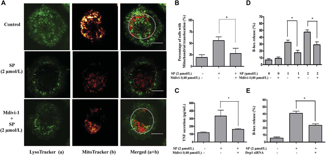
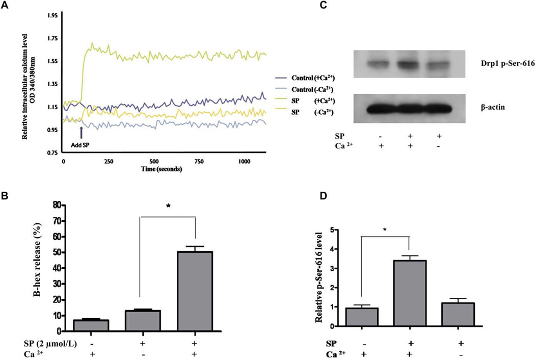
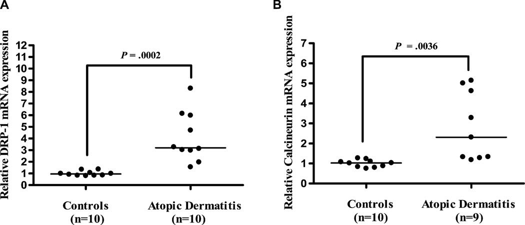
Methods: Human umbilical cord blood-derived mast cells (hCBMCs) and Laboratory of Allergic Diseases 2 (LAD2) mast cells were examined by confocal and differential interference contrast microscopy during activation by IgE/antigen and substance P (SP). Mast cells in control and atopic dermatitis (AD) skin were evaluated by transmission electron microscopy. LAD2 cells were pretreated with mitochondrial division inhibitor, a dynamin-related protein 1 (Drp1) inhibitor, and small interfering RNA for Drp1, which is necessary for mitochondrial fission and translocation. Calcineurin and Drp1 gene expression was analyzed in stimulated LAD2 cells and AD skin biopsies.

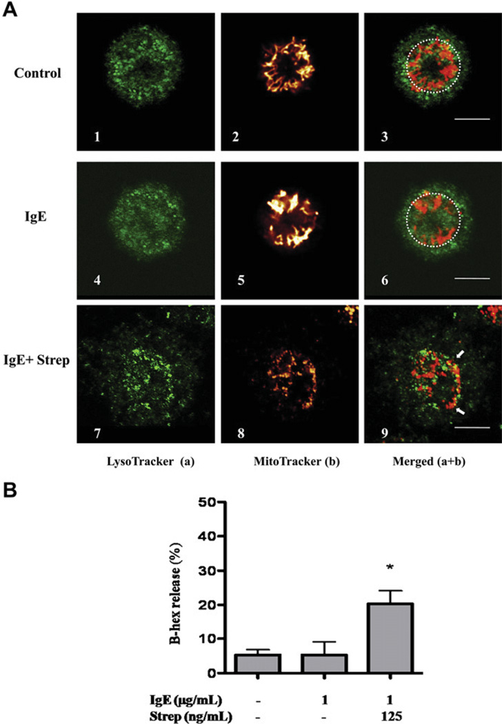
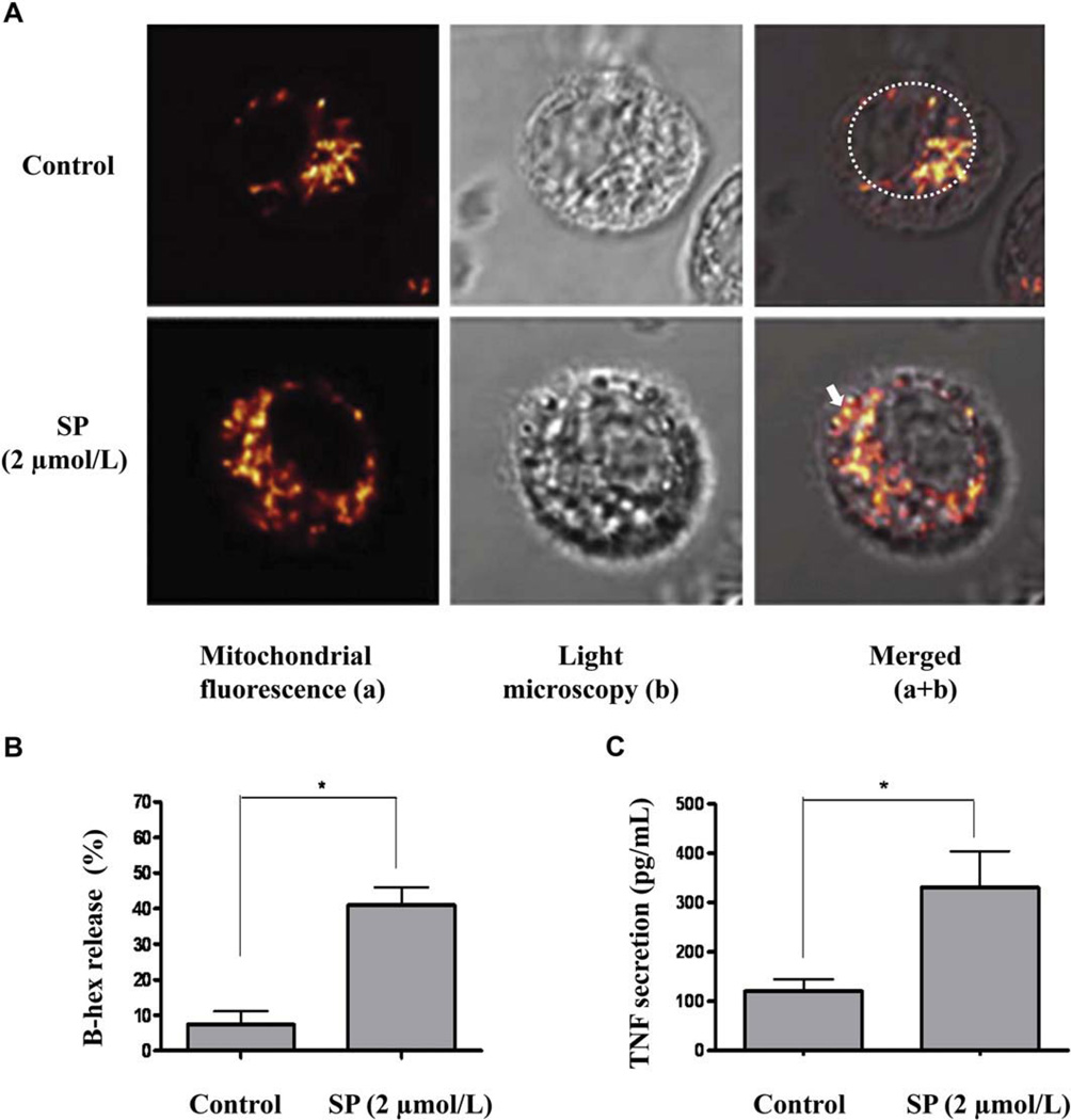
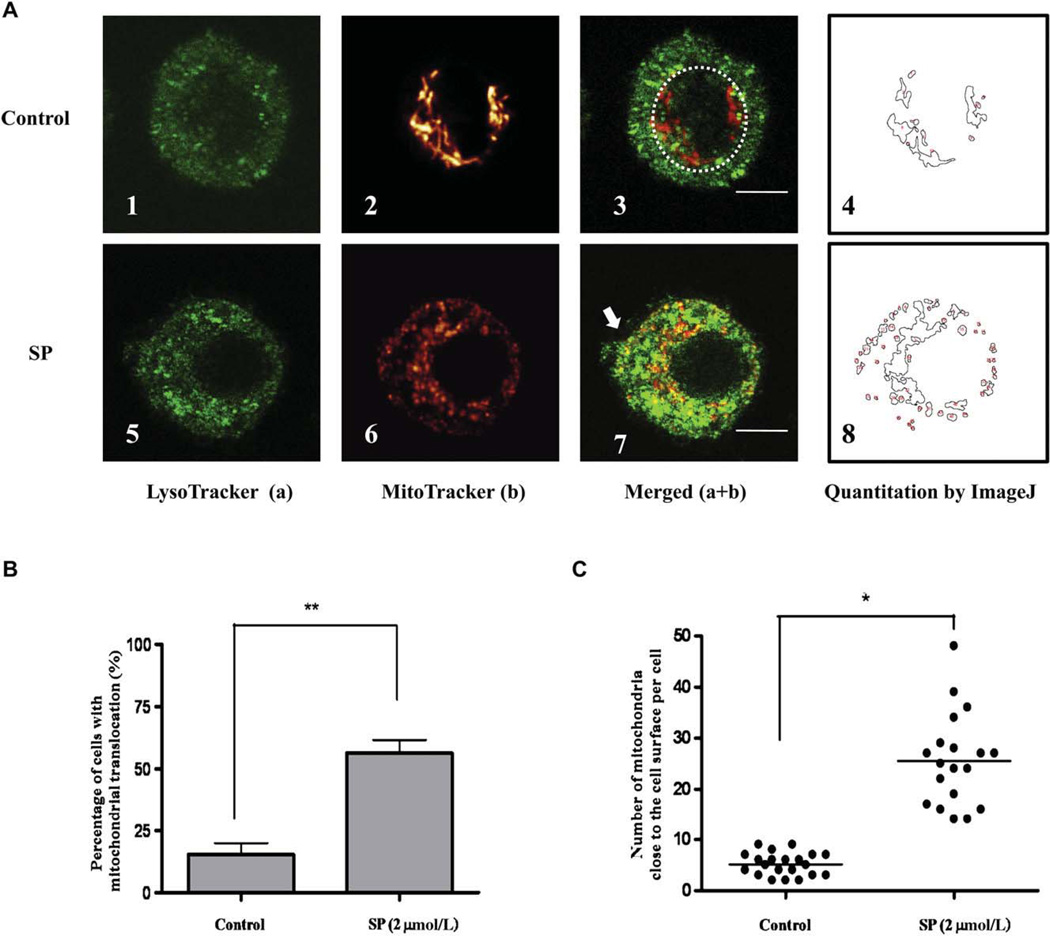
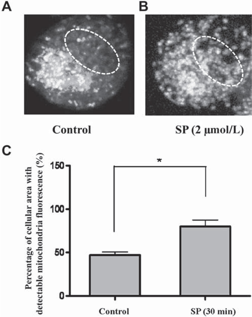
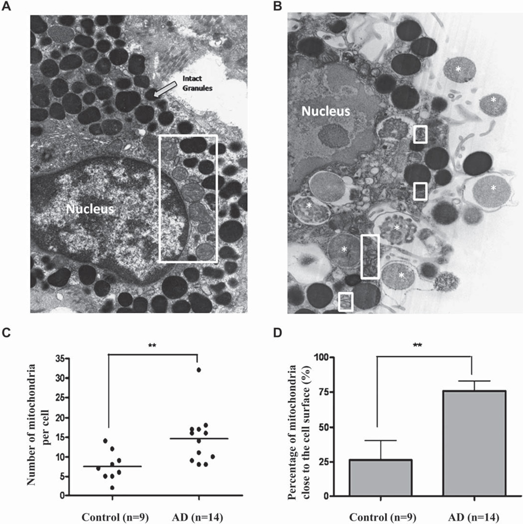
Results: Stimulation of hCBMCs with IgE/antigen or LAD2 cells with SP leads to rapid (30 minutes) secretion of preformed TNF. Degranulation is accompanied by mitochondrial translocation from a perinuclear location to exocytosis sites. Extracellular calcium depletion prevents these effects, indicating calcium requirement. The calcium-dependent calcineurin and Drp1 are activated 30 minutes after SP stimulation. Reduction of Drp1 activity by mitochondrial division inhibitor and decrease of Drp1 expression using small interfering RNA inhibit mitochondrial translocation, degranulation, and TNF secretion. Mitochondrial translocation is also evident by transmission electron microscopy in skin mast cells from AD biopsies, in which gene expression of calcineurin, Drp1, and SP is higher than in normal skin.

Conclusion: Human mast cell degranulation requires mitochondrial dynamics, also implicated in AD.


Conflict of interest statement

Disclosure of potential conflict of interest: T. C. Theoharides and B. Zhang are listed as the inventors of the provisional patent application US 61/405,414. The rest of the authors have declared that they have no conflict of interest.

Figures

FIG 1
FIG 1
Mitochondrial translocation and degranulation in hCBMCs observed by confocal microscopy. A, Mitochondrial distribution in resting (panels 1–3), IgE-incubated (1 µg/mL) (panels 4–6), and IgE (1 µg/mL) + streptavidin (125 ng/mL)–stimulated (panels 7–9) cells. Cells were stained with LysoTracker (a, green) and MitoTracker (b, red). B, β-hex release from hCBMCs treated with IgE or IgE + streptavidin (Strep) for 30 minutes. n = 3; *P < .05 compared with control. Bars equal to 5 µm.
FIG 2
FIG 2
Mitochondrial translocation observed by differential interference contrast, and degranulation and TNF secretion in LAD2 mast cells. A, Mitochondrial distribution of control (upper panels) and degranulated (bottom panels) mast cells stimulated with SP (2 µmol/L) for 30 minutes. Cells were stained with Mito-Tracker (red). Mast cell degranulation by SP was confirmed by β-hex release (B) and TNF secretion (C). n = 3; *P < .05 compared with control.
FIG 3
FIG 3
Quantification of mitochondrial translocation during degranulation in human LAD2 cells. A, Control (n = 19; panels 1–4) and degranulated (n = 20; panels 5–8) mast cells (SP 2 µmol/L, 30 minutes). B, Percentage of translocated mitochondria in resting and stimulated cells (n = 3). C, Number of mitochondria per cell within 1 µm from the cell surface. Cells were randomly selected for analysis (*P < .05; **P < .01; horizontal bars indicate the means). Bars equal to 5 µm.
FIG 4
FIG 4
Z-stack mitochondrial fluorescence projection in control and stimulated human LAD2 mast cells. Control (A) and degranulated (B) LAD2 cell. White dashed lines represent the nuclear region. C, The percentage of the cellular area with detectable mitochondrial fluorescence indicating mitochondrial distribution was calculated from 20 cells (*P < .05).
FIG 5
FIG 5
Electron photomicrographs showing mitochondrial translocation in human skin mast cells from patients with AD. Human skin mast cells from control (3 subjects, 9 mast cells; A) and lesional skin from patients with AD (5 subjects, 14 mast cells; magnification × 13,800; B). Mitochondria are shown within white rectangles. White asterisks represent secreted granular material. C, Number of mitochondria per cell. D, Percentage of mitochondria close to the cell surface in each cell (**P < .01; horizontal bars indicate the means).
FIG 6
FIG 6
Mdivi-1 and Drp1 siRNA inhibit mast cell mitochondrial translocation, degranulation, and preformed TNF secretion. A, Mitochondrial morphology before or after stimulation with SP and pretreatment with mdivi-1. Cells were stained with LysoTracker (green) and MitoTracker (red). B, Percentage of translocated mitochondria in resting, stimulated, and mdivi-1-treated cells. C, TNF secretion. D, β-hex release. E, β-hex release after treatment with Drp1 siRNA before SP stimulation (n = 3; *P < .05). Bars equal to 5 µm.
FIG 7
FIG 7
Intracellular calcium increase and Drp1 phosphorylation at Ser-616 during LAD2 mast cell degranulation. A, Intracellular calciumlevel was measured after SP stimulation. B, LAD2 cell degranulation with or without extracellular calcium, as measured by β-hex release. C, Western blot of Drp1 phosphorylation at Ser-616 after SP (2 µmol/L) stimulation with or without extracellular calcium. D, Drp1 protein density was normalized against β-actin (n = 3; *P < .05). There was no statistical difference in Drp1 phosphorylation at Ser-616 between control and SP stimulation in the absence of calcium.
FIG 8
FIG 8
Drp1 and calcineurin gene expression in skin from patients with AD compared with healthy controls. Gene expression of Drp1 (controls, n = 10; patients, n = 10; A) and calcineurin (controls, n = 10; patients, n = 9; B). The calcineurin patient samples were 1 fewer because the cDNA was exhausted. Relative quantities of mRNA expression were measured by quantitative real-time-PCR and normalized to GAPDH (horizontal bars indicate the means).

References

    1. Galli SJ, Nakae S, Tsai M. Mast cells in the development of adaptive immune responses. Nat Immunol. 2005;6:135–142. - PubMed
    1. Theoharides TC, Kalogeromitros D. The critical role of mast cell in allergy and inflammation. Ann N Y Acad Sci. 2006;1088:78–99. - PubMed
    1. Gordon JR, Galli SJ. Mast cells as a source of both preformed and immunologically inducible TNF-α/cachectin. Nature. 1990;346:274–276. - PubMed
    1. Olszewski MB, Groot AJ, Dastych J, Knol EF. TNF trafficking to human mast cell granules: mature chain-dependent endocytosis. J Immunol. 2007;178:5701–5709. - PubMed
    1. Gibbs BF, Wierecky J, Welker P, Henz BM, Wolff HH, Grabbe J. Human skin mast cell rapidly release preformed and newly generated TNF-alpha and IL-8 following stimulation with anti-IgE and other secretagogues. Exp Dermatol. 2001;10:312–320. - PubMed

Publication types

MeSH terms