. 2011 May 26;117(21):5692-700.
doi: 10.1182/blood-2010-12-323857. Epub 2011 Mar 31.

A novel vascular disrupting agent plinabulin triggers JNK-mediated apoptosis and inhibits angiogenesis in multiple myeloma cells

Affiliations

A novel vascular disrupting agent plinabulin triggers JNK-mediated apoptosis and inhibits angiogenesis in multiple myeloma cells

Ajita V Singh et al. Blood. .

Abstract

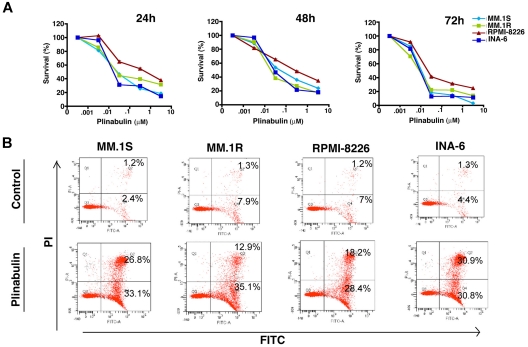
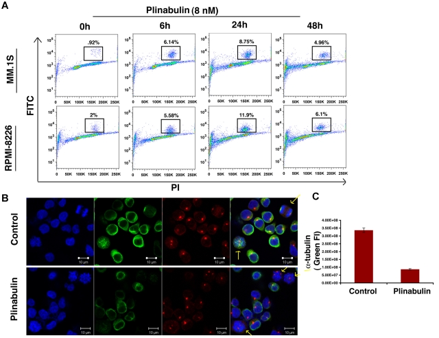
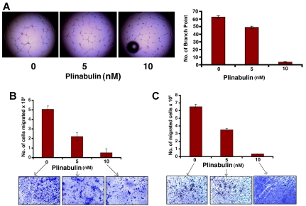
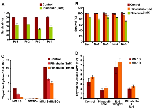
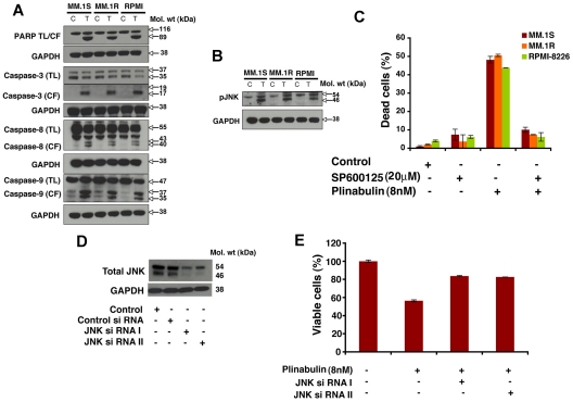
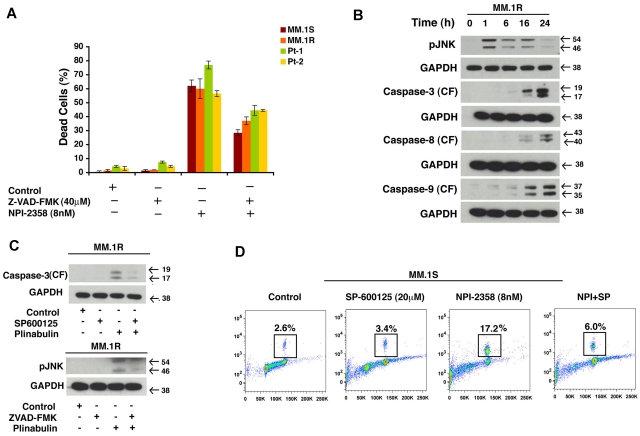
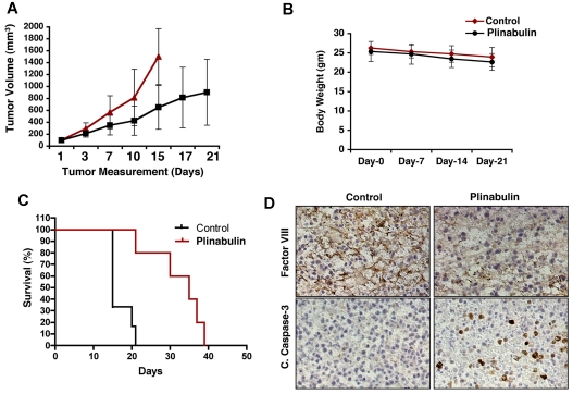
Previous studies have established a role of vascular-disrupting agents as anti- cancer agents. Plinabulin is a novel vascular-disrupting agent that exhibits potent interruption of tumor blood flow because of the disruption of tumor vascular endothelial cells, resulting in tumor necrosis. In addition, plinabulin exerts a direct action on tumor cells, resulting in apoptosis. In the present study, we examined the anti-multiple myeloma (MM) activity of plinabulin. We show that low concentrations of plinabulin exhibit a potent antiangiogenic action on vascular endothelial cells. Importantly, plinabulin also induces apoptotic cell death in MM cell lines and tumor cells from patients with MM, associated with mitotic growth arrest. Plinabulin-induced apoptosis is mediated through activation of caspase-3, caspase-8, caspase-9, and poly(ADP-ribose) polymerase cleavage. Moreover, plinabulin triggered phosphorylation of stress response protein JNK, as a primary target, whereas blockade of JNK with a biochemical inhibitor or small interfering RNA strategy abrogated plinabulin-induced mitotic block or MM cell death. Finally, in vivo studies show that plinabulin was well tolerated and significantly inhibited tumor growth and prolonged survival in a human MM.1S plasmacytoma murine xenograft model. Our study therefore provides the rationale for clinical evaluation of plinabulin to improve patient outcome in MM.


Figures

Figure 1
Figure 1
Plinabulin inhibits growth and triggers apoptosis in MM. (A) Human MM cell lines MM.1S, MM.1R, RPMI-8226, and INA-6 were treated with plinabulin (dose range, 0.001-10μM) for 24, 48, and 72 hours; cell viability was measured with MTT assays. Data presented are mean ± SD of 3 independent experiments (P < .05 for all the cell lines at different time points). (B) MM.1S, MM.1R, RPMI-8226, and INA-6 cell lines were treated with plinabulin (8nM) for 48 hours, and apoptosis was measured with Annexin V/PI binding assay by flow cytometry (P < .05; n = 3). A representative graph from 3 independent experiments is shown. Right quadrant: top panel, Annexin V+/PI+ cells; bottom panel, Annexin V+.
Figure 2
Figure 2
Plinabulin treatment leads to mitotic block in MM. (A) MM cell lines MM.1S and RPMI-8226 were treated with plinabulin (8nM) for 6, 24, and 48 hours, followed by analysis for histone H3 phosphorylation with the use of flow cytometry. Data are representation of 3 independent experiments. (B) MM.1S cells were treated with plinabulin (8nM) for 24 hours and stained for α-tubulin, pericentrin, and Hoechst. (C) Fluorescence intensity (FI) of α-tubulin (green fluorescence) was measured with the use of Velocity software (Improvision). Representative figures (C-D) are from 4 independent experiments. Error bars indicate SD.
Figure 3
Figure 3
Antivascular activity of plinabulin. (A) HUVECs were treated with plinabulin (5nM) for 12 hours and assessed for in vitro vascularization with the use of matrigel capillary-like tube structure formation assays (magnification, 4×/0.10 NA oil; media, EBM-2). (Left) Micrograph images show the effect of plinabulin on capillary tube branch formation. (Right) The bar graph represents quantification of capillary-like tube structure formation in response to plinabulin. Branch points in several random view fields/well were counted; values were averaged; and statistically significant differences were measured with the Student t test. (B-C) For migration assay, HUVECs and MM cells were treated with plinabulin (5nM and 10nM) for 12 hours; cells were > 90% viable at this time point. Cells were washed and cultured in serum-free medium, plated on a fibronectin-coated polycarbonate membrane in the upper chamber of trans-well inserts, and exposed for 2 hours to serum containing medium in the lower chamber. Cells migrating to the bottom face of the membrane were fixed with 90% ethanol and stained with crystal violet (magnification, 10×/0.25 NA oil). A total of 3 randomly selected fields were examined for cells that had migrated from the top to the bottom chambers. (B-C top) Bar graph represents quantification of migrated cells. Data presented are means ± SD (n = 2; P < .05 for control versus plinabulin). (B-C bottom) Image is representative of 2 experiments with similar results.
Figure 4
Figure 4
Plinabulin induces cell death in patient tumor (CD138+) cells and inhibits BMSC-induced MM cell growth. (A) Purified patient MM cells were treated with plinabulin (8nM) for 48 hours, and cell death was measured with Trypan Blue Exclusion assays. Data presented are mean ± SD of triplicate samples (P < .05 for all patients). (B) PBMCs from 5 healthy donors were treated with plinabulin (0.1μM and 1μM) for 48 hours and then analyzed for viability with MTT assay. Data presented are mean ± SD of triplicate samples (P < .05; n = 3). (C) MM.1S cells were treated with plinabulin (8nM, 48 hours) in the presence or absence of 3 different patient BMSCs, and cell growth was measured with thymidine incorporation. Data presented are mean ± SD of triplicate samples (P < .05; n = 3,). (D) MM.1S and MM.1R cells were treated with plinabulin (8nM, 48 hours) in the presence or absence of rhIL-6 (10 ng/mL), and then cell growth was measured with thymidine incorporation. Data presented are mean ± SD of triplicate samples (P < .05 for all the cell lines).
Figure 5
Figure 5
Plinabulin-induced apoptosis in MM cells is associated with activation of caspases and JNK. (A) MM.1S, MM.1R, and RPMI-8226 MM cells were treated with plinabulin (8nM) for 48 hours and harvested, and total protein lysates were subjected to Western blot analysis with the use of antibodies against PARP, caspase-3, caspase-8, caspase-9, or GAPDH. TL indicates total length; CF, cleaved fragment. Blots shown are representative of 2 independent experiments. (B) MM.1S and MM.1R MM cells were treated with plinabulin (8nM) for 48 hours and harvested, and total protein lysates were subjected to Western blot analysis with antibodies against pJNK or GAPDH. Blots shown are representative of 2 independent experiments. (C) MM.1S, MM.1R, and RPMI-8226 MM cells were pretreated with the biochemical inhibitor of JNK (SP600125; 20μM for 30 minutes), followed by plinabulin treatment (8nM, 48 hours). After incubation, cell death was measured with the Trypan Blue assay (n = 3; P < .05). (D) MM.1S cells were transfected with 100nM siRNA JNK I or JNK II or scrambled siRNA with the use of the cell line Nucleofactor Kit V solution (Amaxa Biosystems/Lonza) for 72 hours, and protein expression of JNK-I or JNK-II was examined by immunoblotting with antibodies specific for JNK I and II. (E) Transfected MM.1S cells were treated with plinabulin (8nM, 48 hours), and cell viability was measured with the MTT assay (n = 3; P > .05).
Figure 6
Figure 6
Cell death induced by plinabulin depends on JNK as well as caspases. (A) MM.1S, MM.1R, and primary patients cells were pretreated with PAN caspase inhibitor Z-VAD-FMK (40μM, 2 hours) followed by plinabulin treatment (8nM, 48 hours). After the desired time point cell death were measured with the Trypan Blue assay (n = 2; P > .05). (B) MM.1R cells were treated with higher doses of plinabulin (20nM) for 1, 6, 16, and 24 hours, and Western blot analyses were performed with antibodies against pJNK, caspase-3, caspase-8, caspase-9, or GAPDH. CF indicates cleaved fragment. Results were representative of 2 independent experiments with similar results. (C top) MM.1R cells were treated with PAN caspase inhibitor for 2 hours, and then plinabulin (8nM) was added for an additional 48 hours. Protein lysate was prepared; phosphorylation of JNK was checked with Western blot analysis. (C bottom) MM.1R cells were pretreated with JNK inhibitor SP600125 (20μM, 2 hours), and plinabulin (8nM) was added for another 48 hours. After 48 hours protein lysate was prepared and cleaved caspase-3 expression was checked with Western blot analysis. (D) MM cell line MM.1S was pretreated with SP600125 (20μM) for 2 hours, and plinabulin (8nM) was added for an additional 16 hours followed by analysis for histone H3 phosphorylation with the use of flow cytometry. Data are representation of 3 independent experiments.
Figure 7
Figure 7
In vivo anti-MM activity of plinabulin. (A) MM.1S cells (5 × 10⁶ in 100 μL of serum-free RPMI-1640 medium) were implanted subcutaneously in mice (7 mice/group); average and standard deviation of tumor volume (mm3) were monitored every third day. Mice were treated intraperitoneally with plinabulin (7.5 mg/kg) or vehicle alone twice weekly for 3 weeks. Bars indicate mean ± SD (P = .05). (B) Body weight of plinabulin-treated versus control mice was monitored once a week. Data show ± SD of 6 different mice/group. (C) Kaplan-Meier plot showing survival of mice treated with plinabulin compared with vehicle-treated controls. (D) Tumors from control and plinabulin-treated mice were subjected to immunostaining with antibodies against cleaved caspase-3 and factor VIII. Photographs are representative of similar observations in 2 different mice receiving the same treatment.

References

    1. Strobeck M. Multiple myeloma therapies. Nat Rev Drug Discov. 2007;6(3):181–182. - PubMed
    1. Hideshima T, Mitsiades C, Tonon G, Richardson PG, Anderson KC. Understanding multiple myeloma pathogenesis in the bone marrow to identify new therapeutic targets. Nat Rev Cancer. 2007;7(8):585–598. - PubMed
    1. Dimopoulos MA, Arbuck S, Huber M, et al. Primary therapy of multiple myeloma with paclitaxel (taxol). Ann Oncol. 1994;5(8):757–759. - PubMed
    1. Miller HJ, Leong T, Khandekar JD, Greipp PR, Gertz MA, Kyle RA. Paclitaxel as the initial treatment of multiple myeloma: an Eastern Cooperative Oncology Group Study (E1A93). Am J Clin Oncol. 1998;21(6):553–556. - PubMed
    1. Friedenberg WR, Graham D, Greipp P, Blood E, Winston RD. The treatment of multiple myeloma with docetaxel (an ECOG study). Leuk Res. 2003;27(8):751–754. - PubMed

Publication types

MeSH terms