

The E3 ubiquitin ligase- and protein phosphatase 2A (PP2A)-binding domains of the Alpha4 protein are both required for Alpha4 to inhibit PP2A degradation
- PMID: 21454489
- PMCID: PMC3093842
- DOI: 10.1074/jbc.M111.222414
The E3 ubiquitin ligase- and protein phosphatase 2A (PP2A)-binding domains of the Alpha4 protein are both required for Alpha4 to inhibit PP2A degradation
Abstract
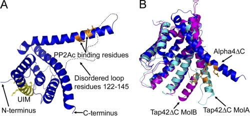
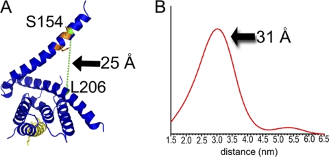
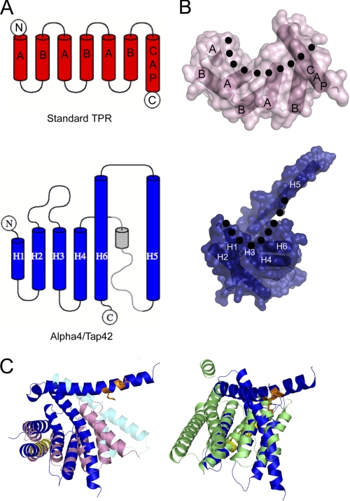
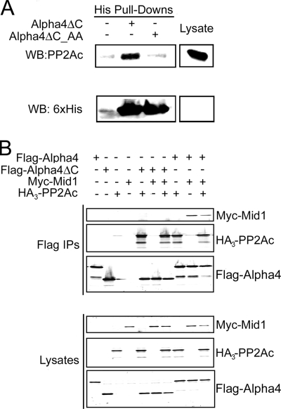
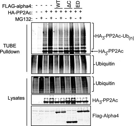
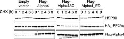
Protein phosphatase 2A (PP2A) is regulated through a variety of mechanisms, including post-translational modifications and association with regulatory proteins. Alpha4 is one such regulatory protein that binds the PP2A catalytic subunit (PP2Ac) and protects it from polyubiquitination and degradation. Alpha4 is a multidomain protein with a C-terminal domain that binds Mid1, a putative E3 ubiquitin ligase, and an N-terminal domain containing the PP2Ac-binding site. In this work, we present the structure of the N-terminal domain of mammalian Alpha4 determined by x-ray crystallography and use double electron-electron resonance spectroscopy to show that it is a flexible tetratricopeptide repeat-like protein. Structurally, Alpha4 differs from its yeast homolog, Tap42, in two important ways: 1) the position of the helix containing the PP2Ac-binding residues is in a more open conformation, showing flexibility in this region; and 2) Alpha4 contains a ubiquitin-interacting motif. The effects of wild-type and mutant Alpha4 on PP2Ac ubiquitination and stability were examined in mammalian cells by performing tandem ubiquitin-binding entity precipitations and cycloheximide chase experiments. Our results reveal that both the C-terminal Mid1-binding domain and the PP2Ac-binding determinants are required for Alpha4-mediated protection of PP2Ac from polyubiquitination and degradation.
© 2011 by The American Society for Biochemistry and Molecular Biology, Inc.
Figures
Similar articles
-
MID1 catalyzes the ubiquitination of protein phosphatase 2A and mutations within its Bbox1 domain disrupt polyubiquitination of alpha4 but not of PP2Ac.PLoS One. 2014 Sep 10;9(9):e107428. doi: 10.1371/journal.pone.0107428. eCollection 2014. PLoS One. 2014. PMID: 25207814 Free PMC article.
-
Alpha4 is a ubiquitin-binding protein that regulates protein serine/threonine phosphatase 2A ubiquitination.Biochemistry. 2010 Mar 2;49(8):1713-8. doi: 10.1021/bi901837h. Biochemistry. 2010. PMID: 20092282 Free PMC article.
-
The MID1 E3 ligase catalyzes the polyubiquitination of Alpha4 (α4), a regulatory subunit of protein phosphatase 2A (PP2A): novel insights into MID1-mediated regulation of PP2A.J Biol Chem. 2013 Jul 19;288(29):21341-21350. doi: 10.1074/jbc.M113.481093. Epub 2013 Jun 5. J Biol Chem. 2013. PMID: 23740247 Free PMC article.
-
Design Principles Involving Protein Disorder Facilitate Specific Substrate Selection and Degradation by the Ubiquitin-Proteasome System.J Biol Chem. 2016 Mar 25;291(13):6723-31. doi: 10.1074/jbc.R115.692665. Epub 2016 Feb 5. J Biol Chem. 2016. PMID: 26851277 Free PMC article. Review.
-
The MID1 gene product in physiology and disease.Gene. 2020 Jul 15;747:144655. doi: 10.1016/j.gene.2020.144655. Epub 2020 Apr 10. Gene. 2020. PMID: 32283114 Free PMC article. Review.
Cited by
-
Identification and characterization of an alternatively spliced isoform of the human protein phosphatase 2Aα catalytic subunit.J Biol Chem. 2012 Feb 10;287(7):4853-62. doi: 10.1074/jbc.M111.283341. Epub 2011 Dec 13. J Biol Chem. 2012. PMID: 22167190 Free PMC article.
-
Discovery of new substrates of the elongation factor-2 kinase suggests a broader role in the cellular nutrient response.Cell Signal. 2017 Jan;29:78-83. doi: 10.1016/j.cellsig.2016.10.006. Epub 2016 Oct 17. Cell Signal. 2017. PMID: 27760376 Free PMC article.
-
Emerging Roles of the TRIM E3 Ubiquitin Ligases MID1 and MID2 in Cytokinesis.Front Physiol. 2019 Mar 19;10:274. doi: 10.3389/fphys.2019.00274. eCollection 2019. Front Physiol. 2019. PMID: 30941058 Free PMC article. Review.
-
TRIM Proteins and Antiviral Microtubule Reorganization: A Novel Component in Innate Immune Responses?Viruses. 2024 Aug 20;16(8):1328. doi: 10.3390/v16081328. Viruses. 2024. PMID: 39205302 Free PMC article. Review.
-
Essential roles of the Tap42-regulated protein phosphatase 2A (PP2A) family in wing imaginal disc development of Drosophila melanogaster.PLoS One. 2012;7(6):e38569. doi: 10.1371/journal.pone.0038569. Epub 2012 Jun 6. PLoS One. 2012. PMID: 22701670 Free PMC article.
References
-
- Kong M., Bui T. V., Ditsworth D., Gruber J. J., Goncharov D., Krymskaya V. P., Lindsten T., Thompson C. B. (2007) J. Biol. Chem. 282, 29712–29720 - PubMed
-
- Kong M., Fox C. J., Mu J., Solt L., Xu A., Cinalli R. M., Birnbaum M. J., Lindsten T., Thompson C. B. (2004) Science 306, 695–698 - PubMed
-
- Goedert M., Jakes R., Qi Z., Wang J. H., Cohen P. (1995) J. Neurochem. 65, 2804–2807 - PubMed
-
- Eichhorn P. J., Creyghton M. P., Bernards R. (2009) Biochim. Biophys. Acta 1795, 1–15 - PubMed
Publication types
MeSH terms
Substances
Associated data
- Actions
Grants and funding
LinkOut - more resources
Full Text Sources
Molecular Biology Databases
