RhoA GTPase is dispensable for actomyosin regulation but is essential for mitosis in primary mouse embryonic fibroblasts
- PMID: 21454503
- PMCID: PMC3083211
- DOI: 10.1074/jbc.C111.229336
RhoA GTPase is dispensable for actomyosin regulation but is essential for mitosis in primary mouse embryonic fibroblasts
Abstract
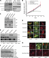
RhoA, the founding member of mammalian Rho GTPase family, is thought to be essential for actomyosin regulation. To date, the physiologic function of RhoA in mammalian cell regulation has yet to be determined genetically. Here we have created RhoA conditional knock-out mice. Mouse embryonic fibroblasts deleted of RhoA showed no significant change in actin stress fiber or focal adhesion complex formation in response to serum or LPA, nor any detectable change in Rho-kinase signaling activity. Concomitant knock-out or knockdown of RhoB and RhoC in the RhoA(-/-) cells resulted in a loss of actin stress fiber and focal adhesion similar to that of C3 toxin treatment. Proliferation of RhoA(-/-) cells was impaired due to a complete cell cycle block during mitosis, an effect that is associated with defective cytokinesis and chromosome segregation and can be readily rescued by exogenous expression of RhoA. Furthermore, RhoA deletion did not affect the transcriptional activity of Stat3, NFκB, or serum response factor, nor the expression of the cell division kinase inhibitor p21(Cip)1 or p27(Kip1). These genetic results demonstrate that in primary mouse embryonic fibroblasts, RhoA is uniquely required for cell mitosis but is redundant with related RhoB and RhoC GTPases in actomyosin regulation.
Figures


Similar articles
-
Mouse macrophages completely lacking Rho subfamily GTPases (RhoA, RhoB, and RhoC) have severe lamellipodial retraction defects, but robust chemotactic navigation and altered motility.J Biol Chem. 2014 Oct 31;289(44):30772-30784. doi: 10.1074/jbc.M114.563270. Epub 2014 Sep 11. J Biol Chem. 2014. PMID: 25213860 Free PMC article.
-
A novel strategy for specifically down-regulating individual Rho GTPase activity in tumor cells.J Biol Chem. 2003 Nov 7;278(45):44617-25. doi: 10.1074/jbc.M308929200. Epub 2003 Aug 25. J Biol Chem. 2003. PMID: 12939257
-
XPLN, a guanine nucleotide exchange factor for RhoA and RhoB, but not RhoC.J Biol Chem. 2002 Nov 8;277(45):42964-72. doi: 10.1074/jbc.M207401200. Epub 2002 Sep 6. J Biol Chem. 2002. PMID: 12221096
-
Why three Rho proteins? RhoA, RhoB, RhoC, and cell motility.Exp Cell Res. 2004 Nov 15;301(1):43-9. doi: 10.1016/j.yexcr.2004.08.012. Exp Cell Res. 2004. PMID: 15501444 Review.
-
A current overview of RhoA, RhoB, and RhoC functions in vascular biology and pathology.Biochem Pharmacol. 2022 Dec;206:115321. doi: 10.1016/j.bcp.2022.115321. Epub 2022 Oct 25. Biochem Pharmacol. 2022. PMID: 36306821 Review.
Cited by
-
A RhoC biosensor reveals differences in the activation kinetics of RhoA and RhoC in migrating cells.PLoS One. 2013 Nov 5;8(11):e79877. doi: 10.1371/journal.pone.0079877. eCollection 2013. PLoS One. 2013. PMID: 24224016 Free PMC article.
-
Gene targeting RhoA reveals its essential role in coordinating mitochondrial function and thymocyte development.J Immunol. 2014 Dec 15;193(12):5973-82. doi: 10.4049/jimmunol.1400839. Epub 2014 Nov 14. J Immunol. 2014. PMID: 25398325 Free PMC article.
-
Mouse macrophages completely lacking Rho subfamily GTPases (RhoA, RhoB, and RhoC) have severe lamellipodial retraction defects, but robust chemotactic navigation and altered motility.J Biol Chem. 2014 Oct 31;289(44):30772-30784. doi: 10.1074/jbc.M114.563270. Epub 2014 Sep 11. J Biol Chem. 2014. PMID: 25213860 Free PMC article.
-
Loss of RhoA Exacerbates, Rather Than Dampens, Oncogenic K-Ras Induced Lung Adenoma Formation in Mice.PLoS One. 2015 Jun 1;10(6):e0127923. doi: 10.1371/journal.pone.0127923. eCollection 2015. PLoS One. 2015. PMID: 26030593 Free PMC article.
-
The incredible journey: From megakaryocyte development to platelet formation.J Cell Biol. 2013 Jun 10;201(6):785-96. doi: 10.1083/jcb.201304054. J Cell Biol. 2013. PMID: 23751492 Free PMC article. Review.
References
-
- Etienne-Manneville S., Hall A. (2002) Nature 420, 629–635 - PubMed
-
- Hall A. (1998) Science 279, 509–514 - PubMed
-
- Just I., Hofmann F., Genth H., Gerhard R. (2001) Int. J. Med. Microbiol. 291, 243–250 - PubMed
-
- Kaibuchi K., Kuroda S., Amano M. (1999) Annu. Rev. Biochem. 68, 459–486 - PubMed
-
- Ridley A. J. (2001) J. Cell Sci. 114, 2713–2722 - PubMed
Publication types
MeSH terms
Substances
Grants and funding
LinkOut - more resources
Full Text Sources
Other Literature Sources
Molecular Biology Databases
Miscellaneous

