. 2011 May 13;286(19):17239-47.
doi: 10.1074/jbc.M110.209015. Epub 2011 Mar 21.

Recognition of gram-positive intestinal bacteria by hybridoma- and colostrum-derived secretory immunoglobulin A is mediated by carbohydrates

Affiliations

Recognition of gram-positive intestinal bacteria by hybridoma- and colostrum-derived secretory immunoglobulin A is mediated by carbohydrates

Amandine Mathias et al. J Biol Chem. .

Abstract

Humans live in symbiosis with 10(14) commensal bacteria among which >99% resides in their gastrointestinal tract. The molecular bases pertaining to the interaction between mucosal secretory IgA (SIgA) and bacteria residing in the intestine are not known. Previous studies have demonstrated that commensals are naturally coated by SIgA in the gut lumen. Thus, understanding how natural SIgA interacts with commensal bacteria can provide new clues on its multiple functions at mucosal surfaces. Using fluorescently labeled, nonspecific SIgA or secretory component (SC), we visualized by confocal microscopy the interaction with various commensal bacteria, including Lactobacillus, Bifidobacteria, Escherichia coli, and Bacteroides strains. These experiments revealed that the interaction between SIgA and commensal bacteria involves Fab- and Fc-independent structural motifs, featuring SC as a crucial partner. Removal of glycans present on free SC or bound in SIgA resulted in a drastic drop in the interaction with gram-positive bacteria, indicating the essential role of carbohydrates in the process. In contrast, poor binding of gram-positive bacteria by control IgG was observed. The interaction with gram-negative bacteria was preserved whatever the molecular form of protein partner used, suggesting the involvement of different binding motifs. Purified SIgA and SC from either mouse hybridoma cells or human colostrum exhibited identical patterns of recognition for gram-positive bacteria, emphasizing conserved plasticity between species. Thus, sugar-mediated binding of commensals by SIgA highlights the currently underappreciated role of glycans in mediating the interaction between a highly diverse microbiota and the mucosal immune system.


Figures
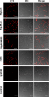
FIGURE 1.
FIGURE 1.
LSCM imaging of the association between fluorescently labeled proteins and LPR. Colocalization of bacteria (visualized by differential interference contrast) with nonspecific proteins including SIgAC5, SIgASal4, SIgAHNK20, mSC, and IgGC20 (seen as red dots) is shown. Control panels are obtained upon differential interference contrast (DIC)-mediated visualization of LPR alone. One representative field obtained from 10 different observations following analysis of 5 different slides is depicted. Scale bars, 10 μm.
FIGURE 2.
FIGURE 2.
Impact of SIgA and SC deglycosylation on the association with LPR. A, Western blot analysis of mSC in reassociated SIgAC5, SIgASal4, or SIgAHNK20 and mSC was performed after 0, 2 h, or 4 h of incubation with 5 units of N-glycosidase F (see “Experimental Procedures”). Samples were separated onto an 8% polyacrylamide gel under reducing conditions. The molecular mass (kDa) of detected protein is marked alongside the lanes. B, immunodetection was performed using anti-α chain Ab of the same samples as in A. Proteins detected at 62 kDa represent nondeglycosylated polypeptides. C, LSCM imaging of the association of LPR formed with native or deglycosylated Cy3-labeled proteins is carried out as in Fig. 1. One representative field obtained from 10 different observations after analysis of 5 different slides is depicted. Scale bars, 10 μm.
FIGURE 3.
FIGURE 3.
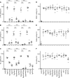
Quantification of bacterial coating by fluorescently labeled proteins. Using ImageJ software, changes in the percentage of bacterial area covered by nonspecific labeled proteins including SIgAC5, SIgAC5dg, pIgAC5, SIgASal4, SIgASal4dg, pIgASal4, SIgAHNK20, SIgAHNK20dg, pIgAHNK20, mSC, mSCdg, or IgGC20 were assessed in the case of LPR (A), ST11 (B), BL (C), D2241 (D), Nissle (E), and Bt (F) bacterial strains (see “Experimental Procedures”). Control quantifications were carried out on pictures obtained with bacteria alone. Bars represent the mean values ± S.E. Statistically significant differences are indicated above the brackets for intragroup tests: ***, p < 0.0001. Data were obtained from 5 different fields of one experiment repeated 5 times.
FIGURE 4.
FIGURE 4.
Purification and characterization of colostrum-derived SIgA and SC. A, fractionation scheme leading to the purification of SIgAcol and SCcol (for details, see “Experimental Procedures”). B, characterization of purified SIgAcol performed by silver staining (lane 1) and by Western blot analysis using antisera specific for human α chain (lane 2), hSC (lane 3), κ chain (lane 4), and J chain (lane 5). C, characterization of purified SCcol performed by silver staining (lane 1) and by Western blot analysis using antisera specific for hSC (lane 2). Denaturing 8% polyacrylamide gels were run under nonreducing conditions.
FIGURE 5.
FIGURE 5.
Deglycosylation of colostrum-derived SIgA or SCcol and defective association with LPR observed by LSCM. A, immunodetection of the hSC was performed after 0, 2 h, 4 h, or 6 h of incubation with 5 units of N-glycosidase F from 0 to 3 h and a further 5 units of enzyme for the last 3 h. Samples were separated onto a 10% polyacrylamide gel under reducing conditions. The molecular mass (kDa) of detected polypeptides is marked alongside the lanes. B, samples were separated as in A with immunodetection of the α chain. The unique signal detected at 62 kDa represents nondeglycosylated α chains. C, LSCM imaging of complexes formed between LPR and native or deglycosylated SIgAcol or SCcol was performed as in Fig. 1. SIgAC5 were used as a positive control for the interaction of bacteria with proteins. One representative field obtained from 10 different observations after analysis of 5 different slides is depicted. Scale bars, 10 μm. D, quantification of LPR coating by fluorescently labeled proteins was carried out on pictures of LPR alone (control) or pictures of complexes with SIgAC5, SIgAcol, SIgAcoldg, Sccol, and SCcoldg. Bars represent the mean values ± S.E. Statistically significant differences are indicated above the brackets for intragroup tests: ***, p < 0.0001. Data were obtained from 5 to 10 different fields of one experiment repeated 5 times.

Similar articles

Cited by

References

    1. Frank D. N., St. Amand A. L., Feldman R. A., Boedeker E. C., Harpaz N., Pace N. R. (2007) Proc. Natl. Acad. Sci. U.S.A. 104, 13780–13785 - PMC - PubMed
    1. Hooper L. V., Gordon J. I. (2001) Science 292, 1115–1118 - PubMed
    1. Mestecky J., Russell M. W., Elson C. O. (1999) Gut 44, 2–5 - PMC - PubMed
    1. Newburg D. S., Walker W. A. (2007) Pediatr. Res. 61, 2–8 - PubMed
    1. Sjögren Y. M., Tomicic S., Lundberg A., Böttcher M. F., Björkstén B., Sverremark-Ekström E., Jenmalm M. C. (2009) Clin. Exp. Allergy 39, 1842–1851 - PubMed

Publication types

LinkOut - more resources