. 2011 May 20;286(20):17714-21.
doi: 10.1074/jbc.M111.223453. Epub 2011 Mar 29.

Sequential establishment of marks on soluble histones H3 and H4

Affiliations

Sequential establishment of marks on soluble histones H3 and H4

Francisca Alvarez et al. J Biol Chem. .

Abstract

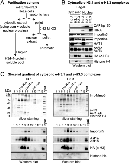
Much progress has been made concerning histone function in the nucleus; however, following their synthesis, how their marking and subcellular trafficking are regulated remains to be explored. To gain an insight into these issues, we focused on soluble histones and analyzed endogenous and tagged H3 histones in parallel. We distinguished six complexes that we could place to account for maturation events occurring on histones H3 and H4 from their synthesis onward. In each complex, a different set of chaperones is involved, and we found specific post-translational modifications. Interestingly, we revealed that histones H3 and H4 are transiently poly(ADP-ribosylated). The impact of these marks in histone metabolism proved to be important as we found that acetylation of lysines 5 and 12 on histone H4 stimulated its nuclear translocation. Furthermore, we showed that, depending on particular histone H3 modifications, the balance in the presence of the different translocation complexes changes. Therefore, our results enabled us to propose a regulatory means of these marks for controlling cytoplasmic/nuclear shuttling and the establishment of early modification patterns.


Figures

FIGURE 1.
FIGURE 1.
Isolation of the cytosolic e-H3.1 and e-H3.3 complexes. A, scheme illustrating the purification procedure for obtaining the cytosolic e-H3.1 and e-H3.3 complexes. B, Western blots of the cytosolic and nuclear e-H3.1 and e-H3.3 complex subunits, as indicated. Input corresponded to 10% of the extract utilized on immunoprecipitation (IP). C, silver staining (top) and Western blot (bottom) analysis of the fractions derived from the glycerol gradient of cytosolic e-H3.1 (left) and e-H3.3 (right) complexes. Fraction 1 corresponded to the top of the gradient and fraction 19 to the bottom. It should be pointed out that histone H4 does not stain well in silver staining, and the HA antibody used to detect e-H3 is more sensitive than the one used to detect histone H4, explaining why histone H4 is only detected in fraction 9 of the glycerol gradient.
FIGURE 2.
FIGURE 2.
Isolation of soluble endogenous histone H3 and H4 complexes. A, scheme illustrating the purification procedure for obtaining the endogenous soluble histone H3 and H4 complexes. B, Western blots of the fractions derived from the DEAE-5PW column, as indicated. The elution of the six different histone complexes is indicated, with their corresponding names. Below, all of the fractions between fraction 17 and fraction 23 were Western blotted to resolve Complex IVa and Complex IVb. C, Western blot analysis of the fractions derived from the gel-filtration column: Complexes Ia, Ib, II, and III, blotted as indicated. On top, elution of the molecular markers is indicated. D, Western blot analysis of fractions derived from the DEAE-5PW column, blotted as indicated. PAR, poly(ADP-ribose). Dotted lines indicate different autoradiograms.
FIGURE 3.
FIGURE 3.
Histones H3 and H4 interact with Importin4. A, top, scheme illustrating the Importin pulldown assay. Bottom, Western blot of the pulldown assay. B, top, scheme illustrating the GST-H4 pulldown assay in the presence of Importin4. Bottom, Western blot of the pulldown assay. Ponceau Red staining provided the loading control for the reaction. C, representative images of the transfection of HeLa cells with the constructs EYFP, H3-NLS-EYFP, and H4-NLS-EYFP, visualized by fluorescence microscopy. Left, images of the cells expressing EYFP and H3-NLS-EYFP or H4-NLS-EYFP quantified to obtain the number of cells that showed enrichment of fluorescence in the nucleus (N) or in the cytoplasm (C). Right, N/C ratio values of H3-NLS-EYFP and H4-NLS-EYFP normalized with the EYFP N/C ratio. The S.D. was derived from the quantification of at least 300 cells for each construct. D, top, scheme illustrating the Importin4 immunoprecipitation assay in the presence of the Ran mutant RanQ69L, which was unable to hydrolyze GTP. Bottom, Western blot of the sample from the immunoprecipitation assay.
FIGURE 4.
FIGURE 4.
Function of the soluble histone H3 and H4 PTMs. A, top, scheme illustrating the Importin pulldown assay. Prebound Ni2+-agarose beads-Importin4 were incubated with increasing amounts of the unmodified or tetraacetylated (K5K8K12K16) histone H4 peptide (residues 2–20), followed by incubation with the H3-H4 histones. Bottom, Western blot of the pulldown assay. B, nuclear import assay performed in the presence of H4-NLS-EYFP (left) and the mutant H4K5K12Q-NLS-EYFP (right), in the absence (top) or presence (bottom) of the reticulocyte extract. Middle, images of the cells incubated with H4-NLS-EYFP (WT) and the mutant H4K5K12Q-NLS-EYFP (K5K12Q) quantified to obtain the mean fluorescence intensity of the nucleus. The enrichment fold was calculated taken the fluorescence intensity of H4-NLS-EYFP as 1. The S.D. was derived from the quantification of at least 50 cells. The graph is a representation of three independent experiments. Right, Coomassie Blue-stained gel of increasing amounts of H4-NLS-EYFP and the mutant H4K5K12Q-NLS-EYFP proteins. C, methylation assay performed using H3-NLS-EYFP and H3K14QK18Q-NLS-EYFP proteins and recombinant SetDB1 and detected by antibodies against H3K9me1. Ponceau Red is shown as the loading control. D, model for the establishment of soluble H3 and H4 histone modifications. PAR, poly(ADP-ribose).

References

    1. Kornberg R. D. (1974) Science 184, 868–871 - PubMed
    1. Campos E. I., Reinberg D. (2009) Annu. Rev. Genet. 43, 559–599 - PubMed
    1. Luger K., Mäder A. W., Richmond R. K., Sargent D. F., Richmond T. J. (1997) Nature 389, 251–260 - PubMed
    1. Rada-Iglesias A., Bajpai R., Swigut T., Brugmann S. A., Flynn R. A., Wysocka J. (2011) Nature 470, 279–283 - PMC - PubMed
    1. He H. H., Meyer C. A., Shin H., Bailey S. T., Wei G., Wang Q., Zhang Y., Xu K., Ni M., Lupien M., Mieczkowski P., Lieb J. D., Zhao K., Brown M., Liu X. S. (2010) Nat. Genet. 42, 343–347 - PMC - PubMed

Publication types