. 2011 May 13;286(19):16596-605.
doi: 10.1074/jbc.M110.186916. Epub 2011 Mar 15.

Differential regulation of dihydroceramide desaturase by palmitate versus monounsaturated fatty acids: implications for insulin resistance

Affiliations

Differential regulation of dihydroceramide desaturase by palmitate versus monounsaturated fatty acids: implications for insulin resistance

Wei Hu et al. J Biol Chem. .

Abstract

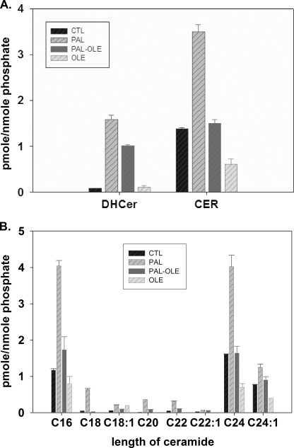
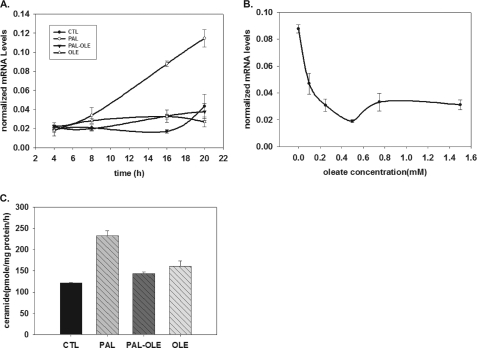
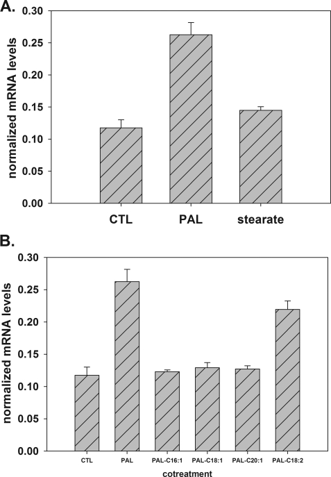
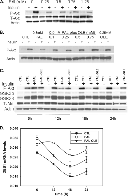
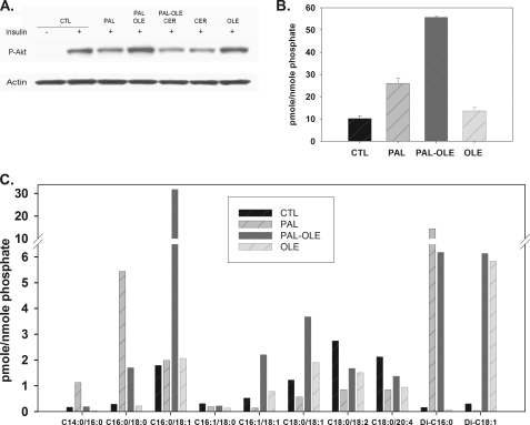
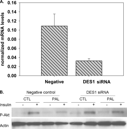
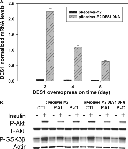
Much data implicate saturated fatty acids in deleterious processes associated with obesity, diabetes, and the metabolic syndrome. Many of these changes may be due to aberrant generation of bioactive lipids when saturated fatty acid availability to tissues is increased. On the other hand, studies are emerging that implicate the monounsaturated fatty acid oleate in protection from saturated fat mediated toxicity; however, the mechanisms are not well understood. Our data demonstrate a novel role for palmitate in increasing mRNA encoding DES1, which is the enzyme responsible for generating ceramide from its precursor dihydroceramide and thus controls synthesis of the bioactive lipid ceramide. Moreover, co-treatment with oleate prevented the increase in ceramide, and this occurred through attenuation of the increase in message and activity of DES1. Knockdown of DES1 also protected from palmitate-induced insulin resistance, and overexpression of this enzyme ameliorated the protective effect of oleate. Together, these findings provide insight into the mechanisms of oleate-mediated protection against metabolic disease and provide novel evidence for fatty acid-mediated regulation of a key enzyme of ceramide biosynthesis.


Figures

FIGURE 1.
FIGURE 1.
Oleate attentuates palmitate-induced ceramide production. Mouse C2C12 myotubes were treated with vehicle control, 1.25 mm palmitate, 1.25 mm palmitate plus 0.75 mm oleate, or 0.75 mm oleate alone. Lipid profiles were determined by LC/MS after 16 h of treatment. A, total dihydroceramide and ceramide. B, N-acyl chain length of ceramides measured. Data are means ± S.E. (n = 2) of three experiments. CTL, control; PAL, palmitate; OLE, oleate; CER, ceramide; DHCer, dihydroceramide.
FIGURE 2.
FIGURE 2.
Oleate prevents palmitate-induced-DES1 up-regulation in a dose- and time-dependent manner. Mouse C2C12 myotubes were treated with vehicle control, 1.25 mm palmitate, 1.25 mm palmitate plus 0.75 mm oleate, or 0.75 mm oleate alone. DES1 mRNA expression was determined by qPCR. Data are presented as means (n = 6) ± S.E. A, on a 4–20-h time course; B, 1.25 mm palmitate co-treatment with 0–1.5 mm oleate for 16 h. C, at 16 h treatment, DES1 enzymatic activities were determined. Data are presented as mean ± S.E. (n = 3). CTL, control; PAL, palmitate; OLE, oleate.
FIGURE 3.
FIGURE 3.
Fatty acid specificity of DES1 message regulation. A, C2C12 myotubes were treated with vehicle control, 1.25 mm palmitate, or 1.25 mm stearate for 16 h. DES1 mRNA expression was determined by qPCR. Data are presented as means (n = 3) ± S.E. B, C2C12 myotubes were treated with vehicle control, 1.25 mm palmitate, or 1.25 mm palmitate plus 0.3 mm C16:1, C18:1, C20:1, and C18:2. At 16 h of treatment, DES1 mRNA expression was determined by qPCR. Data are presented as means (n = 3) ± S.E. CTL, control; PAL, palmitate.
FIGURE 4.
FIGURE 4.
Oleate blocks palmitate-mediated insulin resistance in C2C12 myotubes. A, myotubes were treated with 0.25, 0.5, 0.75, and 1.25 mm palmitate for 16 h and stimulated with 100 nm insulin for 10 min. Akt phosphorylation was measured by Western blot. B, at 0.5 mm palmitate co-treatment with 0.1, 0.25, 0.5, and 0.75 mm oleate for 16 h, cells were stimulated with 100 nm insulin for 10 min. Akt phosphorylation was measured by Western blot. The membranes were stripped and reprobed with an antibody toward β-actin. C, myotubes were treated with 0.75 mm palmitate plus 0.25 mm oleate for 6, 12, 18, and 24 h and then stimulated with 100 nm insulin for 10 min. Akt and GSK 3α/β phosphorylation were measured by Western blot, and the membrane were also reprobed with total Akt and Actin. D, myotubes were treated as the same as C; the DES1 message level was measured by qPCR. Data are representative of two experiments. CTL, control; PAL, palmitate; OLE, oleate; P-Akt, phospho-Akt (Ser473); T-Akt, total Akt; P-GSK3α, phospho-GSK3α (Ser21); P-GSK3β, phospho-GSK 3β (Ser9).
FIGURE 5.
FIGURE 5.
Exogenous ceramide overcomes the protective effect of oleate. A, myotubes were treated with vehicle, 1.25 mm palmitate, 1.25 mm palmitate plus 0.75 mm oleate, or 0.75 mm oleate alone. At 14 h of treatment, the samples co-treated with palmitate plus oleate were incubated with 50 μm C2-ceramide for 2 h and then stimulated with 100 nm insulin for 10 min. The proteins were extracted as described under “Experimental Procedures” and used for Western blot with phospho-Akt antibody. Data are representative of two experiments. B, myotubes were treated with vehicle control, 1.25 mm palmitate, 1.25 mm palmitate plus 0.75 mm oleate, or 0.75 mm oleate alone. Total DAG was determined by LC/MS after 16 h of treatment. C, length chain of DAG Data are represented as mean (n = 6) ± S.E. CTL, control; PAL, palmitate; OLE, oleate.
FIGURE 6.
FIGURE 6.
Effects of knockdown DES1 expression on insulin signaling and protection by oleate. A, DES1 siRNA prevents palmitate-mediated phospho-Akt inhibition. Myotubes were cultured in 60-mm dishes, at 4 days after differentiation, cells were transfected with DES1 siRNA or negative control siRNA, and DES1 mRNA levels were measured at 72 h post-transfection. B, at 72 h post-transfection cells were treated with vehicle control or 1.25 mm palmitate for 14 h and then stimulated with 100 nm insulin for 10 min, and phospho-Akt was measured by Western blot. Data are representative of three experiments; CTL, control; PAL, palmitate.
FIGURE 7.
FIGURE 7.
Overexpression DES1 diminish the protective effect of oleate in insulin signaling. Mouse DES1 expression plasmids were transfected into mouse C2C12 myotubes as described under “Experimental Procedures.” A, at 3, 4, and 5 days post-transfection, DES1 mRNA was measured by qPCR. B, at 72 h post-transfection, the cells were treated for 16 h with vehicle, 0.5 mm palmitate, 0.5 mm palmitate plus 0.25 mm oleate and then stimulated with 100 nm insulin for 10 min. Phospho-Akt and GSK3β were measured by Western blot. Data are representative of three experiments. CTL, control; PAL, palmitate. P-O, palmitate plus oleate; P-Akt, phospho-Akt(Ser473); T-Akt, total Akt; P-GSK3β, phospho-GSK 3β(Ser9).

Similar articles

Cited by

References

    1. Lakka H. M., Laaksonen D. E., Lakka T. A., Niskanen L. K., Kumpusalo E., Tuomilehto J., Salonen J. T. (2002) JAMA 288, 2709–2716 - PubMed
    1. Samuel V. T., Petersen K. F., Shulman G. I. (2010) Lancet 375, 2267–2277 - PMC - PubMed
    1. Berkowitz D. (1966) JAMA 197, 77–80 - PubMed
    1. Hansen B. C. (1999) Ann. N.Y. Acad. Sci. 892, 1–24 - PubMed
    1. Moro C., Galgani J. E., Luu L., Pasarica M., Mairal A., Bajpeyi S., Schmitz G., Langin D., Liebisch G., Smith S. R. (2009) J. Clin. Endocrinol. Metab. 94, 3440–3447 - PMC - PubMed

Publication types