. 2011 May 27;286(21):18562-72.
doi: 10.1074/jbc.M110.184101. Epub 2011 Mar 30.

FXYD proteins reverse inhibition of the Na+-K+ pump mediated by glutathionylation of its beta1 subunit

Affiliations

FXYD proteins reverse inhibition of the Na+-K+ pump mediated by glutathionylation of its beta1 subunit

Stéphanie Bibert et al. J Biol Chem. .

Abstract

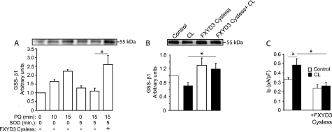
The seven members of the FXYD protein family associate with the Na(+)-K(+) pump and modulate its activity. We investigated whether conserved cysteines in FXYD proteins are susceptible to glutathionylation and whether such reactivity affects Na(+)-K(+) pump function in cardiac myocytes and Xenopus oocytes. Glutathionylation was detected by immunoblotting streptavidin precipitate from biotin-GSH loaded cells or by a GSH antibody. Incubation of myocytes with recombinant FXYD proteins resulted in competitive displacement of native FXYD1. Myocyte and Xenopus oocyte pump currents were measured with whole-cell and two-electrode voltage clamp techniques, respectively. Native FXYD1 in myocytes and FXYD1 expressed in oocytes were susceptible to glutathionylation. Mutagenesis identified the specific cysteine in the cytoplasmic terminal that was reactive. Its reactivity was dependent on flanking basic amino acids. We have reported that Na(+)-K(+) pump β(1) subunit glutathionylation induced by oxidative signals causes pump inhibition in a previous study. In the present study, we found that β(1) subunit glutathionylation and pump inhibition could be reversed by exposing myocytes to exogenous wild-type FXYD3. A cysteine-free FXYD3 derivative had no effect. Similar results were obtained with wild-type and mutant FXYD proteins expressed in oocytes. Glutathionylation of the β(1) subunit was increased in myocardium from FXYD1(-/-) mice. In conclusion, there is a dependence of Na(+)-K(+) pump regulation on reactivity of two specifically identified cysteines on separate components of the multimeric Na(+)-K(+) pump complex. By facilitating deglutathionylation of the β(1) subunit, FXYD proteins reverse oxidative inhibition of the Na(+)-K(+) pump and play a dynamic role in its regulation.


Figures

FIGURE 1.
FIGURE 1.
Glutathionylation of FXYD1 in the heart. A, FXYD1 immunoblot (IB) of myocyte lysate and glutathionylated proteins (GSS-protein) precipitated with streptavidin. Streptavidin precipitate from myocytes not incubated in biotin-GSH was a negative (−ve) control. Glutathionylation was sensitive to DTT. B, mean densitometry of FXYD1 immunoblots in streptavidin pulldown of control myocytes and myocytes exposed to ONOO for 15 min at the nominal concentration of 100 μm. C, glutathionylation of FXYD1 detected by GSH antibody technique in myocytes exposed to angiotensin II (Ang II) for 15 min. D, glutathionylation of FXYD1 in myocytes exposed to 100 nm forskolin (Fsk), with and without incubation with 200 international units/ml PEGylated superoxide dismutase (SOD). E, glutathionylation of FXYD1 from a sheep model of infarction from myocardium remote to the infarct, the peri-infarct zone, and infarct zone. Immunoprecipitations (IP) were performed with FXYD1 antibodies and immunoblots with FXYD1 antibodies (upper panel) or GSH antibodies (lower panel). Densitometry of immunoblots (mean ± S.E.) was normalized against control (n = 3 for each experiment). *, p < 0.05.
FIGURE 2.
FIGURE 2.
Expression of FXYD1, β1 subunit glutathionylation and Na+-K+ pump activity in oocytes. A, FXYD1 (upper panel) and β1 subunit (lower panel) immunoblots of Xenopus oocyte microsomes directly loaded on gels (lanes 7–12) or precipitated with streptavidin beads (GSS-proteins, lanes 1–6). Experiments were performed 2 days after injection with Na+-K+ pump subunit and FXYD1 cRNAs as indicated. Injection of ONOO 15 min before lysis is indicated. Core glycosylated and fully glycosylated β1 subunits are indicated by cg and fg. B, quantification of glutathionylation of Na+-K+ pump β1 subunit by densitometry (normalized by total amount of protein expressed, mean ± S.E.) in five independent experiments. C, FXYD1 abolishes ONOO-induced decrease in mean Na+-K+ pump current of 20 oocytes from two different batches. Currents from oocytes not injected with cRNA (ni) were not subtracted from currents of injected oocytes. *, p < 0.05. IB, immunoblot.
FIGURE 3.
FIGURE 3.
FXYD proteins, β1 subunit glutathionylation, and Na+-K+ pump activity in myocytes. A, immunoblot (IB) of α1 subunit, FXYD1 and FXYD3 of α1 subunit immunoprecipitate (IP) from myocytes incubated with 500 nm recombinant FXYD3, or the mutated Cysless FXYD3 for 15 min. B, glutathionylation detected with GSH antibody in FXYD3 immunoprecipitate of lysed myocytes preincubated with recombinant FXYD3 or Cysless FXYD3 before exposure to ONOO at a nominal concentration of 100 μm. C, glutathionylation of the β1 subunit detected with the GSH technique (GSS-β1) in myocytes preincubated with FXYD3 and Cysless FXYD3 with or without subsequent exposure to ONOO. D, glutathionylation of the β1 subunit in myocytes incubated with FXYD3 and Cysless FXYD3 with or without subsequent exposure to 100 nm angiotensin II (AngII) for 10 min. E, Ip measured with and without 500 nm FXYD1 in pipette solutions (n ≥ 5 in each group). The superfusate was Na+-free, and the test potential was −40 mV. F, Ip measured with or without 100 μm paraquat and 200 nm FXYD1 in the pipette solution (n ≥ 9 in each group). The superfusate contained Na+, and the test potential was −40 mV. G, effect of 500 nm FXYD3 or Cysless FXYD3 on angiotensin II-induced decrease in Ip (n ≥ 5 in each group). Pipette solutions contained l-arginine, and the superfusate was Na+-free, and the test potential was −14 mV. H, β1 subunit glutathionylation in myocardium from FXYD1−/− (KO) mice and WT littermates (three mice per group). TL indicates total lysate. Histograms show mean ± S.E. *, p < 0.05.
FIGURE 4.
FIGURE 4.
Role of native FXYD1 in reversal of oxidative Na+-K+ pump modification. A, myocytes were preincubated with Cysless FXYD3 or control solutions and exposed to 100 μm paraquat for 0, 10, or 15 min. This oxidant signal was quenched with superoxide dismutase (SOD) 10 min after paraquat (PQ) exposure, as indicated, and myocytes were lysed after an additional 5 min. β1 subunit glutathionylation was assessed using the biotin-GSH technique. B, myocytes preincubated with Cysless FXYD3 were exposed to 1 μm CL316,243 (CL) prior to lysis. C, effect of Cysless FXYD3 on the increase in Ip induced by 10 nm CL316,243. The superfusate was Na+-free, and the test potential was −14 mV. Histograms show mean densitometry ± S.E. of immunoblots normalized against control (A and B; n = 3 for each experiment), or mean Ip (C; n = 6–9 in each group). *, p < 0.05.
FIGURE 5.
FIGURE 5.
Reactive cysteine residue in FXYD1, β1 subunit glutathionylation and Na+-K+ pump current. A, sequence alignment of FXYD proteins. Numbering below corresponds to the sequence of FXYD1 and begins at 1 after the signal peptide (not shown). Conserved residues are marked with filled circles. Transmembrane domain is indicated by TM. B–E, Xenopus oocytes expressing Xenopus α1 and β1 pump subunits alone or with WT or mutated FXYD1 (FXYD1C1A, FXYD1C2A, or FXYD1C1AC2A) were injected with ONOO as indicated. B, FXYD1 immunoblot (IB) of oocyte microsomes directly loaded on gels (upper panel) or immunoprecipitated with streptavidin beads (GSS-FXYD1, lower panel). C, mean densitometry ± S.E. of GSS-FXYD1 immunoblot normalized against control from four independent experiments. D, β1 subunit immunoblot of oocyte microsomes (upper panel) or glutathionylated proteins (GSS protein; lower panel). E, mean densitometry of GSS-β1 immunoblots normalized to the total amount of proteins from four independent experiments. Data from oocytes expressing α1, β1, and FXYD1C1C2 subunits were arbitrarily set to 1. F, Na+-K+ pump currents of 20 oocytes from four different batches. Values of oocytes not injected with cRNAs (ni) were not subtracted from values of injected oocytes. *, p < 0.05 versus control. Core glycosylated and fully glycosylated β1 subunits are indicated by cg and fg.
FIGURE 6.
FIGURE 6.
Adjacent basic amino acids and reactivity of FXYD C2-equivalent cysteine. A, projected three-dimensional structure, around the membrane/cytoplasmic interface of FXYD10. The extrapolated model is depicted using PyMOL. The last amino acid residue firmly detected in the crystal structure is Lys42 just before the C1 equivalent (Cys43). The putative trajectory of the FXYD cytoplasmic domain is shown by a dotted red line. M indicates transmembrane helix. B, Na+-K+ pump currents of oocytes expressing WT and mutant FXYD proteins. RCG and RCK refer to C2 (Fig. 5A) and its adjacent residues. Oocytes were injected with ONOO as indicated. Results from 15 oocytes from three different batches are shown. Imax from oocytes not injected with cRNAs (ni) were not subtracted from currents of injected oocytes. C, immunoblots (IB) of WT and mutant FXYD1. Oocyte microsomes were directly loaded on gel (upper panel) and glutathionylated subfraction precipitated by streptavidin (GSS-FXYD1, lower panel). D, β1 Na+-K+ pump subunit immunoblots of oocyte microsomes directly loaded on gel (upper panel); and glutathionylated subfraction precipitated by streptavidin (GSS-β1). E, mean densitometry of GSS-β1 immunoblots from three independent experiments (as shown in D) in arbitrary units normalized by the total amount of proteins expressed. Data from oocytes expressing α1, β1, and FXYD1 RCG were arbitrarily set to 1. *, p < 0.05. Core glycosylated and fully glycosylated β1 subunits are indicated by cg and fg. Histograms show mean ± S.E.
FIGURE 7.
FIGURE 7.
Oxidative stimuli and cardiac FXYD1/α11 Na+-K+ pump subunit interaction. A, α1 subunit immunoblot (IB) of FXYD1 immunoprecipitate (IP) in myocytes exposed to angiotensin II (AngII) or control. B, β1 subunit immunoblot of FXYD1 immunoprecipitate in myocytes exposed to angiotensin II or control. C, α1 subunit immunoblot of FXYD1 immunoprecipitate in sheep myocardial samples taken from regions in and around a zone of myocardial infarction. D, β1 subunit immunoblot of FXYD1 immunoprecipitate in myocardial samples from regions in and around a zone of myocardial infarction. Histograms show mean densitometry ± S.E. of immunoblots normalized against control (n = 3 for each experiment). *, p < 0.05.
FIGURE 8.
FIGURE 8.
Reactivity of FXYD protein and α11 subunit co-immunoprecipitation. A, oocytes expressing α1 and β1 pump subunit cRNAs with or without WT or mutant FXYD1 (FXYD1C2) were injected with ONOO as indicated. Oocyte microsomes were directly loaded on gels (a and b), or immunoprecipitated (IP) with an α1 subunit antibody (c and d) or with streptavidin beads (GSS-β, e). a and c show α1, and b, d, and e show β1 subunit immunoblots (IB). B, densitometry of β1 subunit immunoblot after α1 subunit immunoprecipitation in three independent experiments. Data are normalized by the total amount of protein and by α1 subunit immunoprecipitated, expressed as a ratio compared with control oocyte. C, myocytes preincubated with recombinant FXYD3 or Cysless FXYD3 were exposed to ONOO as indicated, and α11 subunit co-immunoprecipitation was detected in cell lysate. Densitometry of immunoblots from three independent experiments normalized against control are summarized. D, effect of oxidant stress of sheep myocardial infarction on α11 subunit interaction. Densitometry normalized against control in three experiments are summarized. Histograms show mean ± S.E. *, p < 0.05 versus control.

Similar articles

Cited by

References

    1. Sweadner K. J., Rael E. (2000) Genomics 68, 41–56 - PubMed
    1. Crambert G., Fuzesi M., Garty H., Karlish S., Geering K. (2002) Proc. Natl. Acad. Sci. U.S.A. 99, 11476–11481 - PMC - PubMed
    1. Geering K. (2006) Am. J. Physiol. Renal Physiol 290, F241–250 - PubMed
    1. Zhang X. Q., Wang J., Carl L. L., Song J., Ahlers B. A., Cheung J. Y. (2009) Am. J. Physiol. Cell Physiol. 296, C911–921 - PMC - PubMed
    1. Teriete P., Franzin C. M., Choi J., Marassi F. M. (2007) Biochemistry 46, 6774–6783 - PMC - PubMed

Publication types

MeSH terms