. 2011 May 20;286(20):17693-703.
doi: 10.1074/jbc.M110.208306. Epub 2011 Mar 18.

Sequence and generation of mature ribosomal RNA transcripts in Dictyostelium discoideum

Affiliations

Sequence and generation of mature ribosomal RNA transcripts in Dictyostelium discoideum

Carsten Boesler et al. J Biol Chem. .

Abstract

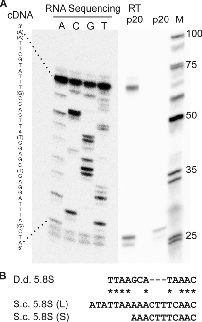
The amoeba Dictyostelium discoideum is a well established model organism for studying numerous aspects of cellular and developmental functions. Its ribosomal RNA (rRNA) is encoded in an extrachromosomal palindrome that exists in ∼100 copies in the cell. In this study, we have set out to investigate the sequence of the expressed rRNA. For this, we have ligated the rRNA ends and performed RT-PCR on these circular RNAs. Sequencing revealed that the mature 26 S, 17 S, 5.8 S, and 5 S rRNAs have sizes of 3741, 1871, 162, and 112 nucleotides, respectively. Unlike the published data, all mature rRNAs of the same type uniformly display the same start and end nucleotides in the analyzed AX2 strain. We show the existence of a short lived primary transcript covering the rRNA transcription unit of 17 S, 5.8 S, and 26 S rRNA. Northern blots and RT-PCR reveal that from this primary transcript two precursor molecules of the 17 S and two precursors of the 26 S rRNA are generated. We have also determined the sequences of these precursor molecules, and based on these data, we propose a model for the maturation of the rRNAs in Dictyostelium discoideum that we compare with the processing of the rRNA transcription unit of Saccharomyces cerevisiae.


Figures

FIGURE 1.
FIGURE 1.
Ribosomal RNA in D. discoideum. A, size determination by denaturing gel electrophoresis. Total RNA (1 μg) isolated from the AX2 wild type strain (bands I–VI) was run together with the Fermentas RiboRuler High Range RNA ladder (M) and visualized by ethidium bromide staining. B, organization of the RNA polymerase I transcription unit for 17 S, 5.8 S, and 26 S rRNA. For clarity, 1000 and 1500 nucleotides are omitted for the 17 S and 26 S rRNA, respectively, as indicated by diagonal breaks. Annotated transcripts 1 and 2 for these three rRNAs are displayed above as thin lines in dark and light gray, respectively. Because of the different ends of these annotated transcripts, the rRNA ends are shown as diffused. Transcription starts at the 5′ external transcribed spacer (5′ETS) and ends with the 3′ETS. The rRNAs are separated by two internal transcribed spacers, ITSI and ITSII, respectively. The position where DNA antisense oligonucleotides p4, p5, p12, and p9 bind are indicated by arrowheads. A size standard is indicated. C–F, sequence analysis by RNase H digest. Each panel displays an ethidium bromide stain of a denaturing gel electrophoresis run on 3.0 μg/lane total RNA alone, after incubation with the oligonucleotide p4, p5, p12, and p9 and upon additional incubation with an enzyme displaying RNase H activity in C–F, respectively. Bands with altered migration behavior are indicated by asterisks.
FIGURE 2.
FIGURE 2.
Principle of the cRT-PCR method used to determine the ends of RNA molecules. Total RNA is dephosphorylated by shrimp alkaline phosphatase (SAP) and then 5′-monophosphorylated by T4 polynucleotide kinase (T4 PNK). The resulting 5′-phosphate and the 3′-hydroxyl moieties (symbolized by up and down triangles, respectively) allow us to circularize individual RNA molecules by means of T4 RNA ligase. With the specific first strand primer 1, a cDNA product that contains the ligated ends of the RNA molecule can be generated using a reverse transcriptase. The 5′ to 3′ junction is then amplified by means of the specific primers 2 and 3 and TaqDNA polymerase, in a PCR, allowing for subsequent sequencing. Scheme was modified from Ref. .
FIGURE 3.
FIGURE 3.
Amplification of the 17 S and 26 S rRNA ends by cRT-PCR. A, schematic representation of the rRNA transcription unit. The indicated oligonucleotides bind to either the rRNA (reverse arrowheads) or its cDNA (forward arrowheads), respectively. All other description are as in Fig. 1B. Gel picture of the cRT-PCR product of the 17 S rRNA (B) and the 26 S rRNA (C) ends. The templates for the final PCRs were water (dH2O), genomic DNA (gDNA), total RNA, circularized total RNA (cRNA), and sequence-specific cDNAs. For the 17 S rRNA, p4 was used as the first strand primer and p6 and p18 in the subsequent PCR, and for the 26 S rRNA p12 was used as the first strand primer and p28 and p32 in the PCR. M denotes the 100-bp plus DNA ladder (Fermentas), and DNA sizes are indicated in base pairs.
FIGURE 4.
FIGURE 4.
5.8 S rRNA species in D. discoideum. A, primer extension (RT p20) analysis on total RNA using radiolabeled DNA oligonucleotide p20, which has been loaded for comparison in a separate lane (p20). RNA sequencing denotes RT reactions that were carried out in the presence of the indicated dideoxynucleotides. The cDNA sequence is stated to the left, in which positions with very weak signal are stated in parentheses. M denotes the radiolabeled ultralow range marker (Fermentas) with fragment sizes indicated in base pairs. To allow for the detection of weak bands in the RNA sequencing, the gel picture has been assembled from two parts using different integration settings (separated by a black line). B, sequence comparison of the 5′ end of 5.8 S rRNA from D. discoideum (D.d.) and the long (L) and short (S) form of S. cerevisiae (S.c.), using published sequences (30).
FIGURE 5.
FIGURE 5.
Detection of the mature rRNAs from D. discoideum. A, size of the mature rRNAs based on the results of the cRT-PCR. The position where DNA antisense oligonucleotides p6, p2, p12, p15, and p29 bind are indicated by arrowheads. All other descriptions are as in Fig. 1B. B, Northern blot analysis of rRNA species in total (T) and nucleus-enriched (N) RNA. Each lane contains 2.0 μg of the indicated RNA preparation, and the gel is either stained with ethidium bromide or subjected to Northern blot hybridization with the indicated radiolabeled oligonucleotides as sequence-specific probes. The asterisk marks RNA species with an apparent migration behavior above the 26 S rRNA.
FIGURE 6.
FIGURE 6.
Analysis of rRNA precursors by cRT-PCR. A, orientation of the indicated oligonucleotides with respect to the rRNA transcription unit, which bind either to the rRNA (reverse arrowheads) or its cDNA (forward arrowheads), respectively. All other descriptions are as in Fig. 1B. Gel pictures with the cRT-PCR products of the 17 S rRNA (B), the 5.8 S rRNA (C), and the 26 S rRNA (D) precursors are shown. The templates for the final PCRs were water (dH2O), genomic DNA (gDNA), total RNA, circularized total RNA (cRNA), and sequence-specific cDNAs. For the 17 S rRNA, p4 was used as the first strand primer and p6 and p7 in the PCR, and for the 5.8 S rRNA p2 was used as the first strand primer together with p17 also in the subsequent PCR. The first strand primer p12 served for generation of the 26 S precursor cDNA, which was amplified with p19 and p28 in the PCR. M denotes the 100-bp plus DNA ladder (Fermentas), and DNA sizes are indicated in base pairs.
FIGURE 7.
FIGURE 7.
RT-PCR on the 17 S rRNA precursor. A, organization of the region downstream of the 17 S rRNA. The position where the first strand primers p37 and p38 bind is indicated, as well as the position of the forward primer p18. A size standard is indicated. B, sequences of the ITSI-rDNA and the complementary first strand primer p37 and its poly(T) version p38. C, gel picture with the RT-PCR product of the 3′ end of the 17 S rRNA precursor. The templates for the final PCRs with the oligonucleotides p18 and p38 were water (dH2O), genomic DNA (gDNA), total RNA, and the two sequence-specific cDNAs generated with the first strand primers p37 and p38, respectively. M denotes the 100-bp plus DNA ladder (Fermentas), and DNA sizes are indicated in base pairs.
FIGURE 8.
FIGURE 8.
Detection of the 17 S rRNA precursors from D. discoideum. A, organization of the regions surrounding the 17 S rRNA. The position where the complementary DNA oligonucleotides p39, p36, p6, and p8 bind are shown by arrowheads. All other descriptions are as in Fig. 1B. B, Northern blot analysis of rRNA species in total (T) and nucleus-enriched (N) RNA. Each lane contains 2.0 μg of the indicated RNA preparation, and the gel is either stained with ethidium bromide or subjected to Northern blot hybridization with the indicated radiolabeled oligonucleotides as sequence-specific probes. The migration of the 26 S and the 17 S rRNAs is indicated from the ethidium bromide staining. C, primer extension analysis using radiolabeled oligonucleotides p6 and p36. Reaction products were analyzed on denaturing polyacrylamide gels of 10% (left panel) and 5% (right panel) to allow for a good separation. M1 denotes the ultralow range marker and M2 the 100-bp plus DNA ladder (Fermentas). DNA sizes are indicated in base pairs. Note that the region between 400 and 500 bp is represented on both gels.
FIGURE 9.
FIGURE 9.
Detection of the primary transcript. A, orientation of the indicated oligonucleotides with respect to the rRNA transcription unit, which bind either to the rRNA (reverse arrowheads) or its cDNA (forward arrowheads), respectively. All other descriptions are as in Fig. 1B. Gel pictures with RT-PCR products for detection of the 5′ETS (B), the internal transcribed spacer I (C), and the 5.8 Spre1 precursor (D) are shown. The templates for the final PCRs were water (dH2O), genomic DNA (gDNA), total RNA and cDNA, which were generated with the indicated first strand primers p19, p32, or p15, respectively. For detection of the transcribed 5′ETS, primers p1 and p36 were used that amplify a PCR product of 240 bp (B); primers p7 and p2 were used to amplify a 333-bp large RT-PCR product of ITSI (C). As a control, primers p31 and p19 were used to amplify a 223-bp large RT-PCR product corresponding to the 26 Spre1 precursor. M denotes the 100-bp plus DNA ladder (Fermentas), and DNA sizes are indicated in base pairs.
FIGURE 10.
FIGURE 10.
Scheme for the rRNA maturation in D. discoideum. The rRNAs and their identified precursors are displayed with the cleavage sites c1–c10 (pink triangles), using the descriptions of Fig. 1B. A, mature 37 S rRNA is initially cleaved at the three sites c2, c3, and c1, generating the precursors 17 Spre2 and 26 Spre2. B, precursor 17 Spre2 is cleaved at c4 to generate the mature 5′ end of the 17 S rRNA and is polyadenylated at position c2, resulting in the precursor 17 Spre1. This in turn is cut at c7 to generate the mature 17 S rRNA. C, precursor 26 Spre2 is processed at c5 and c6. This generates in the resulting precursor 26 Spre1 the mature 5′ end of the 5.8 S rRNA and the mature 3′ end of the 26 S rRNA, respectively. Cleavage of precursor 26 Spre1 at c9 generates the mature 26 S rRNA and at c8 the defined 3′ end of the 5.8 Spre1 species. This is processed at c10 to yield the mature 5.8 S rRNA. D, comparison of the sequences of the cleavage sites determined here for D. discoideum (D.d.) with those of S. cerevisiae (S.c.) (30). Alignments were generated using ClustalW (43). Ribosomal RNAs are highlighted using the color code of A–C.

Similar articles

Cited by

References

    1. Nissen P., Hansen J., Ban N., Moore P. B., Steitz T. A. (2000) Science 289, 920–930 - PubMed
    1. Schluenzen F., Tocilj A., Zarivach R., Harms J., Gluehmann M., Janell D., Bashan A., Bartels H., Agmon I., Franceschi F., Yonath A. (2000) Cell 102, 615–623 - PubMed
    1. Wimberly B. T., Brodersen D. E., Clemons W. M., Jr., Morgan-Warren R. J., Carter A. P., Vonrhein C., Hartsch T., Ramakrishnan V. (2000) Nature 407, 327–339 - PubMed
    1. Noller H. F., Hoffarth V., Zimniak L. (1992) Science 256, 1416–1419 - PubMed
    1. Maizels N. (1976) Cell 9, 431–438 - PubMed

Publication types

MeSH terms

LinkOut - more resources