. 2011 May 6;286(18):16140-9.
doi: 10.1074/jbc.M111.234005. Epub 2011 Mar 16.

Rac1 protein rescues neurite retraction caused by G2019S leucine-rich repeat kinase 2 (LRRK2)

Affiliations

Rac1 protein rescues neurite retraction caused by G2019S leucine-rich repeat kinase 2 (LRRK2)

Diane Chan et al. J Biol Chem. .

Abstract

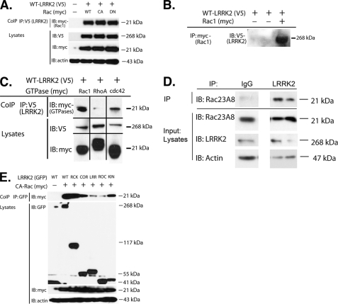
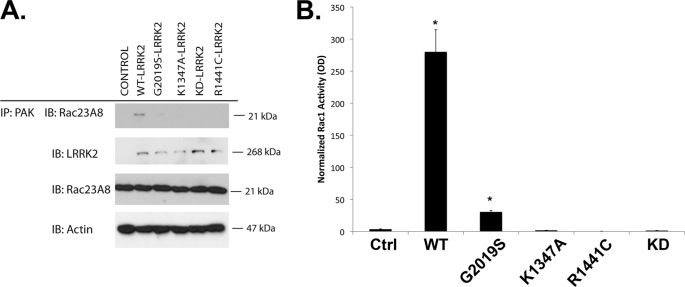
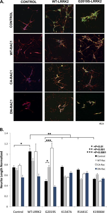
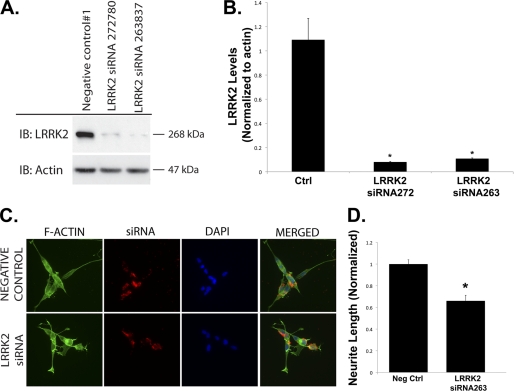
Mutations in leucine-rich repeat kinase 2 (LRRK2) are currently the most common genetic cause of familial late-onset Parkinson disease, which is clinically indistinguishable from idiopathic disease. The most common pathological mutation in LRRK2, G2019S LRRK2, is known to cause neurite retraction. However, molecular mechanisms underlying regulation of neurite length by LRRK2 are unknown. Here, we demonstrate a novel interaction between LRRK2 and the Rho GTPase, Rac1, which plays a critical role in actin cytoskeleton remodeling necessary for the maintenance of neurite morphology. LRRK2 binds strongly to endogenous or expressed Rac1, while showing weak binding to Cdc42 and no binding to RhoA. Co-expression with LRRK2 increases Rac1 activity, as shown by increased binding to the p21-activated kinase, which modulates actin cytoskeletal dynamics. LRRK2 constructs carrying mutations that inactivate the kinase or GTPase activities do not activate Rac1. Interestingly, LRRK2 does not increase levels of membrane-bound Rac1 but dramatically changes the cellular localization of Rac1, causing polarization, which is augmented further when LRRK2 is co-expressed with constitutively active Rac1. Four different disease-related mutations in LRRK2 altered binding to Rac1, with the G2019S and R1441C LRRK2 mutations attenuating Rac1 binding and the Y1699C and I2020T LRRK2 mutations increasing binding. Co-expressing Rac1 in SH-SY5Y cells rescues the G2019S mutant phenotype of neurite retraction. We hypothesize that pathological mutations in LRRK2 attenuates activation of Rac1, causing disassembly of actin filaments, leading to neurite retraction. The interactions between LRRK2 and Rho GTPases provide a novel pathway through which LRRK2 might modulate cellular dynamics and contribute to the pathophysiology of Parkinson disease.


Figures

FIGURE 1.
FIGURE 1.
Co-immunoprecipitation of LRRK2 and Rac1. A, LRRK2 (V5, WT) was co-expressed with Rac1 (Myc, WT, CA, DN) in HEK293 FT cells, and LRRK2 was immunoprecipitated. Rac1 was readily detectable in complex. B, Co-association of LRRK2 and Rac1 was also readily apparent following overexpression of LRRK2 (WT) and Rac1 (CA) followed by immunoprecipitation (IP) of Rac1 and immunoblotting (IB) for LRRK2. C, association of LRRK2 with Rho GTPases appears to be strongest for Rac1. LRRK2 (V5, WT) was co-expressed with Rac1, RhoA, or Cdc42 (Myc, WT for each) in HEK293 cells, and the LRRK2 was immunoprecipitated. Rac1 and Cdc42 were detectable in the complexes following a rank order for detection of Rac1 > Cdc42 ≫ RhoA. D, LRRK2 was immunoprecipitated from human brain striatal lysate and then probed with anti-Rac1 antibody. A robust Rac1 signal was evident in the immunoprecipitate but absent when nonspecific IgG was substituted for the LRRK2 antibody. E, LRRK2 (GFP, WT) deletion constructs were expressed with Rac1 (Myc, CA) to identify which domains are most important for binding. The LRRK2 was immunoprecipitated and immunoblotted for Rac1. Rac1 binding was apparent with the ROC-COR kinase constructs, as well as with constructs carrying the subdomains of COR or kinase. KIN, kinase; RCK, ROC-COR-kinase domains; CoIP, co-immunoprecipitation.
FIGURE 2.
FIGURE 2.
Immunoprecipitation of LRRK2 constructs carrying disease-linked mutations. G2019S and R1441C exhibit weaker binding to CARac1, whereas Y1699C and I2020T exhibit increased binding. Shown is a representative immunoblot (IB). Shown is a quantification of three independent experiments (n = 3). *, p < 0.05; **, p < 0.001. CoIP, coimmunoprecipitation; KD, knockdown.
FIGURE 3.
FIGURE 3.
WT LRRK2 increases Rac1 activity. A, HEK293 cells were transfected with LRRK2 (WT, G2019S, R1441C, K1347A, K1906M, with the latter abbreviated as KD). Endogenous Rac1 was then precipitated using PAK-GST. Levels of Rac1 in the PAK complex and total lysates were immunoblotted (IB). The presence of WT LRRK2 increased the amount of Rac1 with PAK, but did not change total Rac1 levels. B, quantification of Rac1 in the PAK-GST precipitates (n = 4). *, p < 0.01. Ctrl, control; IP, immunoprecipitate.
FIGURE 4.
FIGURE 4.
Cellular distribution of LRRK2 and Rac1. Expression of WT LRRK2 alone produced a broad cellular distribution (a–c). In contrast, WT Rac1 yielded a distribution at or close to membranes (d–f), whereas the distribution of CA-Rac1 (g–i) and DN Rac1 (k–l) was less pronounced. Co-expression of LRRK2 with WT or CA-Rac1 produced striking polarization. WT LRRK2 plus WT-Rac1 yielded asymmetric polarization (m–o), whereas WT LRRK2 plus CA-Rac1 (q and r). However, DN-Rac1 did not elicit changes in LRRK2 distribution (s–u). Higher magnification figures of selected areas of putative co-localization are shown in the boxed insets.
FIGURE 5.
FIGURE 5.
Rac1 does not affect localization of R1441C LRRK2 and vice versa. Expression of R1441C LRRK2 neither co-localized with Rac1 nor modified the distribution of Rac1. CA-Rac1 and DN-Rac1 also were not sensitive to R1441C LRRK2 and did not modify the distribution of R1441C LRRK2.
FIGURE 6.
FIGURE 6.
Rac1 rescues neurite retraction induced by G2019S LRRK2. A, immunocytochemical pictures showing the effects of LRRK2 (green) expression on process outgrowth in differentiated SH-SY5Y neurons, using phalloidin (red) to identify actin filaments. WT LRRK2 increased neurite length, whereas G2019S LRRK2 reduced neurite length. WT and CA-Rac1 rescued neuronal retraction induced by GG2019S LRRK2. DN-Rac1 blocked the effects of LRRK2, suggesting that the two proteins act in same pathway rather than through epistasis. B, quantification of changes in neurite outgrowth associated with LRRK2 and Rac1 expression. Lengths normalized to nontransfected differentiated SH-SY5Y neurons (n = 90 neurons). *, p < 0.01; **, p < 0.001; and ***, p < 0.0001.
FIGURE 7.
FIGURE 7.
Knockdown of LRRK2 by siRNA reduces neurite process length. A and B, siRNA-mediated knockdown of LRRK2 reduced LRRK2 protein levels in HEK293 FT cells. C and D, fluorescent tagged siRNA was used to knockdown LRRK2 in differentiated SH-SY5Y cells. IP, immunoprecipitation; IB, immunoblot. Neg Ctrl, negative control.

References

    1. Dauer W., Przedborski S. (2003) Neuron 39, 889–909 - PubMed
    1. Dawson T. M., Moore D. J., West A. B. (2009) Biochim. Biophys. Acta 1792, 585–586 - PubMed
    1. Greggio E., Cookson M. R. (2009) ASN Neuro. 1, e00002. - PMC - PubMed
    1. Biskup S., Gerlach M., Kupsch A., Reichmann H., Riederer P., Vieregge P., Wüllner U., Gasser T. (2008) J. Neurol. 255, 8–17 - PubMed
    1. Taylor J. P., Mata I. F., Farrer M. J. (2006) Trends Mol. Med. 12, 76–82 - PubMed

Publication types

MeSH terms

Substances