. 2011 May 13;286(19):16681-92.
doi: 10.1074/jbc.M110.205062. Epub 2011 Mar 25.

The cAMP-responsive Rap1 guanine nucleotide exchange factor, Epac, induces smooth muscle relaxation by down-regulation of RhoA activity

Affiliations

The cAMP-responsive Rap1 guanine nucleotide exchange factor, Epac, induces smooth muscle relaxation by down-regulation of RhoA activity

Bartosz J Zieba et al. J Biol Chem. .

Abstract

Agonist activation of the small GTPase, RhoA, and its effector Rho kinase leads to down-regulation of smooth muscle (SM) myosin light chain phosphatase activity, an increase in myosin light chain (RLC(20)) phosphorylation and force. Cyclic nucleotides can reverse this process. We report a new mechanism of cAMP-mediated relaxation through Epac, a GTP exchange factor for the small GTPase Rap1 resulting in an increase in Rap1 activity and suppression of RhoA activity. An Epac-selective cAMP analog, 8-pCPT-2'-O-Me-cAMP ("007"), significantly reduced agonist-induced contractile force, RLC(20), and myosin light chain phosphatase phosphorylation in both intact and permeabilized vascular, gut, and airway SMs independently of PKA and PKG. The vasodilator PGI(2) analog, cicaprost, increased Rap1 activity and decreased RhoA activity in intact SMs. Forskolin, phosphodiesterase inhibitor isobutylmethylxanthine, and isoproterenol also significantly increased Rap1-GTP in rat aortic SM cells. The PKA inhibitor H89 was without effect on the 007-induced increase in Rap1-GTP. Lysophosphatidic acid-induced RhoA activity was reduced by treatment with 007 in WT but not Rap1B null fibroblasts, consistent with Epac signaling through Rap1B to down-regulate RhoA activity. Isoproterenol-induced increase in Rap1 activity was inhibited by silencing Epac1 in rat aortic SM cells. Evidence is presented that cooperative cAMP activation of PKA and Epac contribute to relaxation of SM. Our findings demonstrate a cAMP-mediated signaling mechanism whereby activation of Epac results in a PKA-independent, Rap1-dependent Ca(2+) desensitization of force in SM through down-regulation of RhoA activity. Cyclic AMP inhibition of RhoA is mediated through activation of both Epac and PKA.


Figures

FIGURE 1.
FIGURE 1.
007-induced relaxation, RLC20 dephosphorylation, and disinhibition of MLCP activity as well as a decrease in RhoA activity. A, 007 (30 μm) significantly relaxed phenylephrine (PE)-induced force in intact rabbit portal vein. B, in α-toxin-permeabilized rabbit pulmonary artery precontracted with pCa 6.5, 007 (100 μm) relaxed the thromboxane analog U46619-induced Ca2+-sensitized force. C and D, Ca2+-sensitized force induced by carbachol (Carb), endothelin-1 (ET), and bradykinin (Brad) was completely relaxed by 007 (50 μm) in α-toxin-permeabilized mouse bronchi and mouse fundus in the presence of 2 μm GTP and [Ca2+]i clamped at pCa 6.3 or 6.0. E, shown is a dose-response curve for 007-mediated relaxation of U46619-induced Ca2+-sensitized force in rabbit pulmonary artery. Agonist-induced Ca2+-sensitized force was taken as 100% (mean ± S.E., n = 8). F, Western blot analysis showed a decrease in carbachol-induced RLC20 phosphorylation and MYPT1 phosphorylation at the Rho kinase site Thr-853 after treatment of mouse fundus with 007 (30 μm). G, 007-induced relaxation of U46619-induced Ca2+-sensitized force is associated with a decrease in U46619-induced RhoA activity in rabbit pulmonary artery. H, shown is a summary of normalized RLC20 phosphorylation and MYPT1 phosphorylation at Thr-853 and Thr-696 after treatment of rabbit pulmonary artery with U46619 and 007 (50 or 100 μm). 007 significantly decreased RLC20 phosphorylation and MYPT1 phosphorylation at Thr-853 (mean ± S.E.; #, p < 0.01; *, p < 0.001).
FIGURE 2.
FIGURE 2.
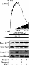
Cicaprost (15 μm) and IBMX (10 μm) significantly relaxed force induced by an EC50 dose of U46619 (0.2 μm) in intact rabbit pulmonary artery. U46619-induced force development coincided with a significant increase in active RhoA, but no changes in Rap1 activity were observed. Subsequent addition of cicaprost and IBMX increased Rap1-GTP and decreased of RhoA activity almost to base line.
FIGURE 3.
FIGURE 3.
Expression profiles of Rap1, RhoA and Epac1 in rabbit, rat, and mouse SM tissues. Western blot analysis of Rap1, RhoA, and Epac1 in various rabbit tissues and cultured rat aortic cells (R518) and Epac1 in various mouse tissues is shown. Note the high Epac content of mouse fundus.
FIGURE 4.
FIGURE 4.
cAMP analogs discriminate between PKA and Epac activation in permeabilized smooth muscle. A, U46619 (300 nm) in the presence of 2 μm GTP for 10 min induced Ca2+-sensitized force in α-toxin-permeabilized rabbit pulmonary artery precontracted in pCa 6.7. The PKA-specific activator 6-Bnz-cAMP (1 μm) significantly relaxed U46619-induced Ca2+-sensitized force. Both 6-Bnz-cAMP- and 007 (30 μm)-induced relaxation were significantly inhibited by the RP-8CPT-cAMPS (50 μm) (mean ± S.E., n = 4–26). B and C, the PKA-specific inhibitory peptide (PKI; 10 μm) inhibited the PKA-specific relaxation but not Epac-mediated relaxation of PE-induced Ca2+-sensitized force. In these experiments rabbit portal veins were permeabilized with β-escin to allow PKA-specific inhibitory peptide access to the cytoplasm in the presence of 1 μm calmodulin and subsequently relaxed with 6-Bnz-cAMP (1 μm) or 007 (30 μm). Force was normalized to the pCa plus agonist-induced Ca2+-sensitized force taken as 100% (mean ± S.E., n = 10). The pCa -induced force at the onset of each force trace has been truncated in C. PE, phenylephrine; N.S., not significant.
FIGURE 5.
FIGURE 5.
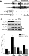
Activation of Epac does not lead to phosphorylation of the PKA specific target VASP. A, both 6-Bnz-cAMP and forskolin increased VASP phosphorylation in intact pulmonary artery. 007, 50 μm (2 min, 10 min), did not increase phosphorylation of VASP at Ser-157 in permeabilized pulmonary arteries precontracted with U46619. Surprisingly, VASP phosphorylation tended to decrease under these conditions. VASP phosphorylation in permeabilized SM before and after U46619 stimulation did not differ. B, both forskolin (FK) and 6-Bnz-cAMP (p < 0.001 and p < 0.03, respectively), but not 007, increased VASP phosphorylation at Ser-157 in rat aortic SMCs. H89 significantly decreased control and forskolin-, 007-, and 6-Bnz-cAMP-induced VASP phosphorylation (p < 0.001; p < 0.002; p < 0.01; and p < 0.03, respectively, n = 4 for each condition. Changes in VASP phosphorylation in the presence of 007 with and without H89 did not differ from control samples. VASP phosphorylation was normalized to total Rap1 (lower panel). Data are shown as a percentage of basal Ser-157-VASP phosphorylation.
FIGURE 6.
FIGURE 6.
A and B, 007 increases Rap1 activity independently of PKA. A, 007 (50 μm, 15 min) significantly increased Rap1 activity in rat aortic SMCs in the presence and absence of H89. H89 (5 μm) alone had no significant effect. Rap1-GTP levels were normalized to total Rap1 and are shown as a % of control (mean ± S.E., n = 3); control versus 007: #, p < 0.0001; 007 versus 007 + H89: *, p < 0.001. B, 007 activates wild type Epac1 but not a mutant dominant negative Epac1. Overexpression of wild type Epac1 in NIH-3T3 significantly increased Rap1 activity upon 007 (50 μm, 15 min) stimulation but had no effect on basal Rap1 activity, whereas overexpression of dominant-negative Epac1 totally blocked Rap1 activation after 007 treatment as well as decreased basal Rap1 activity (mean ± S.E., n = 3–5; *, p < 0.008; #, p < 0.001 versus Epac1-WT plus 007; +, p < 0.03 versus non-stimulated control). C, forskolin, IBMX and isoproterenol (iso) increase Rap1 activity in aortic cells. Serum-starved rat aortic SMCs were treated with 10 μm forskolin (FK), 1 mm IBMX, or 100 μm isoproterenol for 15 min and subsequently assayed for Rap1 activation. p < 0.05, n = 4–5.
FIGURE 7.
FIGURE 7.
007 activates Rap1 and decreases RhoA activity in SMCs. A, a Western blot shows 007-induced increased Rap1 and decreased RhoA activity for serum-starved rat aortic, human airway, and mouse fundus SMCs. B, LPA (1 μm) significantly increased RhoA activity in all three cell types (p < 0.01, p < 0.003, and p < 0.001, respectively) that were reduced by treatment with 007 (50 μm) (p < 0.007 n = 3, p < 0.04 n = 3, p < 0.01, respectively). A, C, and D, 007 significantly increased Rap1 activity in rat aortic (n = 3), human airway (n = 3), and fundus (n = 8) SMCs compared with control cells (p < 0.03 (*), p < 0.001(#), and p < 0.02 (+) respectively). B, statistical significance for each comparison designated by brackets was p < 0.05.
FIGURE 8.
FIGURE 8.
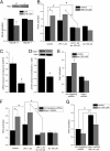
A, 007 decreases LPA-induced RhoA activity in WT but not Rap1B null mouse embryonic fibroblasts. Serum-starved cells were treated with 1 μm LPA ± 50 μm 007 for 15 min. In Rap1B+/+ cells, 007 significantly decreased RhoA-GTP levels (*, p < 0.02, n = 3) but not in the Rap1B−/− cells. The inset shows the absence of Rap1B protein in Rap1B−/− cells. B, isoproterenol (iso, 100 μm) decreases LPA-induced RhoA activity in a PKA-independent manner. LPA stimulation (1 μm, 15 min) of serum-starved rat aortic SMCs significantly increased RhoA activity that was decreased to control levels by co-treatment with isoproterenol. Pretreatment of cells with H89 (1 μm, 1 h) significantly increased basal RhoA activity that was not further increased by LPA stimulation. H89 did not inhibit the isoproterenol-induced decrease in RhoA activity (mean ± S.E.; *, p < 0.006; #, p < 0.02, n = 4). C, D, and E, Epac1 shRNA decreases both Epac1 mRNA and protein levels and totally abolished isoproterenol-induced increases in Rap1 activity. Rat aortic SMCs were infected with either Epac1 shRNA- or non-targeting shRNA-carrying virus and incubated for 36 h followed by serum-free medium for another 16 h. Shown are mRNA (C) and Epac1 (D) mRNA and protein levels, respectively (*, p < 0.05; #, p < 0.0005, n = 3). E, isoproterenol (100 μm, 15 min) significantly increased Rap1 activity in non-targeting shRNA infected cells but not in Epac1 shRNA-infected cells (*, p < 0.0005, n = 4). F, isoproterenol decreased LPA-induced RhoA activity in an Epac1-independent manner. LPA stimulation (1 μm, 15 min) of serum-starved rat aortic SMCs significantly increased RhoA activity that was subsequently decreased to basal levels by co-treatment with isoproterenol (100 μm). A 50% knockdown of Epac1 significantly increased basal RhoA-GTP that did not change after LPA addition. However, knockdown of Epac1 did not prevent the isoproterenol-induced decrease in RhoA activity (*, p < 0.02; #, p < 0.04, n = 4). G, simultaneous inhibition of both Epac1 and PKA pathways significantly inhibits the isoproterenol-induced decrease of RhoA-GTP levels in rat aortic SMCs. Note that basal RhoA activity is significantly increased by treatment with both H89 and Epac1 shRNA (*, p < 0.0005, n = 4). Isoproterenol treatment = 15 min.
FIGURE 9.
FIGURE 9.
Scheme illustrating cAMP induced relaxation of RhoA-mediated Ca2+-sensitized force in smooth muscle via two signaling pathways, PKA and Epac. PKA induces SM relaxation through decreasing [Ca2+]i by inhibitory phosphorylation of RhoA at Ser-188 (–16) by phosphorylation of telokin (18, 19), which activates MLCP activity or by binding to and activating MYPT1 by an unknown mechanism. cAMP activation of Epac leads to GTP exchange onto Rap1, which possibly by activation of a Rap1-activated RhoGAP results in a decrease in RhoA activity and relaxation through disinhibition of MLCP and a fall in RLC20 phosphorylation and Ca2+-sensitized force. GEF, guanine nucleotide exchange factor. AC, adenyl cyclase.

Similar articles

Cited by

References

    1. Somlyo A. P., Somlyo A. V. (2003) Physiol. Rev. 83, 1325–1358 - PubMed
    1. Somlyo A. P., Kitazawa T., Himpens B., Matthijs G., Horiuti K., Kobayashi S., Goldman Y. E., A. V. S. (1989) Adv. Prot. Phosphatases 5, 181–195
    1. Matsui T., Amano M., Yamamoto T., Chihara K., Nakafuku M., Ito M., Nakano T., Okawa K., Iwamatsu A., Kaibuchi K. (1996) EMBO J. 15, 2208–2216 - PMC - PubMed
    1. Uehata M., Ishizaki T., Satoh H., Ono T., Kawahara T., Morishita T., Tamakawa H., Yamagami K., Inui J., Maekawa M., Narumiya S. (1997) Nature 389, 990–994 - PubMed
    1. Kitazawa T., Masuo M., Somlyo A. P. (1991) Proc. Natl. Acad. Sci. U.S.A. 88, 9307–9310 - PMC - PubMed

Publication types

LinkOut - more resources