. 2011 Jun 10;286(23):21013-22.
doi: 10.1074/jbc.M110.189274. Epub 2011 Mar 21.

Impairment of human immunodeficiency virus type-1 integrase SUMOylation correlates with an early replication defect

Affiliations

Impairment of human immunodeficiency virus type-1 integrase SUMOylation correlates with an early replication defect

Alessia Zamborlini et al. J Biol Chem. .

Abstract

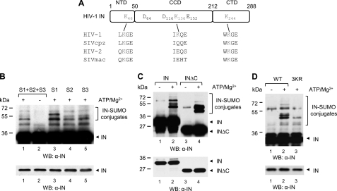
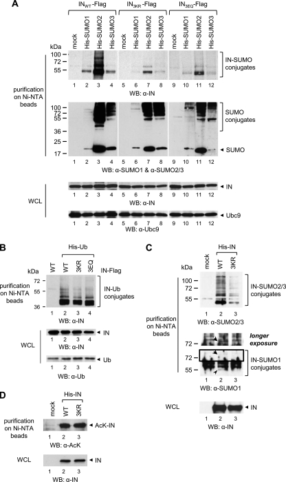
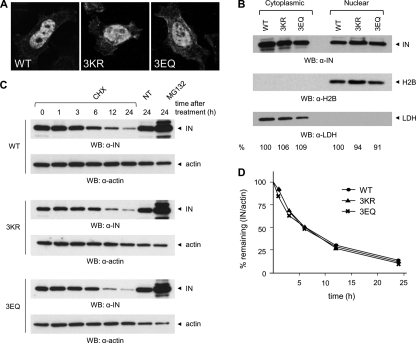
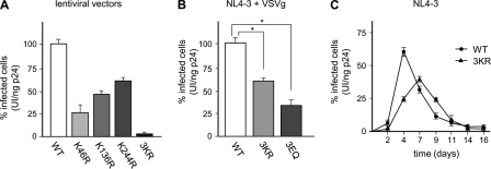
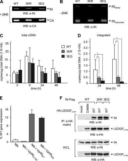
HIV-1 integrase (IN) orchestrates the integration of the reverse transcribed viral cDNA into the host cell genome and participates also in other steps of HIV-1 replication. Cellular and viral factors assist IN in performing its multiple functions, and post-translational modifications contribute to modulate its activities. Here, we show that HIV-1 IN is modified by SUMO proteins and that phylogenetically conserved SUMOylation consensus motifs represent major SUMO acceptor sites. Viruses harboring SUMOylation site IN mutants displayed a replication defect that was mapped during the early stages of infection, before integration but after reverse transcription. Because SUMOylation-defective IN mutants retained WT catalytic activity, we hypothesize that SUMOylation might regulate the affinity of IN for co-factors, contributing to efficient HIV-1 replication.


Figures

FIGURE 1.
FIGURE 1.
HIV-1 IN is modified by the three SUMO paralogues in vitro on conserved Lys residues. A, schematic representation of IN protein. Amino acid sequence alignment of candidate SUMOylation motifs of HIV-1 (HXB2 reference sequence), SIVcpz, HIV-2rod, and SIVmac are shown. DDE, active site residues. B, purified recombinant His-IN (200 nm) was incubated with purified SUMO-specific E1 and E2 enzymes, and the three SUMO proteins (S1, S2, and S3) were added simultaneously or individually. The reaction was conducted in the presence or in the absence of ATP/Mg2+, as indicated. Modified and unmodified forms of IN were visualized with an anti-IN antibody. Incubation of purified His-tagged full-length and C-terminal truncation mutant IN (INΔC) (C) or WT and 3KR mutant IN proteins (D) with the three SUMO proteins simultaneously was performed as in B. The lower panels from B–D show shorter exposure times. WB, Western blot.
FIGURE 2.
FIGURE 2.
HIV-1 IN is SUMOylated by overexpressed or endogenous SUMO proteins in the cell. A, 293T cells were co-transfected with plasmids encoding FLAG-tagged INWT, or IN3KR, or IN3EQ and Ubc9; and His-tagged SUMO-1, or SUMO-2, or SUMO-3 or an empty vector (mock) and, 40 h later, were lysed in denaturing conditions followed by purification on nickel-NTA beads. B, 293T cells co-expressing WT or 3KR or 3EQ IN-FLAG and His-tagged ubiquitin were treated as in A. C, 293T cells expressing His-INWT or His-IN3R were treated as in A, and modification by endogenous SUMO-1 or SUMO-2/3 was assessed. A longer exposure time of the inset enclosed in the black square is shown (middle). The arrows indicate SUMO-1-conjugated IN forms. D, acetylated forms of His-INWT or His-IN3R were analyzed following purification as in A. Proteins expressed in the cells or enriched on nickel-NTA beads in A–D were visualized by Western blot with the indicated antibodies. WB, Western blot; WCL, whole cell lysate.
FIGURE 3.
FIGURE 3.
Subcellular distribution and stability of HIV-1 IN are not affected by mutation of SUMOylation consensus motifs. A, HeLa cells expressing WT or 3KR or 3EQ IN-FLAG were fixed with 4% PFA and then stained with an antibody anti-FLAG, followed by an Alexa488-conjugated secondary antibody. Cells were visualized on a confocal microscope, using a ×63 magnification. Representative images are shown. B, 24 h after transfection, 293T cells expressing FLAG-tagged INWT, IN3R, or IN3Q were subject to cell fractionation followed by Western blot with anti-IN. Purity of the fractions was verified with a nuclear marker, H2B, and a cytoplasmic marker, LDH. Following quantification of ECL signals with ImageJ, the distribution of WT or mutant IN proteins in the nucleus and the cytoplasm was determined by dividing the intensity of IN signals for the corresponding H2B or LDH signals. The values for INWT were arbitrarily set to 100. C, 24 h after transfection, 293T cells expressing FLAG-tagged INWT, IN3KR, or IN3EQ were treated with cycloheximide (CHX) or MG132 or left untreated (NT). At the indicated times, cells were lysed, and total proteins (25 μg/lane) were separated by SDS-PAGE followed by Western blot with anti-IN or anti-actin antibodies. D, ECL signals from Western blots of B were quantified using ImageJ, and values were plotted as the percentage of the signal at t = 0 (given an arbitrary value of 100%) remaining at the indicated time points. Results shown are representative of three independent experiments. WB, Western blot.
FIGURE 4.
FIGURE 4.
HIV-1 virions harboring SUMOylation site mutant IN display reduced infectivity. A, equivalent p24CA amounts (5 ng) of VSVg-pseudotyped lentiviral vectors harboring WT or SUMOylation site mutant IN and encoding GFP were used to infect 293T cells. Infectivity of each viral stock, expressed as the percentage of GFP-positive cells at 48 h postinfection, was measured by flow cytometry. The infectivity of WT viruses was arbitrarily set to 100. B, the infectivity of equal p24CA amounts of VSVg-pseudotyped HIV-1WT, HIV-13KR, or HIV-13EQ (5 ng) was determined as described in A. *, p < 0.05 (paired t test). C, for spreading infection experiments, CEM-GFP cells were infected with equal amounts of HIV-1WT or HIV-13KR (20 ng of p24CA, an approximate multiplicity of infection of 0.15). Mock infections were performed using heat-inactivated WT viruses. Plotted values in A–C represent the mean ± S.D. (error bars) from three independent experiments.
FIGURE 5.
FIGURE 5.
HIV-1 harboring SUMOylation-defective IN is impaired in provirus formation but retains WT protein composition, catalytic activity, and LEDGF/p75-binding. Proteins contained in HIV-1WT or HIV-13KR or HIV-13EQ viral preparations (100 or 300 ng of p24CA) were separated by SDS-PAGE in reducing (with β-mercaptoethanol (+βME)) (A) or non-reducing (without β-mercaptoethanol (−βME)) (B) conditions and were revealed by Western blot with anti-CA or anti-IN antibodies. Emission signal intensities relative to CA and IN were quantified by laser scanning of corresponding bands on an Odyssey infrared imaging system. C, 293T cells were infected with the indicated single-round viruses (100 ng of p24CA, corresponding to a multiplicity of infection of 0.5 for HIV-1WT), and at the indicated time points, late reverse transcripts were quantified by real-time PCR and normalized for total DNA content. Signals detected in parallel infections with VSVg-minus viruses were subtracted from envelope-mediated infections to correct for input plasmid DNA carry over. D, integrated proviruses were quantified by Alu-PCR at 24 and 48 h postinfection. Results shown in A and B represent the mean ± S.D. of two independent experiments performed in duplicates. *, p < 0.05 (paired t test). E, analysis of the ability of WT, D116A or 3KR IN expressed in trans as Vpr fusion proteins to rescue the replication defect of integration-defective virions (NN), expressed as a percentage of WT HIV-1NLX.Luc(R−) activity (56). F, lysates from 293T cells expressing FLAG-tagged INWT, IN3KR, or IN3EQ and HA-tagged WT or D366N mutant LEDGFCter were immunoprecipitated with an HA affinity matrix. Bound proteins were analyzed by Western blot with anti-FLAG and anti-HA antibodies. WCL, whole cell lysate.

References

    1. Craigie R. (2001) J. Biol. Chem. 276, 23213–23216 - PubMed
    1. Esposito D., Craigie R. (1999) Adv. Virus. Res. 52, 319–333 - PubMed
    1. Vandegraaff N., Engelman A. (2007) Expert Rev. Mol. Med. 9, 1–19 - PubMed
    1. Craigie R., Fujiwara T., Bushman F. (1990) Cell 62, 829–837 - PubMed
    1. Katz R. A., Merkel G., Kulkosky J., Leis J., Skalka A. M. (1990) Cell 63, 87–95 - PubMed

Publication types