Comparative Study
. 2011 May 20;286(20):18149-60.
doi: 10.1074/jbc.M110.194258. Epub 2011 Mar 23.

Effect of kallikrein 4 loss on enamel mineralization: comparison with mice lacking matrix metalloproteinase 20

Affiliations
Comparative Study

Effect of kallikrein 4 loss on enamel mineralization: comparison with mice lacking matrix metalloproteinase 20

Charles E Smith et al. J Biol Chem. .

Abstract

Enamel formation depends on a triad of tissue-specific matrix proteins (amelogenin, ameloblastin, and enamelin) to help initiate and stabilize progressively elongating, thin mineral ribbons of hydroxyapatite formed during an appositional growth phase. Subsequently, these proteins are eradicated to facilitate lateral expansion of the hydroxyapatite crystallites. The purpose of this study was to investigate changes in enamel mineralization occurring in mice unable to produce kallikrein 4 (Klk4), a proteinase associated with terminal extracellular degradation of matrix proteins during the maturation stage. Mice lacking functional matrix metalloproteinase 20 (Mmp20), a proteinase associated with early cleavage of matrix proteins during the secretory stage, were also analyzed as a frame of reference. The results indicated that mice lacking Klk4 produce enamel that is normal in thickness and overall organization in terms of layers and rod/inter-rod structure, but there is a developmental defect in enamel rods where they first form near the dentinoenamel junction. Mineralization is normal up to early maturation after which the enamel both retains and gains additional proteins and is unable to mature beyond 85% mineral by weight. The outmost enamel is hard, but inner regions are soft and contain much more protein than normal. The rate of mineral acquisition overall is lower by 25%. Mice lacking functional Mmp20 produce enamel that is thin and structurally abnormal. Relatively high amounts of protein remain throughout maturation, but the enamel is able to change from 67 to 75% mineral by weight during maturation. These findings reaffirm the importance of secreted proteinases to enamel mineral acquisition.


Figures

FIGURE 1.
FIGURE 1.
Mineral content in enamel organ cells from maxillary and mandibular incisors of wild-type (Klk4+/+, Mmp20+/+), heterozygous (Klk4+/−, Mmp20+/−), and null (Klk4−/−, Mmp20−/−) mice. Mean plots ± 95% confidence intervals of the percent mineral by weight in 1-mm-long freeze-dried cell strips dissected across the portion of the incisor embedded in bone (S+M). Most cell samples are associated with little mineral except in the case of mandibular incisors from Mmp20−/− mice (yellow diamonds). The large deviations indicate there is considerable tooth-to-tooth variation in mineral-promoting responses by enamel organ cells to loss-of-function of Mmp20 across the mid- to late stages of maturation. S, secretory stage; M, maturation stage; E, erupted portion.
FIGURE 2.
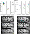
FIGURE 2.
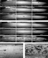
Scanning electron microscopy in backscatter mode at low (A and B) and high (C and D) magnifications showing developing enamel surfaces along the labial side of incisors in the apical (secretory to early maturation), middle (nearly mature), and incisal (erupted) ends of teeth from wild-type (A, Klk4+/+; B, Mmp20+/+), heterozygous (A, Klk4+/-; B, Mmp20+/−), and null (A, Klk4−/−; B, Mmp20−/−) mice. In this technique, whiter areas contain more mineral than darker areas. The enamel surfaces of incisors in wild-type (+/+) mice appear relatively smooth and mostly devoid of cell debris (cd) across their length (A and B; top rows, all panels). Enamel surfaces of incisors in Klk4+/− (A) and Mmp20+/− (B) mice appear similar to wild-type mice except in early maturation where cracks (c) from freeze drying are sometimes more prominent (A and B; top and middle rows, left panels). The incisors of Mmp20+/− mice also consistently show regularly spaced surface undulations (stripes) in enamel running perpendicular to the long axis of the tooth across the erupted portion of the tooth (B, middle row, right panel). The enamel surfaces on incisors from Klk4−/− mice are relatively smooth, although irregular pitted areas (ipa) are seen across regions where the enamel becomes nearly mature (A, bottom row, middle panel, and C). The enamel along the erupted portions of these incisors often shows faint stripes and is consistently fractured (f) near the incisal tips exposing the underlying dentin (d) (A, bottom row, right panel). The enamel surfaces of incisors in Mmp20−/− mice are thin, rough, and obviously malformed (B, bottom row). Nodules (n) and other elongated calcified masses and associated cells debris (cd) project from the surface starting initially at the central labial aspect of the incisor in early maturation (B, bottom row, left panel, and D) and later across the whole labial surface of the tooth (B, bottom row, middle panel). The poorly formed mature enamel (e) is frail and easily fractures off (f) the erupted portions of these incisors exposing underlying dentin (d) (B, bottom row, right panel).
FIGURE 3.
FIGURE 3.
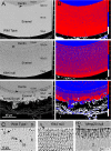
Scanning electron microscopy in backscatter mode at low (A and B) and higher (C) magnifications of a portion of dentin and the entire enamel layer as seen in transverse ground sections of incisors from wild-type and Klk4−/− and Mmp20−/− mice cut at a level close to where the teeth erupt. The images in B are pseudo colored maps of the same four gray level intensities for corresponding images in A (red, highest or most mineral). Arrowheads in A indicate approximate locations near the DEJ for images shown in C. The numbers in a column to the right of each image in A represent the mean gray level intensity ± S.D. counted within a 10 × 10 μm grid over dentin (top numbers) and the inner, middle, and outer regions of the enamel layer. In the bottom panel of A, the numbers 1–3 indicate the tri-layered organization of enamel in Mmp20−/− mice; n, nodule of calcified material projecting from outer surface. C, D is dentin; E is enamel; S is normal developmental enamel spaces; R is enamel rod; IR is inter-rod enamel; IL is initial layer of enamel; HD is hypermineralized dentin. In wild-type and both Klk4−/− and Mmp20−/− mice, the outermost part of the enamel layer always appears more mineralized than the innermost part closest to dentin (B, red versus blue colors). Weaknesses in enamel observed in Klk4−/− and Mmp20−/− mice appear to be due in part to structural defects present near the DEJ (C) and to a less uniform distribution of mineral especially within inter-rod areas of the enamel (B, blue color compared with red).
FIGURE 4.
FIGURE 4.
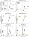
Mineral content in enamel. Absolute (A and D) and relative (B and E) mineral content and mineral acquisition rates (C and F) for developing enamel on incisors from wild-type (A–C, Klk4+/+; D–F, Mmp20+/+), heterozygous (A–C, Klk4+/−; D–F, Mmp20+/−), and null (A–C, Klk4−/−; D–F, Mmp20−/−) mice. Each graph represents mean ± 95% confidence intervals and shows after ashing weight (A and D), percent mineral by weight (B and E), and mineral gain per mm (C and F) for 1-mm-long enamel strips microdissected from maxillary (left graph each panel, Mx) and mandibular (right graph each panel, Mn) incisors. Stages of amelogenesis are illustrated by the dashed lines in A (S, secretory stage; M, maturation stage; E, erupted portion). Enamel in Klk4−/− mice is mildly hypomineralized across the maturation stage (A–C), whereas enamel in Mmp20−/− mice is both hypoplastic and hypomineralized (D–F). At its peak in early to mid-maturation (strip 4 on maxillary and strip 4–5 on mandibular incisors), Klk4−/− mice acquire mineral about 25% slower and Mmp20−/− mice about 70% slower than wild-type or heterozygous mice.
FIGURE 5.
FIGURE 5.
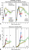
Protein content in enamel. Total dry weight of volatiles (A) and mineral-to-volatiles ratio (B) for developing enamel on incisors from wild-type (Klk4+/+; Mmp20+/+), heterozygous (Klk4+/−; Mmp20+/−), and null (Klk4−/−; Mmp20−/−) mice. The graphs in A and B represent mean ± 95% confidence intervals for 1-mm-long enamel strips microdissected from maxillary (left graph each panel, Mx) and mandibular (right graph each panel, Mn) incisors. Stages of amelogenesis are illustrated by the dashed lines in B (S, secretory stage; M, maturation stage; E, erupted portion). Weight data for Mmp20−/− mice in A were normalized so that genotypes are compared relative to enamel having the same overall thickness. The enamel in both Klk4−/− and Mmp20−/− mice contains excess amounts of volatiles (mostly protein) throughout the maturation stage (A). Klk4−/− mice show an initial loss of volatiles in early to mid-maturation as occurs in normal enamel development, but the decline is less and is followed by a rise in volatiles during late maturation and decline across the erupted portion of the tooth (A, green diamonds). Mmp20−/− mice maintain relatively high amounts of volatiles throughout the maturation stage (A, yellow diamonds). A mild heterozygous effect in volatile content is evident at mid-maturation on mandibular incisors of both Klk4+/− and Mmp20+/− mice (A, strip 5, squares). Mineral-to-volatiles ratios are consistently low throughout maturation in Klk4−/− mice and show a trend to rise across the erupted portion of the incisor (B). Mineral-to-volatiles ratios are even lower in Mmp20−/− mice but show a trend to rise across the maturation stage and onto the erupted portion of the tooth (B).
FIGURE 6.
FIGURE 6.
Mineral content in molars. A, mean plots ± 95% confidence intervals of the before ashing dry weights (A) of the 1st, 2nd, and 3rd maxillary (Mx) and mandibular (Mn) molars in wild-type (Klk4+/+; Mmp20+/+), heterozygous (Klk4+/−; Mmp20+/−), and null (Klk4−/−; Mmp20−/−) mice. Alterations in enamel mineralization and excessive coronal wear due to loss-of-function of Klk4 and Mmp20 are detectable as reduced gross weight of the molars. B, mean plots ± 95% confidence intervals of the percent mineral by weight for molars in the six genotypes. A molar without enamel would have a percent mineral content of 71–73% by weight (see Table 1). Third and to a lesser extent second molars show the greatest relative changes in mineral content. C and D, scanning electron microscopy in backscatter mode of the 1st, 2nd, and 3rd mandibular molar crowns from wild-type (C, Klk4+/+; D, Mmp20+/+), heterozygous (C, Klk4+/−; D, Mmp20+/−), and null (C, Klk4−/−; D, Mmp20−/−) mice. Cracks within some molar crowns are artifacts from freeze drying. The enamel covering the crowns of molars contributes to the overall weight of these teeth and is subject to change as the molar crowns wear down by occlusal attrition. Developmentally weakened and hypomineralized enamel in homozygous mice fractures (f) off the crowns and abrades more easily thereby exposing areas of dentin (d) to severe attrition.

Similar articles

Cited by

References

    1. Simmer J. P., Papagerakis P., Smith C. E., Fisher D. C., Rountrey A. N., Zheng L., Hu J. C. (2010) J. Dent. Res. 89, 1024–1038 - PMC - PubMed
    1. Lacruz R. S., Nanci A., Kurtz I., Wright J. T., Paine M. L. (2010) Calcif. Tissue Int. 86, 91–103 - PMC - PubMed
    1. Hu J. C., Chun Y. H., Al Hazzazzi T., Simmer J. P. (2007) Cells Tissues Organs 186, 78–85 - PubMed
    1. Bei M. (2009) Curr. Opin. Genet. Dev. 19, 504–510 - PMC - PubMed
    1. Wright J. T., Hart T. C., Hart P. S., Simmons D., Suggs C., Daley B., Simmer J., Hu J., Bartlett J. D., Li Y., Yuan Z. A., Seow W. K., Gibson C. W. (2009) Cells Tissues Organs 189, 224–229 - PMC - PubMed

Publication types

MeSH terms