. 2011 May 20;286(20):17992-8001.
doi: 10.1074/jbc.M110.216176. Epub 2011 Mar 23.

Regulatory circuits of the AAA+ disaggregase Hsp104

Affiliations

Regulatory circuits of the AAA+ disaggregase Hsp104

Titus M Franzmann et al. J Biol Chem. .

Abstract

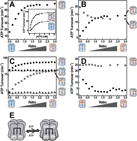
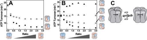
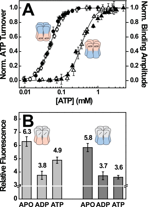
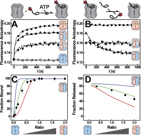
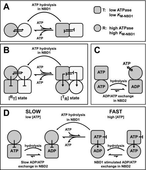
Yeast Hsp104 is an AAA+ chaperone that rescues proteins from the aggregated state. Six protomers associate to form the functional hexamer. Each protomer contains two AAA+ modules, NBD1 and NBD2. Hsp104 converts energy provided by ATP into mechanical force used to thread polypeptides through its axial channel, thereby disrupting protein aggregates. But how the action of its 12 AAA+ domains is co-ordinated to catalyze disaggregation remained unexplained. Here, we identify a sophisticated allosteric network consisting of three distinct pathways that senses the nucleotide state of AAA+ modules and transmits this information across the Hsp104 hexamer. As a result of this communication, NBD1 and NBD2 each adopt two distinct conformations (relaxed and tense) that are reciprocally regulated. The key element in the network is the NBD1-ATP state that enables Hsp104 to switch from a barely active [(T)(R)] state to a highly active [(R)(T)] state. This concerted switch involves both cis and trans protomer interactions and provides Hsp104 with the mechanistic scaffold to catalyze disaggregation. It prepares the chaperone for polypeptide binding and activates NBD2 to generate the power strokes required to resolve protein aggregates. ATP hydrolysis in NBD1 resolves the high affinity [(R)(T)] state and switches the chaperone back into the low affinity [(T)(R)] state. Our model integrates previously unexplained observations and provides the first comprehensive map of nucleotide-related allosteric signals in a class-1 AAA+ protein.


Figures

FIGURE 1.
FIGURE 1.
Dissecting allosteric interactions using mixed hexamers. A, cryo-EM reconstruction of hexameric Hsp104ΔN (32) is shown. B, upon mixing, an active Hsp104 protein such as wild-type Hsp104 (blue) will form hetero hexamers with an inactive Hsp104 protein (red) by exchanging subunits. C, the resulting hetero hexamer distribution depends on the mixing ratio. The larger the mixing ratio, the more inactive subunits a hexamer contains on average. At a mixing ratio of 1, the most abundant hexamer contains three active and three inactive subunits. D, whether the presence of inactive subunits (red) changes the behavior of active subunits (blue) in a hexamer can be assessed by monitoring a property of the active subunits such as ATPase activity. These experiments were carried out by mixing a constant amount of an active hexamer with an increasing amount of an inactive hexamer. Three possible scenarios are depicted. Inactive subunits may not affect active subunits (●), they could stimulate active subunits (green circle) or they could inhibit active subunits (red circle). E, three types of NBD variants were used in this study. Wild-type NBDs (blue) bind and hydrolyze ATP. Variants with a mutated Walker B motif (red) no longer hydrolyze ATP, but still bind it. Variants with a mutated Walker A motif (white) are unable to bind nucleotide. In the text, the three types of NBDs are identified by the symbols [+], [b], and [−], respectively.
FIGURE 2.
FIGURE 2.
ATP binding to NBD1 stimulates ATP hydrolysis in an adjacent NBD2. A, ATPase activity of [++] mixed with [bb] at various ratios (■). Inset, ATPase rate plot of [++] (○) and a [bb·++] mixture at a ratio of 1 (●). B, ATPase activities of [+] (●) and [+] (○) mixed with [bb] at the indicated mixing ratios. C, ATPase activities of [++] (filled symbols) and [+] (open symbols) mixed with [b] (triangles) or [b] (circles) at the indicated ratios. D, ATPase activity of [b+] mixed with [bb] (○) or [b] (●) at the indicated ratios. E, model of Hsp104 showing the stimulatory effect of ATP binding to NBD1 on ATP hydrolysis in NBD2. ATP hydrolysis rates were determined at 30 °C and 2 mm ATP. The concentration of active subunits was 1 μm, whereas the concentration of inactive subunits varied. Hetero complexes were formed by mixing both subunits in the absence of nucleotide and incubation for 1 h at 30 °C prior to measurement.
FIGURE 3.
FIGURE 3.
ATP binding to NBD1 inhibits ATP hydrolysis in an adjacent NBD1. A, ATPase activities of [+] (○) and [+b] (●) mixed with [bb] at the indicated ratios. B, ATPase activities of [+b] (filled symbols) and [+] (open symbols) after mixing with [b] (triangles) or [b] (circles) at the indicated ratio. Experiments were carried out as described in the legend for Fig. 2. C, model of Hsp104 showing the inhibitory effect of ATP binding to NBD1 on ATP hydrolysis in NBD1.
FIGURE 4.
FIGURE 4.
The nucleotide state of NBD2 modulates the affinity of NBD1 toward ATP. A, correlation between ATPase activity and polypeptide binding. ATPase rates of [+b] (●) and [b+] (▴) were recorded in the presence of 3 μm RCMLa. Binding of f-RCMLa (160 nm) to [+b] (○) and [b+] (△) was monitored by fluorescence anisotropy at 515 nm. Datasets were recorded at 30 °C and a hexamer concentration of 80 nm and normalized. Error bars reflect the S.D. of three independent experiments. B, ANS fluorescence of [b] and [+] in the absence and presence of saturating concentrations of ADP and ATP. 5 μm Hsp104 was preincubated with 100 μm ANS and mixed with buffer or 500 μm nucleotide. Error bars reflect the S.D. of three independent experiments.
FIGURE 5.
FIGURE 5.
Substrate binding and processing by Hsp104 hetero complexes. A, binding of 160 nm f-RCMLa to Hsp104 [++] (○), [bb] (●), [bb·++] (▴), and [bb·+] (▾) (both at a mixing ratio of 1) in the presence of 1 mm ATP as monitored by fluorescence anisotropy at 515 nm. The Hsp104 concentrations were 160 nm for homo and 320 nm for hetero hexamers. B, release of bound f-RCMLa from Hsp104, induced by the addition of a 20-fold excess of unlabeled RCMLa. Symbols and conditions are as in A. C, quantitative analysis of f-RCMLa binding to [bb·++]. The lines in color represent the predicted amplitude for scenarios in which at least three (red), two (green), and one (blue) [bb] subunit(s) per hexamer are required for f-RCMLa binding. The observed amplitudes (●) are the means of three independent measurements. f-RCMLa (100 nm) and Hsp104 hexamer (160 nm) concentrations were kept constant. D, quantitative analysis of f-RCMLa release from [bb·++]. The lines in color represent the predicted amplitudes for scenarios in which at least three (red), two (green), and one (blue) [++] subunit(s) per hexamer are required for f-RCMLa release. The observed amplitudes are shown as (●).
FIGURE 6.
FIGURE 6.
Reactivation of chemically denatured luciferase by Hsp104. Disaggregation of 55 nm FFL by [bb·++] mixtures (●) is shown at the indicated ratios. The concentration of [++] subunits was kept constant at 1 μm, whereas the concentration of [bb] subunits varied. Disaggregation was carried out at 3 mm ATP and 25 °C in the absence of Hsp70/40. Activity of renatured FFL was monitored continuously. The initial slope of FFL activity versus time was used as a measure of disaggregation velocity in FFL activity after 70 min (A) was used to assess the yield of the disaggregation reaction in B.
FIGURE 7.
FIGURE 7.
Model for the allosteric communication between AAA+ modules in Hsp104. A, conformational coupling between NBD1 domains is the mechanistic basis for the trans interaction of protomers and propagates nucleotide-induced structural rearrangements across the hexamer. NBD1 can adopt two states that differ in their ATPase activity: T (square, low activity) and R (circle, high activity). The transition between the two states is concerted, i.e. it occurs simultaneously in a number of NBD1s, two of which are depicted here in white and gray. ATP binding to NBD1 stabilizes the T state, whereas ATP hydrolysis induces the transition to the R state. B, the trans interaction of NBD1 co-ordinates the behavior of NBD2 in the Hsp104 hexamer. Depicted are two of six protomers in gray and white. Similar to NBD1 (top), NBD2 (bottom) can adopt T and R states with differing ATPase activity. However, the T↔ R transition in NBD2 is inversely linked to the T↔ R transition in NBD1. ATP binding to NBD1 induces a concerted transition of NBD1 to the T state. Adoption of the T state is propagated to NBD2 using a NBD1→NBD2 cis pathway. It triggers a conformational change of NBD2 to the R state and stimulates nucleotide exchange, thereby strongly activating ATP turnover in this domain. ATP hydrolysis in NBD1 triggers the co-operative transition of NBD1 to the R state and concomitantly causes NBD2 to revert to the T state. C, nucleotide occupancy in NBD2 modulates the affinity of NBD1 toward ATP. When NBD2 is occupied with ADP (or empty), NBD1 adopts an open conformation with a low affinity toward ATP (R state). Binding of ATP to NBD2 induces a conformation of NBD1 to which ATP binds more tightly (T state). ATP hydrolysis in NBD2 facilitates the transition of NBD1 to the R state. D, allosteric model for ATP hydrolysis in Hsp104 is shown. For details, see “Discussion.”

Similar articles

Cited by

References

    1. Ogura T., Wilkinson A. J. (2001) Genes Cells 6, 575–597 - PubMed
    1. Erzberger J. P., Berger J. M. (2006) Annu. Rev. Biophys. Biomol. Struct. 35, 93–114 - PubMed
    1. Parsell D. A., Kowal A. S., Singer M. A., Lindquist S. (1994) Nature 372, 475–478 - PubMed
    1. Sanchez Y., Lindquist S. L. (1990) Science 248, 1112–1115 - PubMed
    1. Motohashi K., Watanabe Y., Yohda M., Yoshida M. (1999) Proc. Natl. Acad. Sci. U.S.A. 96, 7184–7189 - PMC - PubMed

MeSH terms