. 2011 May 13;286(19):17205-16.
doi: 10.1074/jbc.M111.229419. Epub 2011 Mar 18.

Critical role of the beta-subunit CDC50A in the stable expression, assembly, subcellular localization, and lipid transport activity of the P4-ATPase ATP8A2

Affiliations

Critical role of the beta-subunit CDC50A in the stable expression, assembly, subcellular localization, and lipid transport activity of the P4-ATPase ATP8A2

Jonathan A Coleman et al. J Biol Chem. .

Abstract

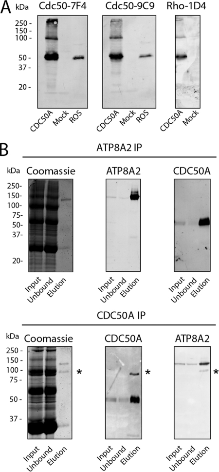
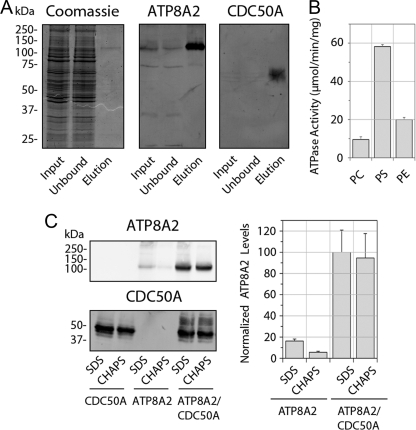
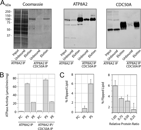
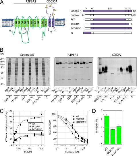
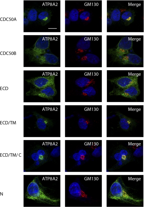
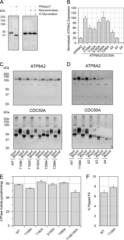
P(4)-ATPases have been implicated in the transport of lipids across cellular membranes. Some P(4)-ATPases are known to associate with members of the CDC50 protein family. Previously, we have shown that the P(4)-ATPase ATP8A2 purified from photoreceptor membranes and reconstituted into liposomes catalyzes the active transport of phosphatidylserine across membranes. However, it was unclear whether ATP8A2 functioned alone or as a complex with a CDC50 protein. Here, we show by mass spectrometry and Western blotting using newly generated anti-CDC50A antibodies that CDC50A is associated with ATP8A2 purified from photoreceptor membranes. ATP8A2 expressed in HEK293T cells assembles with endogenous or expressed CDC50A, but not CDC50B, to generate a heteromeric complex that actively transports phosphatidylserine and to a lesser extent phosphatidylethanolamine across membranes. Chimera CDC50 proteins in which various domains of CDC50B were replaced with the corresponding domains of CDC50A were used to identify domains important in the formation of a functional ATP8A2-CDC50 complex. These studies indicate that both the transmembrane and exocytoplasmic domains of CDC50A are required to generate a functionally active complex. The N-terminal cytoplasmic domain of CDC50A appears to play a direct role in the reaction cycle. Mutagenesis studies further indicate that the N-linked oligosaccharide chains of CDC50A are required for stable expression of an active ATP8A2-CDC50A lipid transport complex. Together, our studies indicate that CDC50A is the β-subunit of ATP8A2 and is crucial for the correct folding, stable expression, export from endoplasmic reticulum, and phosphatidylserine flippase activity of ATP8A2.


Figures

FIGURE 1.
FIGURE 1.
CDC50A monoclonal antibodies and immunoprecipitation of the ATP8A2-CDC50A complex of rod outer segments. A, Western blots of extracts from HEK293T cells transfected with Cdc50a-1D4 plasmid (CDC50A) or empty plasmid (Mock) and bovine ROS membranes were labeled with monoclonal antibodies to CDC50A (Cdc50–7F4 and Cdc50–9C9) and Rho 1D4. B, Western blots of the ATP8A2-CDC50A complex immunoprecipitated (IP) from ROS membranes. CHAPS-solubilized ROS membranes (Input) were incubated with an immunoaffinity support consisting of either the ATP8A2 Atp6C11 antibody (ATP8A2 IP) or the CDC50A Cdc50–7F4 antibody (CDC50A IP) coupled to a Sepharose matrix. The fraction that did not bind to the column (Unbound) and the fraction that was eluted from the column (Elution) using either competing peptide 6C11 peptide (ATP8A2 IP) or 2% SDS (CDCC50A IP) were analyzed on SDS gels stained with Coomassie Blue and Western blots labeled with either the ATP8A2 or CDC50A specific antibodies. The asterisks indicate the presence of antibody present in the SDS-eluted fraction. Approximately 50 μg of input and unbound and 200 ng of elution were applied to the gel.
FIGURE 2.
FIGURE 2.
Immunofluorescence localization of CDC50A in the retina and gene expression of CDC50 variants in the retina and other tissues. A, retinal cryosections labeled with Atp2F6 or Cdc50–7F4 antibodies to ATP8A2 and CDC50A, respectively (green), and counterstained with the nuclear stain 4′,6-diamidino-2-phenylindole (blue). In the control sample, the CDC50A antibody was treated with excess 7F4 peptide prior to immunolabeling. CDC50A is abundantly localized in photoreceptor outer segments and other retinal layers. ATP8A2 is primarily restricted to the photoreceptor outer segment layer. OS, outer segments; IS, inner segments; ONL, outer nuclear layer; OPL, outer plexiform layer; INL, inner nuclear layer; IPL, inner plexiform layer; GCL, ganglion cell layer. Bar, 25 μm. B, gene expression of the cdc50 family members in the retina and other tissues of adult mice by RT-PCR. Gapdh was used as a loading control. Cdc50a is the only member of the cdc50 family detectable in the retina.
FIGURE 3.
FIGURE 3.
ATP8A2 expressed in HEK293T cells interacts with endogenous CDC50A to form a catalytically active ATP8A2-CDC50A complex. A, immunoaffinity purification of ATP8A2-CDC50A complex from HEK293T cells transfected with the Atp8A2 plasmid. Solubilized HEK293T cell extract (Input) was incubated with the Atp6C11 immunoaffinity matrix, and the unbound fraction (Unbound) and the 6C11 peptide eluted fraction (Elution) were analyzed on SDS gels stained with Coomassie Blue, and Western blots were labeled with the Atp6C11 antibody (ATP8A2) and the Cdc50–7F4 antibody (CDC50A). B, ATPase activity of the purified ATP8A2-CDC50A complex in 100% phosphatidylcholine (PC) or 10% phosphatidylserine (PS) or 40% phosphatidylethanolamine (PE) each containing the corresponding concentrations of PC. C, quantitative analysis of expressed and solubilized ATP8A2 and CDC50A. ATP8A2 and CDC50A containing a 1D4 tag were expressed individually (ATP8A2 or CDC50A) or together (ATP8A2/CDC50A) in HEK293T cells. The amounts of ATP8A2 and CDC50A solubilized with either SDS or CHAPS were measured by Western blotting (left), and the relative quantity of solubilized ATP8A2 was determined from Western blots of three experiments (right). Approximately, 30 μg of protein was loaded into each lane.
FIGURE 4.
FIGURE 4.
ATPase activity and phospholipid flippase activity of the expressed and purified ATP8A2-CDC50A complex. A, Coomassie Blue-stained gel and Western blots of the immunoaffinity-purified ATP8A2-CDC50A complex from HEK293T cells co-expressing both ATP8A2 and CDC50A-1D4. The complex was purified on either an Atp6C11 column alone (ATP8A2 IP) or sequentially on Atp6C11 and Rho-1D4 columns (ATP8A2/CDC50A IP). B, ATP8A2 and CDC50A containing a 1D4 tag were co-expressed in HEK293T cells, and the complex was purified by the dual immunoaffinity procedure. The ATPase activity of the complex was measured in the presence of 100% PC or 10% PS or 40% PE lipids each containing the appropriate amount of PC. C, the ATP8A2-CDC50A complex purified by the dual immunoaffinity procedure was reconstituted into liposomes for phospholipid flippase measurements. The percentage of lipid flipped by the ATP8A2-CDC50A complex was measured in phosphatidylcholine proteoliposomes for 2.5% NBD-labeled PC, PE, or PS. Right panel, the PS flippase activity of the ATP8A2-CDC50A complex was measured for decreasing amounts of reconstituted protein. IP, immunoprecipitation.
FIGURE 5.
FIGURE 5.
Interaction and functional activity of ATP8A2-CDC50 complexes containing CDC50A/B chimera proteins. A, topological model of the ATP8A2-CDC50A complex (left panel). ATP8A2 is shown in green, and CDC50A is in purple. The phosphorylated motif (DKTG) is indicated along with the positions of the N-linked glycosylation sites on CDC50A. Right panel, schematic showing various chimera CDC50A/B proteins used in these studies. B, interaction of CDC50A/B chimera proteins containing a 1D4 tag with ATP8A2. Proteins were purified on an ATP8A2 immunoaffinity column and analyzed on SDS gels stained with Coomassie Blue or Western blots labeled with an ATP8A2 antibody (ATP8A2) or Rho-1D4 antibody (CDC50). CDC50A co-purified with ATP8A2, whereas CDC50B did not. Chimera proteins containing only the extracellular domain (ECD) or N-terminal domain (N) of CDC50A did not co-purify with ATP8A2. Chimeras containing the ECD and TM domain consisting of both M1 and M2 segments (ECD/TM) or the ECD, TM, and C-terminal domains (ECD/TM/C) of CDC50A co-purified with ATP8A2. C, left panel, ATPase activity of ATP8A2 associated with either WT CDC50A (WT) or chimera CDC50A proteins (ECD/TM or ECD/TM/C) as a function of PS concentration in the presence of PC. Right panel, vanadate inhibition of ATPase activity of ATP8A2-CDC50 complexes in 1000 μm PS in the presence of PC. D, flippase activity of ATP8A2-CDC50 complexes containing WT CDC50A and the ECD/TM and ECD/TM/C chimeras reconstituted in phosphatidylcholine in the presence of 2.5% NBD-PS.
FIGURE 6.
FIGURE 6.
Immunofluorescence localization of ATP8A2 co-expressed with WT CDC50A, WT CDC50B, or chimera CDC50A/B containing a 1D4 tag in Cos-7 cells. The cells were double labeled for ATP8A2 with the Atp2F6 monoclonal antibody (green) and the GM130 polyclonal antibody as a Golgi marker (red). Co-expression of ATP8A2 with CDC50B or chimeric CDC50A proteins containing the ECD or N-terminal domain (N) domains resulted in a reticular staining pattern of ATP8A2 characteristic of ER localization. Co-expression of ATP8A2 with either WT CDC50A or chimera proteins containing the ECD/TM or ECD/TM/C domains of CDC50A resulted in localization of ATP8A2 to the Golgi as shown in merged images. Expression of CDC50 proteins was confirmed independently using the Rho 1D4 antibody (data not shown). The nuclei (blue) were labeled with 4′,6-diamidino-2-phenylindole. Bar, 10 μm.
FIGURE 7.
FIGURE 7.
N-Linked glycosylation of CDC50A is necessary for stabilization of ATP8A2. A, approximately 50 μg of rod outer segment membranes were treated with PNGase F or neuraminidase with or without O-glycosidase and labeled with the Cdc50–9C9 antibody. B, expression levels of ATP8A2 in the presence of CDC50A glycosylation mutants containing a 1D4 tag. ATP8A2 levels were normalized to β-actin as a loading control. C, co-immunoprecipitation of single CDC50A glycosylation mutants with ATP8A2 on Atp6C11-Sepharose. ATP8A2 was detected with Atp6C11, and the CDC50A-1D4 mutants were detected with the Rho 1D4 antibody. Solubilized HEK293T cells (Input) were incubated in the column, and the bound proteins (Bound) were eluted with 6C11 peptide. D, immunoprecipitation of multiple CDC50A glycosylation mutants with ATP8A2 on Atp6C11-Sepharose. Multiple site CDC50A glycosylation mutants expressed at low levels relative to WT CDC50A. Approximately 30 μg of input and 5–200 ng of the bound fractions were applied to the gel. E, specific ATPase activity of ATP8A2 expressed with various CDC50A N-linked glycosylation mutants and purified on an immunoaffinity column. ATPase activity was measured in the presence of 10% PS and 90% PC. F, lipid flippase activity of WT and T109A mutant reconstituted in PC in the presence of 2.5% NBD-PS. The difference in activities between WT and T109A is not significant.

References

    1. Alder-Baerens N., Lisman Q., Luong L., Pomorski T., Holthuis J. C. (2006) Mol. Biol. Cell 17, 1632–1642 - PMC - PubMed
    1. Darland-Ransom M., Wang X., Sun C. L., Mapes J., Gengyo-Ando K., Mitani S., Xue D. (2008) Science 320, 528–531 - PubMed
    1. Natarajan P., Wang J., Hua Z., Graham T. R. (2004) Proc. Natl. Acad. Sci. U.S.A. 101, 10614–10619 - PMC - PubMed
    1. Wang L., Beserra C., Garbers D. L. (2004) Dev. Biol. 267, 203–215 - PubMed
    1. Coleman J. A., Kwok M. C., Molday R. S. (2009) J. Biol. Chem. 284, 32670–32679 - PMC - PubMed

Publication types