. 2011 May 6;286(18):16052-62.
doi: 10.1074/jbc.M111.223602. Epub 2011 Mar 16.

Mitochondrial c-Jun N-terminal kinase (JNK) signaling initiates physiological changes resulting in amplification of reactive oxygen species generation

Affiliations

Mitochondrial c-Jun N-terminal kinase (JNK) signaling initiates physiological changes resulting in amplification of reactive oxygen species generation

Jeremy W Chambers et al. J Biol Chem. .

Abstract

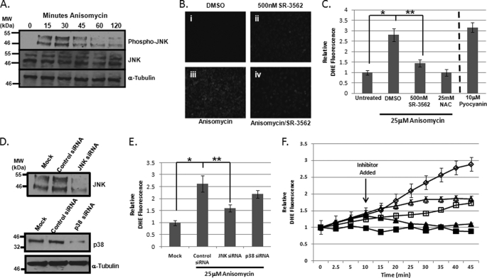
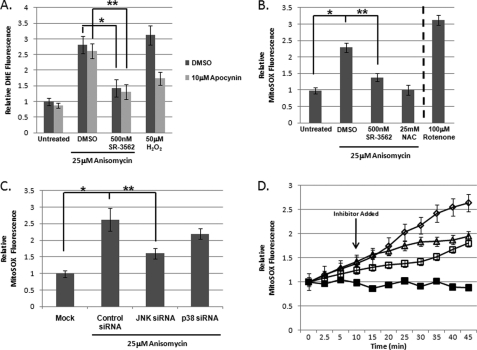
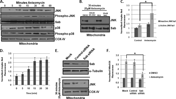
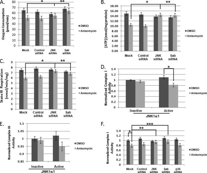
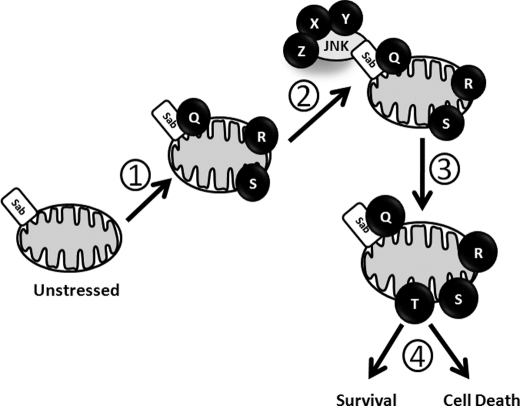
The JNK signaling cascade is critical for cellular responses to a variety of environmental and cellular stimuli. Although gene expression aspects of JNK signal transduction are well studied, there are minimal data on the physiological impact of JNK signaling. To bridge this gap, we investigated how JNK impacted physiology in HeLa cells. We observed that inhibition of JNK activity and JNK silencing with siRNA reduced the level of reactive oxygen species (ROS) generated during anisomycin-induced stress in HeLa cells. Silencing p38 had no significant impact on ROS generation under anisomycin stress. Moreover, JNK signaling mediated amplification of ROS production during stress. Mitochondrial superoxide production was shown to be the source of JNK-induced ROS amplification, as an NADPH oxidase inhibitor demonstrated little impact on JNK-mediated ROS generation. Using mitochondrial isolation from JNK null fibroblasts and targeting the mitochondrial scaffold of JNK, Sab, we demonstrated that mitochondrial JNK signaling was responsible for mitochondrial superoxide amplification. These results suggest that cellular stress altered mitochondria, causing JNK to translocate to the mitochondria and amplify up to 80% of the ROS generated largely by Complex I. This work demonstrates that a sequence of events exist for JNK mitochondrial signaling whereby ROS activates JNK, thereby affecting mitochondrial physiology, which can have effects on cell survival and death.


Figures

FIGURE 1.
FIGURE 1.
Anisomycin-induced stress initiates JNK-dependent ROS generation. A, JNK is activated by anisomycin in HeLa cells. JNK activation over the course of 2 h of 25 μm anisomycin stress was determined by Western blotting for bisphosphorylated JNK on Thr183 and Tyr185 (top). JNK abundance was monitored by Western blotting for total JNK (middle). α-Tubulin served as a loading control bottom. B, fluorescence microscopy of cellular ROS as detected by DHE. DMSO-treated cells (i) were compared with cells treated with 500 nm SR-3562 for 30 min (ii), cells stressed with 25 μm anisomycin for 45 min (iii), and cells pretreated for 30 min with 500 nm SR-3562 then stressed with 25 μm anisomycin (iv). C, fluorometric quantitation of cells stained with DHE to detect cellular ROS. Cells were stained with DHE following treatment with DMSO, 500 nm SR-3562 (30 min), 25 μm anisomycin (45 min), and 500 nm SR-3562 (30 min prior to anisomycin) and 25 μm anisomycin (45 min). 25 mm NAC, an antioxidant, was used as a negative control for ROS generation, and 10 μm pyocyanin, an inducer of ROS generation, was used as a positive control. D, Western blot analysis of JNK and p38 siRNA knockdowns of gene expression. Relative percent knockdown was estimated using densitometry. E, quantitation of cellular ROS in anisomycin-treated HeLa cells with JNK or p38 knockdown. After a 72-h siRNA transfection, cells were treated with 25 μm anisomycin for 45 min. ROS was detected using DHE staining and fluorescence was measured. F, kinetic profile of ROS generation in anisomycin-stressed HeLa cells. Cells were prestained with DHE prior to treatment with 25 mm NAC (black squares), 25 μm anisomycin (open diamonds), or 500 nm SR-3562 (open squares). Fluorescence was monitored for 45 min following the addition of the treatment. In two experiments, 25 mm NAC (black triangles) and 500 nm SR-3562 (open triangles) were added 10 min after 25 μm anisomycin.
FIGURE 2.
FIGURE 2.
JNK signaling amplifies mitochondrial superoxide production. A, JNK-dependent ROS is not generated by NADPH oxidases. Prior to anisomycin stress, cells were treated with either DMSO or 10 μm apocynin, and DHE fluorescence was measured 45 min following the addition of anisomycin. 50 μm Hydrogen peroxide (H2O2) was used as a positive control for NADPH oxidase-generated superoxide production. B, MitoSOX staining indicated that the JNK-mediated ROS was mitochondrial superoxide. MitoSOX fluorescence was used to determine mitochondrial superoxide production following 45 min of anisomycin-induced stress. Fluorescence was monitored in DMSO controls, cells were treated with 500 nm SR-3562, 25 μm anisomycin, and cells were pre-treated with 500 nm SR-3562 for 30 min prior to anisomycin addition. 25 mm NAC was included as a negative control for superoxide generation, whereas 100 μm rotenone served as a positive control for mitochondrial generated superoxide. C, JNK signaling, not p38, was required for mitochondrial superoxide generation during anisomycin stress. Following 72 h of gene silencing, mitochondrial superoxide levels were observed using MitoSOX Red. Mock transfected cells and cells containing JNK, p38, or control siRNAs were treated with anisomycin for 45 min prior to measurement. D, kinetic superoxide profiles demonstrate JNK signaling amplifies mitochondrial ROS generation. Mitochondrial superoxide production (MitoSOX fluorescence) was monitored over 45 min of anisomycin stress. Cells were pre-treated with 25 mm NAC (black squares) or 500 nm SR-3562 (open squares) 30 min prior to stress or 10 min (500 nm SR-3562, open triangles) following 25 μm anisomycin addition (25 μm anisomycin, open diamonds).
FIGURE 3.
FIGURE 3.
Mitochondrial JNK signaling specifically enhances superoxide production. A, anisomycin stress-induced JNK translocation to the mitochondria of HeLa cells. Mitochondria were isolated from HeLa cells at up to 60 min following anisomycin treatment. Mitochondria enrichments were analyzed by Western blotting for the presence of JNK, pJNK, and p38. Cyclooxygenase IV (COX IV) served as a loading control for mitochondrial preaparations. Sab was used as an additional mitochondrial control. B, 500 nm SR-3562 and 25 mm NAC prevented JNK translocation to the mitochondria. Following 30 min of 25 μm anisomycin treatment, mitochondria were isolated from cells that were pretreated with DMSO, 500 nm SR-3562, PBS, and 25 mm NAC. Western blot analysis was performed for the presence of JNK and Sab was used as a mitochondrial loading control. C, addition of active human JNK1α1 to mitochondria isolated from JNK null fibroblasts induced ROS production. Mitochondria from DMSO or anisomycin-treated JNK null fibroblasts were incubated with inactive or active recombinant human JNK1α1. ROS generation was monitored after 30 min of incubation at 30 °C by Amplex Red hydrogen peroxide detection assay. D, anisomycin stress altered mitochondria to promote JNK-mediated ROS production. Mitochondrial enrichments were obtained from JNK null fibroblasts treated with 25 μm anisomycin for up to 30 min. Mitochondria from each time point were then incubated with active JNK1α1 and ROS generation was detected by Amplex Red assay. E, siRNA-mediated knockdown of Sab expression. HeLa cells were transfected with siRNAs specific for Sab or a scrambled control siRNA. Following 72 h of siRNA treatment, protein abundance was monitored by Western blotting for Sab. α-Tubulin served as a loading control. Following mock, control siRNA, or Sab siRNA silencing, cells were treated with 25 μm anisomycin for 30 min, and then mitochondria were harvested. Western blots were performed for JNK. F, mitochondrial superoxide levels were decreased in anisomycin-stressed HeLa cells with reduced levels of Sab. HeLa cells were transfected with control or Sab-specific siRNAs for 72 h. Cells were then treated with 25 μm anisomycin for 45 min and mitochondrial superoxide was monitored using MitoSOX Red.
FIGURE 4.
FIGURE 4.
Mitochondrial JNK activity altered mitochondrial physiology promoting superoxide production. A, mitochondrial JNK decreased cellular oxygen consumption. HeLa cells were plated in assay media and treated with 25 μm anisomycin 30 min prior to oxygen consumption measurement. Seventy-two h prior to the experiment siRNAs were used to silence JNK, Sab, or p38. Oxygen consumption was normalized to cell viability immediately following the assay. B, mitochondrial JNK signaling induced decreased ATP production. ATP was extracted from cells following 2 h of anisomycin-induced stress. ATP was quantitated using a luciferase-based assay from Invitrogen. C, mitochondrial JNK signaling decreased State III respiration. Sab and JNK knockdown were achieved by a 72-h incubation with siRNAs specific for each gene. State III respiration was measured following 45 min of anisomycin stress using the Seahorse XF-96 Analyzer with media supplemented with mitochondrial substrates (glutamate/malate/ADP (8 mm) or pyruvate/malate/ADP (8 mm)). D, mitochondrial JNK signaling induced inhibition of respiratory Complex I. Mitochondria were isolated from DMSO and 25 μm anisomycin-treated JNK null fibroblasts. Mitochondria were then incubated with inactive and active JNK1α1. Complex I activity was detected spectrophotometrically. E, mitochondrial JNK is responsible for a decrease in Complex III activity. Mitochondria were isolated from DMSO and 25 μm anisomycin-treated JNK null fibroblasts. Mitochondria were incubated with inactive and active JNK1α1. Complex III activity was monitored by spectrophotometric methods. F, knockdown of JNK or Sab prevents Complex I inhibition in anisomycin-treated HeLa cells. Following gene knockdowns, mitochondria were isolated after 45 min of anisomycin stress. Complex I enzyme activity was then measured.
FIGURE 5.
FIGURE 5.
Model of JNK mitochondrial signaling. (1) early cellular stress alters mitochondria physiologically and architecturally, such as the recruitment of yet unidentified proteins (illustrated as Q, R, and S) and translocation of the mitochondrial JNK signaling complex. (2) stress-induced mitochondrial changes promote JNK mitochondrial signaling leading to a stress responsive mitochondrial phenotype (illustrated as T) (3). (4) pro-oxidant metabolism and oxidative stress could contribute to cell death or metabolic alterations and may be essential to cellular survival.

Similar articles

Cited by

References

    1. Weston C. R., Davis R. J. (2007) Curr. Opin. Cell Biol. 19, 142–149 - PubMed
    1. Dhanasekaran D. N., Reddy E. P. (2008) Oncogene 27, 6245–6251 - PMC - PubMed
    1. Lee J. J., Lee J. H., Ko Y. G., Hong S. I., Lee J. S. (2010) Oncogene 29, 561–575 - PubMed
    1. Lei K., Nimnual A., Zong W. X., Kennedy N. J., Flavell R. A., Thompson C. B., Bar-Sagi D., Davis R. J. (2002) Mol. Cell. Biol. 22, 4929–4942 - PMC - PubMed
    1. Wiltshire C., Gillespie D. A., May G. H. (2004) Biochem. Soc. Trans. 32, 1075–1077 - PubMed

Publication types

MeSH terms