. 2011 May 20;286(20):18181-9.
doi: 10.1074/jbc.M110.214825. Epub 2011 Mar 28.

A genetic screen targeted on the FO component of mitochondrial ATP synthase in Saccharomyces cerevisiae

Affiliations

A genetic screen targeted on the FO component of mitochondrial ATP synthase in Saccharomyces cerevisiae

François Godard et al. J Biol Chem. .

Abstract

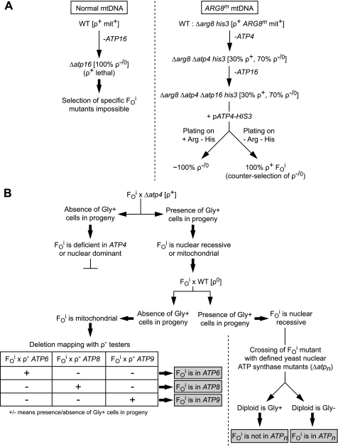
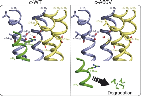
In yeast, the two main F(O) proton-translocating subunits of the ATP synthase (subunits 6/a and 9/c) are encoded by mitochondrial DNA (mtDNA). Unfortunately, mutations that inactivate the F(O) typically result in loss of mtDNA under the form of ρ(-)/ρ(0) cells. Thus, we have designed a novel genetic strategy to circumvent this problem. It exploits previous findings that a null mutation in the nuclear ATP16 gene encoding ATP synthase subunit δ results in massive and lethal F(O)-mediated protons leaks across the inner mitochondrial membrane. Mutations that inactivate the F(O) can thus, in these conditions, be selected positively as cell viability rescuing events. A first set of seven mutants was analyzed and all showed, as expected, very severe F(O) deficiencies. Two mutants carried nuclear mutations in known genes (AEP1, AEP2) required for subunit c expression. The five other mutations were located in mtDNA. Of these, three affect synthesis or stability of subunit a transcripts and the two last consisted in a single amino acid replacement in subunit c. One of the subunit c mutations is particularly interesting. It consists in an alanine to valine change at position 60 of subunit c adjacent to the essential glutamate of subunit c (at position 59) that interacts with the essential arginine 186 of subunit a. The properties of this mutant suggest that the contact zone between subunit a and the ten subunits c-ring structure only involves critical transient interactions confined to the region where protons are exchanged between the subunit a and the c-ring.


Figures

FIGURE 1.
FIGURE 1.
Procedure for the isolation and mapping of yeast mutants with an inactive FO (FOi). Panel A describes the procedure for the isolation of FOi mutants using a host strain where the nuclear ARG8 gene has been replaced by a mitochondrial version of that gene (ARG8m) integrated into an intergenic region of the mitochondrial genome (mtDNA); see text for details. Panel B describes the procedure for mapping the mutations in nuclear or mitochondrial DNA, see text for details. Gly+/− refers to yeast cells able/unable to grow on glycerol; WT, wild-type; ρ/0 refers to large (>50%) deletions (ρ) or complete loss (ρ0) of the mtDNA; ρ+, complete mtDNA; ATPn is a known nuclear gene required for ATP synthase expression.
FIGURE 2.
FIGURE 2.
Test crosses for the mapping of the FOi mutants. Panel A, the nuclear FOi mutants SDC25/1 and SDC25/5 were crossed with a series of yeast strains each carrying a null mutation in one of known nuclear genes (indicated on the left) required for ATP synthase expression. The various strains were crossed drop on drop on rich glucose medium (YPGA) for 2 days at 28 °C, and then replica plated on glycerol medium (N3). The N3 plate was incubated at 28 or 36 °C, as indicated, for 4 days and photographed. As controls the non-mated strains were spotted on the YPGA plate. Panel B, the mitochondrial FOi mutants SDC25/2, SDC25/4, SDC25/6, SDC25/14 and SDC25/26 were crossed with synthetic ρ testers carrying only the ATP6 ATP6, strain SDC30/2a), the ATP8 ATP8, strain FG8) or the ATP9 ATP9, strain FG9) gene in their mitochondria. The various strains were crossed drop on drop on YPGA for 2 days at 28 °C, with as controls the non-mated strains. A N3 replicate of the YPGA plate was incubated for 4 days at 28 °C and then photographed.
FIGURE 3.
FIGURE 3.
Topological location and evolutionary conservation of the Ala-60 and Met-67 residues of yeast subunit c. Panel A, three-dimensional structure of yeast subunit c monomer according to (48); the view is perpendicular to the membrane plane. The positions of the c-E59, c-A60, and c-M67 residues are indicated by arrowheads. H1 and H2 helices of subunit c are colored in bright and dark gray, respectively. The gray bars mark the membrane borders. IMS, intermembrane space. Panel B, alignments of subunits c from various species around helix H2: S. cerevisiae (NP_009319.1), Candida glabrata (NP_818784.1), Schizosaccharomyces pombe (NP_039507.1), Neurospora crassa (0808299A), Arabidopsis thaliana (NP_651852.1), Nicotiana tabacum (NC_006581.1), Mus musculus (NP_778180.1), Xenopus laevis (NP_001080083.1), Drosophila melanogaster (NP_651852.1), and Homo sapiens (NP_001002031.1). The Ala-60 and Met-67 residues of yeast subunit c are replaced by Val and Lys in mutants SDC25/2 and SDC25/14, respectively.
FIGURE 4.
FIGURE 4.
Influence of the c-A60V and c-M67K mutations on mitochondrial protein synthesis and ATP synthase assembly. Panel A, pulse labeling of mtDNA-encoded proteins. Freshly grown cells of the mutants SDC25/14 (c-M67K) and SDC25/2 (c-A60V), and corresponding parental strain SDC17–31b, were incubated for 20 min in the presence of a mixture of [35S]methionine + [35S]cysteine and cycloheximide to inhibit cytosolic protein synthesis. Total cellular protein extracts were prepared from the radiolabeled cells and run in SDS-PAGE gels containing either 12% acrylamide-4 m urea (for a good separation of Cox3p and subunit a) or 17% acrylamide (for good resolution of Atp8 and subunit c). Each lane was loaded with 30000 disintegrations/min, which in this experiment corresponded to about the same numbers of cells for each analyzed strain. The gel was dried and analyzed with a PhosphorImager. The mitochondrial translation products corresponding to the various radioactive bands are indicated on the left. Panels B and C, effects of the c-M67K and c-A60V mutations on ATP synthase assembly. Mitochondria were extracted from the mutants FG2 (c-M67K) and FG13 (c-A60V) and the corresponding wild-type strain (FG4) grown in a synthetic (CSM) galactose medium lacking arginine. Panel B, 15 μg of mitochondrial proteins were separated by SDS-PAGE, transferred onto a nitrocellulose membrane, and probed with antibodies against the indicated proteins. Prior to loading on the gel, the samples were preincubated in presence or absence of chloroform as indicated. In absence of chloroform, some subunit c proteins migrate as c-ring oligomers whereas in the presence of chloroform all c-rings dissociate into monomeric subunit c. Panel C, the mitochondria were treated with 2 g or 10 g of digitonin (digi) per g of protein, as indicated, and then centrifuged to remove insoluble materials. The mitochondrial complexes were separated by BN-PAGE and probed in-gel for ATPase activity (left) or transferred to polyvinylidene difluoride membranes and probed with antibodies against subunit c (right). V1 and V2 correspond to monomeric and dimeric ATP synthase, respectively.
FIGURE 5.
FIGURE 5.
The loss of the interaction between the essential c-E59 and a-R186 residues is hypothesized to be responsible for the degradation of subunit a in the yeast c-A60V mutant. The figure on the left shows a ribbon diagram built with PyMOL of two adjacent c subunits (c1 in blue and c2 in yellow) around the proton binding site in wild-type yeast ATP synthase, according to the recently solved structure of the yeast c-ring/F1 subcomplex (48). The two transmembrane segments of subunit c are labeled H1 and H2. The proton translocating activity of the ATP synthase depends on the interaction of the glutamate residue 59 of subunit c (E59) with the arginine residue 186 of subunit a (a-R186). The latter residue belongs to the fourth transmembrane segment of subunit a (a-H4, represented in green color). It is believed (48) that proper interaction of c-E59 and a-R186 depends on a water molecule (represented by a red sphere), like in the H+ and Na+ ATP synthases of B. pseudofirmus OF4 (49) and I. tartaricus (54), respectively. In one of the FO inactive mutants isolated in this study, the alanine 60 of yeast subunit c is replaced by valine (right panel). The valine substituting c-A60 is shown in two rotameric conformations (solid and dashed line). The c-A60V mutation does not compromise the ability of subunit c to form stable oligomers but leads to elimination from the cells of the subunit a, presumably because of disruption of the interaction between c-E59 and a-R186 (see text for details). The question mark indicates that the water molecule is perhaps prevented to enter the proton binding site.

Similar articles

Cited by

References

    1. Boyer P. D. (1997) Annu. Rev. Biochem. 66, 717–749 - PubMed
    1. Ackerman S. H., Tzagoloff A. (2005) Prog. Nucleic Acids Res. Mol. Biol. 80, 95–133 - PubMed
    1. Senior A. E., Nadanaciva S., Weber J. (2002) Biochim. Biophys. Acta 1553, 188–211 - PubMed
    1. Bianchet M. A., Hullihen J., Pedersen P. L., Amzel L. M. (1998) Proc. Natl. Acad. Sci. U.S.A. 95, 11065–11070 - PMC - PubMed
    1. Abrahams J. P., Leslie A. G., Lutter R., Walker J. E. (1994) Nature 370, 621–628 - PubMed

Publication types

MeSH terms