. 2011 May 13;286(19):16716-25.
doi: 10.1074/jbc.M110.180844. Epub 2011 Mar 21.

DiGeorge critical region 8 (DGCR8) is a double-cysteine-ligated heme protein

Affiliations

DiGeorge critical region 8 (DGCR8) is a double-cysteine-ligated heme protein

Ian Barr et al. J Biol Chem. .

Abstract

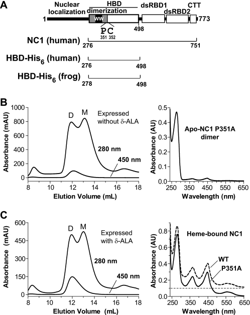
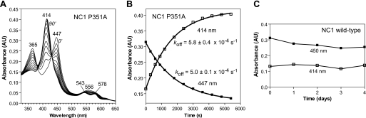
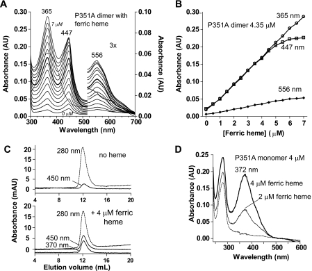
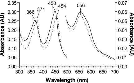
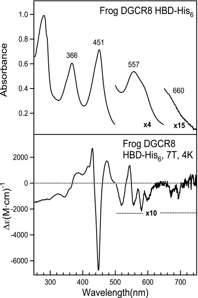
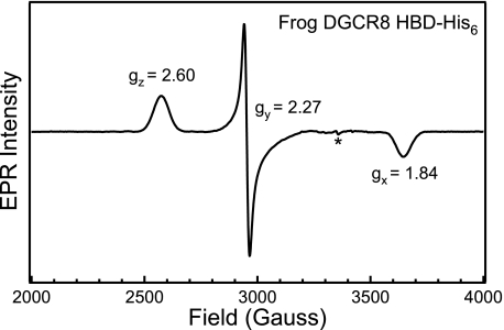
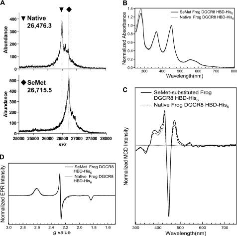
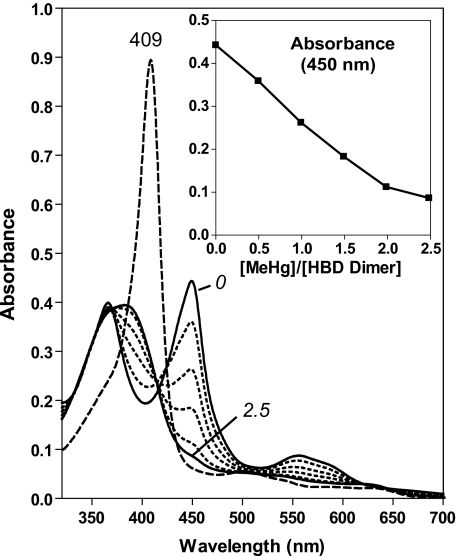
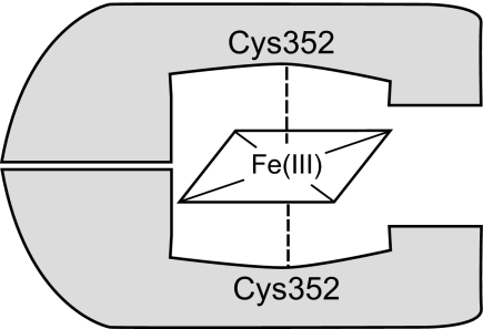
All known heme-thiolate proteins ligate the heme iron using one cysteine side chain. We previously found that DiGeorge Critical Region 8 (DGCR8), an essential microRNA processing factor, associates with heme of unknown redox state when overexpressed in Escherichia coli. On the basis of the similarity of the 450-nm Soret absorption peak of the DGCR8-heme complex to that of cytochrome P450 containing ferrous heme with CO bound, we identified cysteine 352 as a probable axial ligand in DGCR8. Here we further characterize the DGCR8-heme interaction using biochemical and spectroscopic methods. The DGCR8-heme complex is highly stable, with a half-life exceeding 4 days. Mutation of the conserved proline 351 to an alanine increases the rate of heme dissociation and allows the DGCR8-heme complex to be reconstituted biochemically. Surprisingly, DGCR8 binds ferric heme without CO to generate a hyperporphyrin spectrum. The electronic absorption, magnetic circular dichroism, and electron paramagnetic resonance spectra of the DGCR8-heme complex suggest a ferric heme bearing two cysteine ligands. This model was further confirmed using selenomethionine-substituted DGCR8 and mercury titration. DGCR8 is the first example of a heme-binding protein with two endogenous cysteine side chains serving as axial ligands. We further show that native DGCR8 binds heme when expressed in eukaryotic cells. This study provides a chemical basis for understanding the function of the DGCR8-heme interaction in microRNA maturation.


Figures

FIGURE 1.
FIGURE 1.
Characterization of the NC1 P351A mutant. A, domain structure of human DGCR8. The double-stranded RNA-binding domains (dsRBD) and C-terminal tail (CTT) are required and sufficient for cooperative association with pri-miRNAs and for triggering cleavage by the Drosha nuclease. The human NC1, HBD-His6 and frog HBD-His6 protein constructs used in this study are represented by the brackets. B, size exclusion chromatogram of NC1 P351A overexpressed in E. coli. The 367-nm absorption trace overlaps with the 450-nm one and thus is omitted from the chromatogram drawing. D and M indicate the dimer and monomer peaks. An electron absorption spectrum of the dimer peak is shown (right). C, same as B, except that 1 mm δ-ALA was added to the bacterial culture at induction.
FIGURE 2.
FIGURE 2.
Heme dissociates from the wild-type NC1 slowly, and P351A increases the rate. Heme-bound NC1, either P351A (A and B) or wild-type (C), was incubated with a 6-fold excess of apomyoglobin at 25 °C. A, electronic absorption spectra obtained at 0, 5, 10, 15, 20, 30, 40, 50, 60, 70, 80, and 90 min. B, the 414-nm (metmyoglobin) and 447-nm (P351A-heme complex) values shown in A were fit to a single exponential function. The rates shown are mean ± S.D. of three independent repeats. C, wild-type NC1 showed no significant changes of absorbance at 414 nm and 450 nm at room temperature over 4 days.
FIGURE 3.
FIGURE 3.
The P351A-heme complex can be reconstituted using ferric heme. A, electronic absorption spectra of titration of ferric heme to heme-free P351A dimer (4.35 μm) in 0.5 μm intervals. The spectrum of ferric heme (5.0 μm) free from proteins is shown using a dashed line. B, the absorbance values at peak wavelengths are plotted over the concentration of ferric heme added. C, SEC of 4 μm heme-free P351A dimer incubated without (top panel) or with (bottom panel) equal molar of ferric heme. D, the P351A monomer shows neither 447- nor 556-nm peaks when incubated with ferric heme.
FIGURE 4.
FIGURE 4.
The hyperporphyrin spectrum of DGCR8 likely results from double-cysteine ligation to ferric heme. The electronic absorption spectrum of ferric heme-bound NC1 dimer (solid line) is strikingly similar to that of the ferric chloroperoxidase complexed with ethyl-2-mercaptoacetate (dashed line). The latter spectrum was reproduced from an earlier work (31).
FIGURE 5.
FIGURE 5.
Electronic absorption and MCD spectra of the ferric heme-bound frog DGCR8 HBD-His6. The electronic absorption spectrum (top panel) was taken at room temperature using the dimeric frog HBD-His6 at 12.2 μm concentration in a buffer containing 50 mm EPPS (pH 8.0) and 400 mm NaCl. As a control, an electronic absorption spectrum was also recorded at 4 K and displayed no differences in peak positions and intensity (data not shown). The MCD spectrum (bottom panel) was recorded using a protein sample at 24.5 μm in 22.5 mm EPPS (pH 8.0), 180 mm NaCl, and 55% (v/v) glycerol.
FIGURE 6.
FIGURE 6.
EPR spectrum of the ferric heme-bound frog DGCR8 HBD-His6 protein. The frog HBD-His6 protein (153 μm) was in 50 mm EPPS (pH 8.0) and 400 mm NaCl. The spectrum represents an average of 10 scans taken at 10 K, with 9.383 GHz microwave frequency, 8.000 G modulation amplitude, 100 kHz modulation frequency, 60 dB receiver gain, 163.84 ms time constant, and a power of 1.002 milliwatt. The asterisk represents a signal present in the cavity.
FIGURE 7.
FIGURE 7.
The sixth ligand to ferric heme in DGCR8 is not a methionine. A, mass spectra of native and SeMet-labeled frog DGCR8 HBD-His6 indicates nearly complete substitution of Met by SeMet. The difference in their m/z peaks, 239, is consistent with all five methionine residues substituted with SeMet. B, the normalized electronic absorption spectrum of the SeMet-substituted frog HBD-His6 is nearly identical to that of the native protein (Fig. 5, top panel). C, the normalized MCD spectrum of the SeMet-substituted frog HBD-His6 is nearly identical to that of the native protein (Fig. 5, bottom panel). D, comparison of the EPR spectra of the native and SeMet-labeled frog HBD-His6. The spectral parameters for the SeMet-labeled frog HBD-His6 were the same as those of the native listed in Fig. 6, except that the ferric heme concentration of the sample was 142 μm and the spectrum was acquired with a 9.384 GHz microwave frequency.
FIGURE 8.
FIGURE 8.
Cys-352 from both subunits in a DGCR8 dimer serve as the axial ligands to ferric heme. Electronic absorption spectra of hHBDC430S (10 μm) titrated with MeHgAc. The MeHgAc was added at steps of 0.5 molar equivalent of ferric heme-bound HBD dimer. The spectra of the starting point (0 molar equivalent of MeHgAc) and end point (2.5 molar equivalents) are shown in solid traces and the intermediate steps in dotted lines. After the MeHgAc titration, 1.2 molar equivalents of apomyoglobin (relative to the HBD dimer) were added, and the electronic absorption spectrum was immediately recorded (dashed line). The inset shows absorbance at 450 nm over the course of the titration.
FIGURE 9.
FIGURE 9.
DGCR8 binds heme in insect cells. A, electronic absorption spectrum of His6-NC1 (human) expressed in Sf9 cells using baculovirus. His6-NC1 was purified using Ni affinity chromatography followed by cation exchange chromatography. The image of a silver-stained SDS-polyacrylamide gel is shown. B, analytical size exclusion chromatogram of the His6-NC1 sample used in A, with the silver-stained gel of the SEC fractions shown above. The peak indicated by the asterisk contains mainly nucleic acids. C, His6-NC1 expressed in insect cells was used in reconstituted pri-miRNA processing assays. A pri-miR-30a fragment was uniformly labeled with α-32P-UTP and was incubated with recombinant Drosha and NC1 at indicated concentrations. The reactions were analyzed using denaturing gel electrophoresis and autoradiography.
FIGURE 10.
FIGURE 10.
Schematic of how the DGCR8 HBD binds ferric heme.

References

    1. Perutz M. F., Wilkinson A. J., Paoli M., Dodson G. G. (1998) Annu. Rev. Biophys. Biomol. Struct. 27, 1–34 - PubMed
    1. Derbyshire E. R., Marletta M. A. (2009) Handb. Exp. Pharmacol. 191, 17–31 - PubMed
    1. Ortiz de Montellano P. R. (2000) Curr. Opin. Chem. Biol. 4, 221–227 - PubMed
    1. Ortiz de Montellano P. R. (2004) Cytochrome P450: Structure, Mechanism, and Biochemistry, 3rd Ed., Springer, New York
    1. Dawson J. H., Sono M. (1987) Chem. Rev. 87, 1255–1276

Publication types

LinkOut - more resources