. 2011 May 27;286(21):18890-902.
doi: 10.1074/jbc.M110.206110. Epub 2011 Mar 30.

Hormonally active vitamin D3 (1alpha,25-dihydroxycholecalciferol) triggers autophagy in human macrophages that inhibits HIV-1 infection

Affiliations

Hormonally active vitamin D3 (1alpha,25-dihydroxycholecalciferol) triggers autophagy in human macrophages that inhibits HIV-1 infection

Grant R Campbell et al. J Biol Chem. .

Abstract

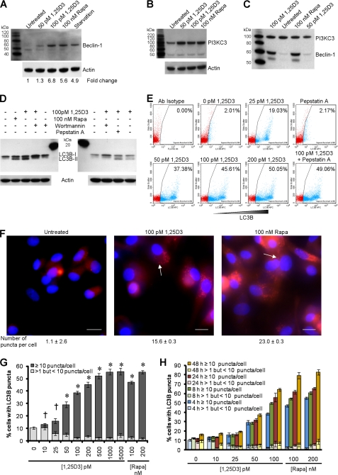
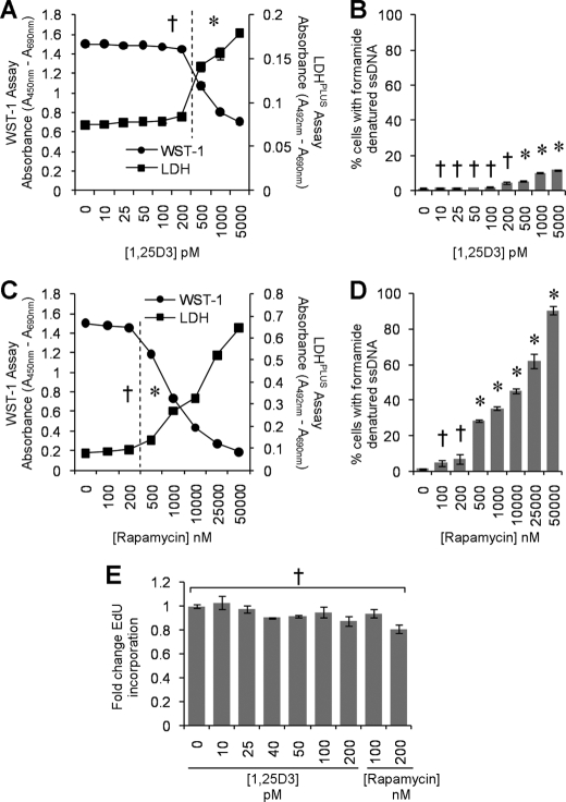
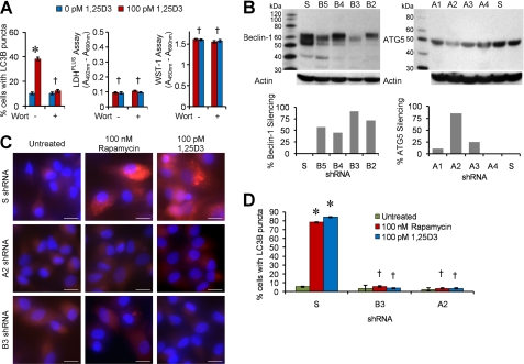
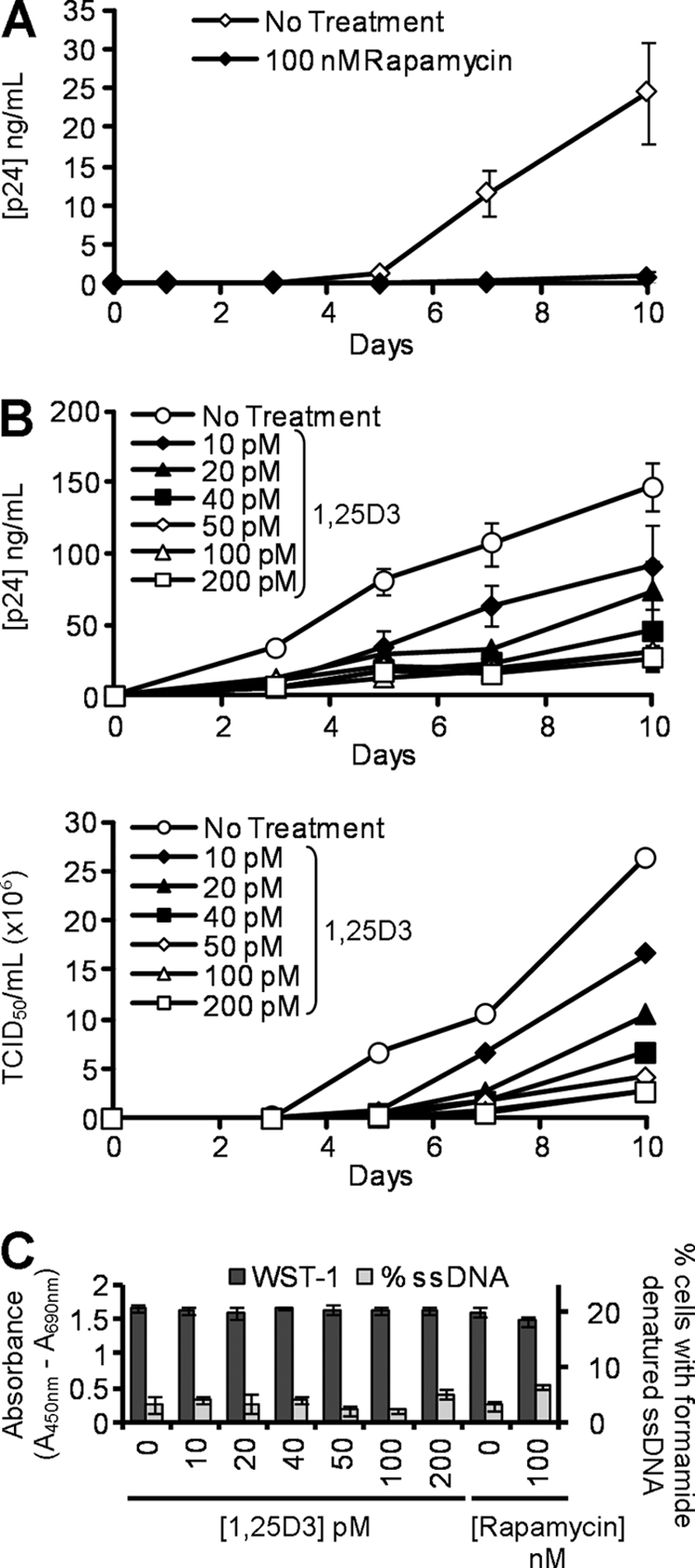
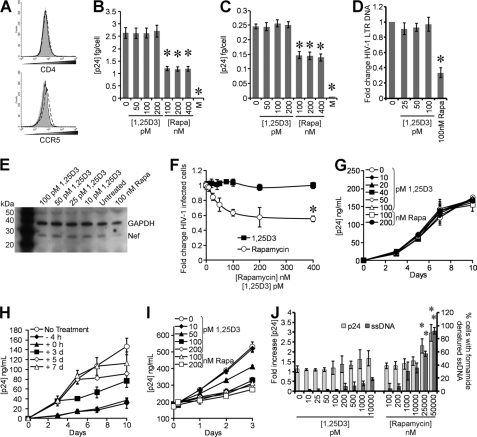
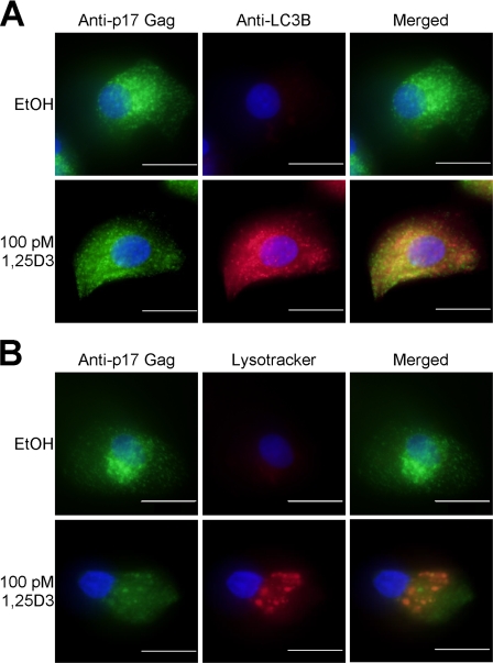
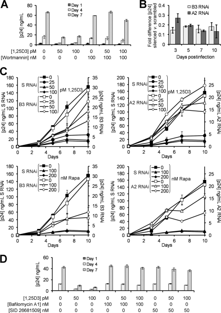
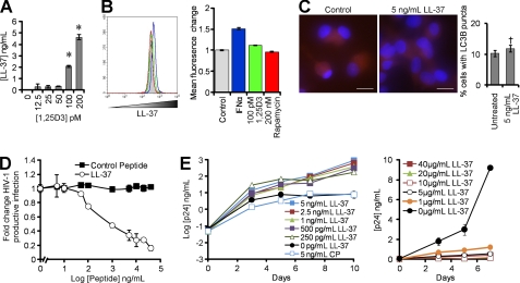
Autophagy is a self-digestion pathway essential for maintaining cellular homeostasis and cell survival and for degrading intracellular pathogens. Human immunodeficiency virus-1 (HIV-1) may utilize autophagy for replication as the autophagy-related protein-7 (ATG-7), microtubule-associated protein 1 light chain 3, ATG-12, and ATG-16L2 are required for productive HIV-1 infection; however, the effects of autophagy induction on HIV-1 infection are unknown. HIV-1-infected individuals have lower levels of 1α,25-dihydroxycholecalciferol, the hormonally active form of vitamin D, than uninfected individuals. with the lowest concentrations found in persons with AIDS. Using human macrophages and RNA interference for ATG-5 and Beclin-1 and chemical inhibition of phosphatidylinositol 3-kinase, we have found that physiologically relevant concentrations of 1α,25-dihydroxycholecalciferol induce autophagy in human macrophages through a phosphatidylinositol 3-kinase-, ATG-5-, and Beclin-1-dependent mechanism that significantly inhibits HIV-1 replication in a dose-dependent manner. We also show that the inhibition of basal autophagy inhibits HIV-1 replication. Furthermore, although 1α,25-dihydroxycholecalciferol induces the secretion of human cathelicidin, at the concentrations produced in vitro, cathelicidin does not trigger autophagy. Our findings support an important role for autophagy during HIV-1 infection and provide new insights into novel approaches to prevent and treat HIV-1 infection and related opportunistic infections.


Figures

FIGURE 1.
FIGURE 1.
1,25D3 induces autophagy in human macrophages. A, macrophages were left untreated or were incubated with 1,25D3, rapamycin (Rapa) or starved by incubation in Earle's balanced salt solution for 4 h, lysed, then subjected to immunoblotting with antibody to Beclin-1 and β-actin. B, shown is an immunoblot using antibody to PI3KC3 and β-actin. C, shown is an immunoblot of co-immunoprecipitation with PI3KC3-specific antibody using antibody to Beclin-1 and antibody to PI3KC3 as loading control. D, shown are immunoblots of LC3B isoforms using antibody to LC3B or β-actin using macrophages pretreated with 100 nm wortmannin, 10 μg/ml pepstatin A, or vehicle control before 1,25D3 or rapamycin treatment. E, shown is a flow cytometry analysis of saponin-resistant LC3B-II in macrophages after 1,25D3 and/or pepstatin A treatment for 4 h. Representative dot blots with the percentage of cells displaying saponin-resistant LC3B-II from three donors are shown. Ab, antibody. F, representative fluorescence microscopy images of macrophages from three donors after 1,25D3 or rapamycin treatment for 4 h are shown. Cells were fixed and permeabilized, then stained with antibody to LC3B (red) and Hoechst 33342 (blue). LC3B puncta are indicated by the white arrows. When puncta were not present, LC3B staining gives rise to a diffuse cytoplasmic pattern. The number of puncta per cell is indicated as the mean ± S.D. Scale bars indicate 10 μm. G, percentage of cells with LC3B puncta after designated treatments for 4 h is shown. H, quantification of cells with LC3B puncta after designated treatments at the time points indicated. Results are the means ± S.E. of three independent experiments performed in triplicate. †, p ≥ 0.05; *, p < 0.001 compared with untreated control.
FIGURE 2.
FIGURE 2.
Physiological concentrations of 1,25D3 are not cytotoxic. Macrophages were incubated with increasing concentrations of 1,25D3 for 96 h. A, aliquots of supernatant taken before the addition of WST-1 were tested for lactate dehydrogenase (LDH) spectrophotometrically using the LDHPLUS assay (black squares). For the last hour cells were incubated with WST-1, and the reduction of the WST-1 reagent to its formazan product was monitored spectrophotometrically (black circles). B, quantification of the number of cells with apoptotic ssDNA using the ssDNA ELISA 96 h after 1,25D3 treatment is shown. C, lactate dehydrogenase (black squares) and WST-1 (black circles) assay after 96 h of incubation of macrophages with increasing concentrations of rapamycin is shown. D, quantification of the number of cells with apoptotic ssDNA using the ssDNA ELISA 96 h after rapamycin treatment is shown. E, effect of 1,25D3 and rapamycin on the uptake of EdU by macrophages is shown. Cells were incubated with 1,25D3 or rapamycin for 78 h, after which cells were incubated a further 18 h in the presence of 10 μm EdU. Cells were harvested, fixed, permeabilized, then probed for EdU incorporation and analyzed for the percentage of EdU-positive cells by flow cytometry. Physiological concentrations of 1,25D3 and rapamycin had no cytotoxic effects. All results are the means ± S.E. of three donors performed in triplicate. †, p ≥ 0.05; *, p < 0.001 compared with untreated control cells.
FIGURE 3.
FIGURE 3.
1,25D3-induced autophagy requires PI3K, ATG5, and Beclin-1. A, macrophages were incubated with wortmannin or vehicle control then treated with 100 pm 1,25D3 for 4 h. Left, quantification of the percentage of LC3B-positive autophagosome-forming cells is shown. Middle, cell supernatants were tested spectrophotometrically for lactate dehydrogenase (LDH) release as a measure of plasma membrane rupture. Right, for the last hour cells were incubated with WST-1, and the reduction of the WST-1 reagent to its formazan product was monitored spectrophotometrically. B, MonoMac1 cells were transfected with scrambled shRNA (S), Beclin-1-specific (B2–B5), or ATG5-specific (A1–A4) shRNA. Top, shown is an immunoblot performed with antibodies to Beclin-1, ATG5, and β-actin. Bottom, quantification of silencing effect on protein content is shown. C, shown are representative microscopy images of LC3B staining in cells transfected with scrambled (S), ATG5 (A2), or Beclin-1 (B3) shRNA treated with 1,25D3 or rapamycin for 4 h. Cells were stained with antibody to LC3B (red) and Hoechst 33342 (blue). Scale bars indicate 10 μm. D, shown is quantification of the percentage of LC3B-positive autophagosome-forming cells transfected with scrambled (S), ATG5 (A2), or Beclin-1 (B3) shRNA and treated with 1,25D3 or rapamycin. Results are the means ± S.E. of three independent experiments performed in triplicate. †, p ≥ 0.05; *, p < 0.001 compared with untreated control cells.
FIGURE 4.
FIGURE 4.
1,25D3 and rapamycin inhibit HIV-1 replication. A, macrophages were incubated with rapamycin before exposure to HIV-1Ba-L. ELISAs were performed for HIV-1 p24 antigen release over 10 days. B, macrophages were incubated with increasing concentrations of 1,25D3 before exposure to HIV-1Ba-L. Top, ELISAs were performed for HIV-1 p24 antigen over 10 days. Bottom, TCID50 is calculated from the supernatants of infected cells. 50 pm 1,25D3 was the minimum concentration required to significantly inhibit HIV-1 replication (p = 0.01). C, after 10 days of HIV-1 infection, cells were incubated with WST-1 for 1 h, and the reduction of the WST-1 reagent to its formazan product was monitored spectrophotometrically (dark gray histograms). Cells were then fixed and permeabilized, and the percentage of cells with apoptotic ssDNA was quantified by ELISA. At all concentrations tested, neither 1,25D3 nor rapamycin induced apoptotic ssDNA formation or reduced cell viability by day 10 of HIV-1 infection. Results are the means ± S.E. of three independent experiments performed in triplicate.
FIGURE 5.
FIGURE 5.
1,25D3 does not affect HIV-1 entry or release. A, macrophages were left untreated (solid gray histogram) or treated with 100 pm 1,25D3 (dashed line) or 100 nm rapamycin (solid line). After 4 h, cells were harvested and stained for surface CD4 and CCR5. B, macrophages treated with 1,25D3, rapamycin, or maraviroc (M) for 4 h were infected with replication competent HIV-1Ba-L. Binding was measured at 3 h postinfection by washing cells extensively, then lysing and analyzing p24 by ELISA. C, entry was measured at 5 h postinfection by washing cells extensively, then trypsinization, lysing, and subsequently analyzing intracellular p24 by ELISA. D, DNA was extracted from cells at 8 h postinfection for PCR analysis of pre-integration strong-stop HIV-1 DNA. RNA polymerase II was amplified as a control. Results are expressed as the ratio between the target gene and the RNA polymerase II and normalized so that HIV-1 LTR in untreated cells equals 1.00. E, macrophages lysed at 5 h postinfection were subjected to immunoblotting for both Nef and GAPDH. F, percentage of TZM-bl cells were productively infected with HIV-1Ba-L after 4 h treatment with 1,25D3 or rapamycin. G, macrophages were incubated with 1 ng p24 antigen from the 10-day aliquots of cell-free supernatants post-1,25D3 or rapamycin treatment for 3 h then cultured for 10 days with ELISA performed for HIV-1 p24 antigen. H, macrophages were incubated with 100 pm 1,25D3 at different time points with respect to infection with HIV-1Ba-L. ELISAs were performed for HIV-1 p24 antigen over 10 days. I, 20-day-old HIV-1-infected macrophages were incubated with rapamycin or 1,25D3 with ELISA performed for HIV-1 p24 antigen over 3 days. J, shown is the percentage of cells with apoptotic ssDNA combined with p24 release after 4 h of treatment with rapamycin or 1,25D3. All data are the means ± S.E. of three independent experiments performed in triplicate. *, p < 0.001 compared with untreated control cells.
FIGURE 6.
FIGURE 6.
LC3B and acidic vacuoles colocalize with HIV-1 Gag. Macrophages were treated with 100 pm 1,25D3 or vehicle control (EtOH) then infected with HIV-1Ba-L for 10 d. A, cells were fixed, permeabilized, and probed for LC3B, Gag-p17, and Hoechst 33342 (blue). Green dots indicate Gag-p17, and red dots indicate LC3B positive structures. B, after 10 days, cells were incubated with Lysotracker Red for 4 h, then fixed, permeabilized, and probed for Gag-p17. Green dots indicate Gag-p17, and red dots indicate acidic vacuoles. Scale bars indicate 10 μm.
FIGURE 7.
FIGURE 7.
1,25D3-mediated induction of autophagy inhibits HIV-1 replication. A, macrophages were incubated with wortmannin or vehicle control before treatment with 50–100 pm 1,25D3 and infection with HIV-1Ba-L. ELISA performed for HIV-1 p24 antigen release over 7 days. B, shown is quantification of p24 antigen secreted by HIV-1-infected ATG5 shRNA (A2) and Beclin-1 shRNA (B3) transduced cells compared with nonspecific scrambled shRNA-transduced cells. C, ATG5 shRNA (A2), Beclin-1 shRNA (B3), and nonspecific scrambled shRNA (S)-transduced cells were incubated with 0–100 pm 1,25D3 or 0–200 nm rapamycin then infected with HIV-1Ba-L. ELISAs were performed for HIV-1 p24 antigen over 10 days. D, macrophages were incubated with bafilomycin A1, SID 26681509, or vehicle control before treatment with 50–100 pm 1,25D3 and infection with HIV-1Ba-L. ELISAs were performed for HIV-1 p24 antigen release over 7 days. Results are the means ± S.E. of three independent experiments performed in triplicate.
FIGURE 8.
FIGURE 8.
1,25D3-induced cathelicidin does not induce autophagy or inhibit HIV-1. A, macrophages were treated with 1,25D3 and secreted LL-37 measured by ELISA. B, shown is a flow cytometry analysis of LL-37 in macrophages left unstimulated (gray histograms) or stimulated with 2000 units/ml IFNα (blue histograms), 100 pm 1,25D3 (green histograms), or 200 nm rapamycin (red histograms). Left, representative flow cytometry histograms are shown. Right, mean fluorescence change is shown. C, macrophages were left untreated or treated with LL-37, fixed, permeabilized, then probed for LC3B and analyzed by fluorescence microscopy. Scale bars indicate 10 μm. Left, representative microscopy images are shown. Right, quantification of cells with LC3B puncta is shown. D, the percentage of TZM-bl cells productively infected with HIV-1Ba-L after pretreatment with increasing concentrations of LL-37 or scrambled control peptide is shown. E, macrophages were incubated with varying concentrations of LL-37 or a scrambled control peptide (CP) for 4 h before exposure to HIV-1Ba-L. ELISAs were performed for HIV-1 p24 antigen release over time. Results are the means ± S.E. of three independent experiments performed in triplicate.

References

    1. Joint United Nations Programme on HIV/AIDS (2008) Report on the Global HIV/AIDS Epidemic 2008, Executive Summary, Joint United Nations Programme on HIV/AIDS, Geneva, Switzerland
    1. Martineau A. R., Honecker F. U., Wilkinson R. J., Griffiths C. J. (2007) J. Steroid Biochem. Mol. Biol. 103, 793–798 - PubMed
    1. Sly L. M., Lopez M., Nauseef W. M., Reiner N. E. (2001) J. Biol. Chem. 276, 35482–35493 - PubMed
    1. Yuk J. M., Shin D. M., Lee H. M., Yang C. S., Jin H. S., Kim K. K., Lee Z. W., Lee S. H., Kim J. M., Jo E. K. (2009) Cell Host. Microbe 6, 231–243 - PubMed
    1. Haug C., Müller F., Aukrust P., Frøland S. S. (1994) J. Infect. Dis. 169, 889–893 - PubMed

Publication types

MeSH terms