. 2011 May 13;286(19):17047-59.
doi: 10.1074/jbc.M110.212571. Epub 2011 Mar 15.

Architecture of a full-length retroviral integrase monomer and dimer, revealed by small angle X-ray scattering and chemical cross-linking

Affiliations

Architecture of a full-length retroviral integrase monomer and dimer, revealed by small angle X-ray scattering and chemical cross-linking

Ravi S Bojja et al. J Biol Chem. .

Abstract

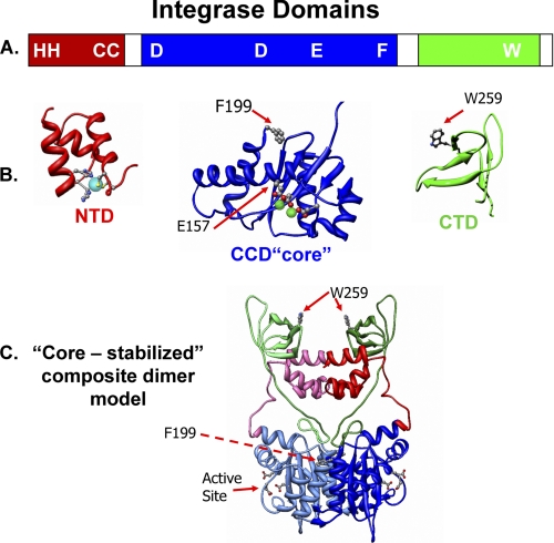
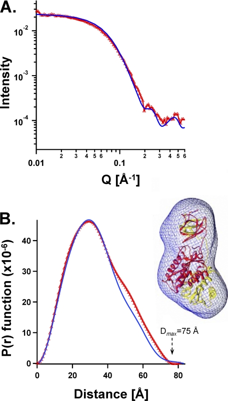
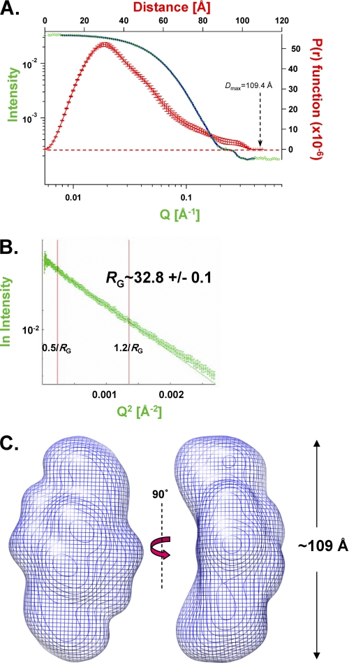
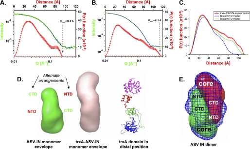
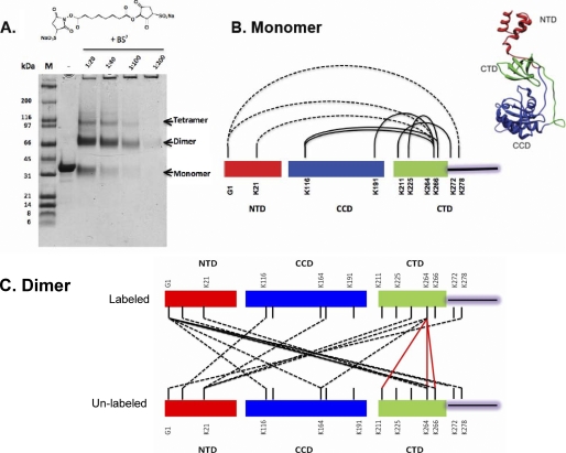
We determined the size and shape of full-length avian sarcoma virus (ASV) integrase (IN) monomers and dimers in solution using small angle x-ray scattering. The low resolution data obtained establish constraints for the relative arrangements of the three component domains in both forms. Domain organization within the small angle x-ray envelopes was determined by combining available atomic resolution data for individual domains with results from cross-linking coupled with mass spectrometry. The full-length dimer architecture so revealed is unequivocally different from that proposed from x-ray crystallographic analyses of two-domain fragments, in which interactions between the catalytic core domains play a prominent role. Core-core interactions are detected only in cross-linked IN tetramers and are required for concerted integration. The solution dimer is stabilized by C-terminal domain (CTD-CTD) interactions and by interactions of the N-terminal domain in one subunit with the core and CTD in the second subunit. These results suggest a pathway for formation of functional IN-DNA complexes that has not previously been considered and possible strategies for preventing such assembly.


Figures

FIGURE 1.
FIGURE 1.
Integrase protein domains and a core-stabilized dimer model. A, linear representation of IN, indicating the borders of the three domains and the locations of several important residues. The color code, red for the NTD, blue for the CCD (core), and green for the CTD, is used throughout; linkers and the unstructured CTD tail are white. B, ribbon models of the isolated core and CTD of ASV IN are based on published data (PDB code 1C1A). The helical NTD is modeled from the HIV-1 IN domain (PDB code 1K6Y). The conserved HHCC residues, in ball-and-stick representation, bind a zinc ion (cyan sphere). The three conserved active site residues in the CCD (core) are shown in ball-and-stick coordinating the metal co-factors (green spheres) required for catalysis. Two additional residues of relevance to the present studies (Phe-199 and Trp-259) are also shown in ball-and-stick. C, a core-core stabilized ASV IN dimer model based on the HIV-1 IN model of Wang et al. (14). One subunit is depicted in muted colors.
FIGURE 2.
FIGURE 2.
Comparison of the crystal structure with solution SAXS dimensions and shapes of the same NTD-lacking two-domain ASV IN fragment (IN(49–286) F199K at 1.1 mg/ml). A, experimentally determined SAXS scattering is shown in red triangles. Values calculated from the crystal structure of the same IN fragment (PDB coordinate file 1C1A) using the Crysol program are represented by a blue line. B, experimentally derived plot of P(r) function for the SAXS data is compared with values calculated from the same crystal structure. Color code is the same as in A. Right, SAXS envelope shape derived from the experimental data is portrayed as a blue wire mesh, and the atomic resolution coordinates of 1C1A are shown within the SAXS envelope, with one monomer colored red and the other yellow.
FIGURE 3.
FIGURE 3.
SAXS analyses of the full-length, wild type ASV IN dimer. A, experimentally determined SAXS data for ASV IN at 2.3 mg/ml, combining scattering data (green data points and blue fit line) and P(r) function (red points with error bars) into a four axis plot. B, Guinier plot of the natural log of the scattering intensity I(Q) versus Q2. A linear fit with data from 0.5/Rg to 1.2/Rg was used to approximate a radius of gyration (Rg) of 32.8 Å. C, shape of the wild type ASV IN dimer in solution, based on the SAXS data and modeled with the program GASBOR. The dimer envelope is shown in a blue mesh representation and two views.
FIGURE 4.
FIGURE 4.
SAXS analyses of monomeric ASV IN proteins and relative positioning of the terminal domains. A, SAXS data obtained with the ASV IN C23S/C125S/F199K/W259A monomer at 0.7 mg/ml. Four axis plot is as described in Fig. 3. B, SAXS data obtained with the thioredoxin-ASV IN chimeric derivative, trxA-IN-C23S/C125S/F199K/W259A at 2.3 mg/ml. C, comparison of experimentally derived P(r) function for trxA-IN protein in B (red circles) with two possible P(r) plots calculated from alternate arrangements of a distal NTD (green line) or distal CTD domain (blue line). D, left, ASV IN monomer envelope indicating possible alternate arrangements of the NTD and CTD. Middle, trxA-IN monomer envelope, Right, ribbon model of a trxA-IN chimeric protein with the trxA domain (magenta) in a distal position from the core domain; IN coloring as in Fig. 1. E, envelope of the dimeric wild type ASV IN is shown in blue mesh representation, with two monomeric envelopes positioned (red and green mesh representations) to fit within the dimer envelope.
FIGURE 5.
FIGURE 5.
Monomer and dimer proximities uncovered by protein cross-linking coupled with mass spectrometry. A, SDS-PAGE showing the separation of ASV multimers after cross-linking with increasing concentrations of BS3. Positions of cross-linked monomers, dimers, and tetramers, which migrate slightly faster than the non cross-linked forms, are indicated at the right of the gel. B, cross-link map of the ASV IN monomer and a model structure. Residues involved in cross-linking between NTD and CTD in labeled wild type monomers are joined with dashed lines; solid lines identify cross-links within CTD residues or between CTD and CCD residues. Similar cross-links were observed with unlabeled monomers (data not shown). Right, HADDOCK-generated monomer IN structure, using the monomer cross-linking data and the SAXS envelope derived from the W259A IN derivative. C, map of dimer cross-links between labeled and unlabeled IN subunits. CTD to CTD links are shown with red lines; some included in supplemental Table S2 are omitted here for clarity. NTD to core or NTD to CTD links are denoted with dashed black lines.
FIGURE 6.
FIGURE 6.
Cross-linking evidence for core-core interactions in tetramers and their functional relevance. A, summary of core-core cross-link data. Red dashed lines show cross-links that were unique to protein in the tetramer band. B, reciprocal interactions in the core-core dimer interface in the crystal structure of the isolated ASV domain (PDB code 1VSH) are mediated predominantly by side chains in α-helices 1 and 5 of this domain; potential electrostatic interactions between Arg-114′ and Glu-200, as well as His-103′ and Glu-187 are highlighted; the prime designation distinguishes subunits. C, single end processing assays. Times of incubation were 5, 10, 20, and 30 min. The arrow labeled2 shows the position of the normal processing product. The position of the 5′-32P-labeled viral DNA end substrate is indicated by S at the right of the gel; the control reaction in the lane marked N contained no IN protein. D, concerted integration assays. An arrowhead marks the position of a half-site reaction, in which a single end is joined to the plasmid target; the product of concerted integration is identified with an asterisk. Minutes of incubation are shown above each lane. The reaction in lane T contained no donor DNA, and in lane No Me2+, the divalent metal cofactor, Mg2+, was omitted. Lane M contains molecular markers, and positions of the supercoiled and nicked circular forms of the target DNA are marked sc and nc, respectively.
FIGURE 7.
FIGURE 7.
A reaching dimer model of the ASV IN apoprotein. A, dimer model of ASV IN that satisfies both distance constraints from the cross-linking experiments and the envelope shape and dimensions from SAXS experiments. Two orthogonal views are shown with our standard color coding for the three IN domains in one monomer, and the second IN monomer in muted colors. B, comparison of the experimentally determined P(r) function (blue line) of dimeric wild type ASV IN, with a P(r) function calculated from the core-core stabilized dimer model shown in Fig. 1C (red line) and the reaching dimer structure in A (green line). C, details of the CTD-CTD interface in the ASV IN dimer showing how stacking between proximal Trp-259 side chains from each CTD is a prominent feature of this interface. D, model of a reaching dimer of HIV-1 IN reveals the potential for conservation of CTD-CTD interface interactions.
FIGURE 8.
FIGURE 8.
Conformational change required for the transition from a reaching dimer to the intasome complex with DNA. A, conformation of a single ASV IN subunit in the apo-IN reaching dimer. B, conformation predicted from the inner dimer of an intasome complex that includes the viral substrate DNA. The subunit structure is modeled from the PFV intasome (PDB code 3OYA (38)). The change in orientation of the CTD residue Trp-259, shown in ball-and-stick, is highlighted with arrows. Active site residues are also shown in ball-and-stick fashion. A supplemental movie that simulates the conformational change between these two states is provided.

References

    1. Jaskolski M., Alexandratos J. N., Bujacz G., Wlodawer A. (2009) FEBS J. 276, 2926–2946 - PMC - PubMed
    1. Katz R. A., Merkel G., Kulkosky J., Leis J., Skalka A. M. (1990) Cell 63, 87–95 - PubMed
    1. Bujacz G., Jaskólski M., Alexandratos J., Wlodawer A., Merkel G., Katz R. A., Skalka A. M. (1996) Structure 4, 89–96 - PubMed
    1. Bujacz G., Alexandratos J., Wlodawer A., Merkel G., Andrake M., Katz R. A., Skalka A. M. (1997) J. Biol. Chem. 272, 18161–18168 - PubMed
    1. Maignan S., Guilloteau J. P., Zhou-Liu Q., Clément-Mella C., Mikol V. (1998) J. Mol. Biol. 282, 359–368 - PubMed

Publication types

LinkOut - more resources