. 2011 May 6;286(18):16218-28.
doi: 10.1074/jbc.M110.214585. Epub 2011 Mar 22.

Mutant p53 disrupts MCF-10A cell polarity in three-dimensional culture via epithelial-to-mesenchymal transitions

Affiliations

Mutant p53 disrupts MCF-10A cell polarity in three-dimensional culture via epithelial-to-mesenchymal transitions

Yanhong Zhang et al. J Biol Chem. .

Abstract

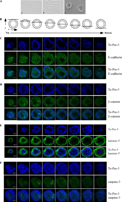
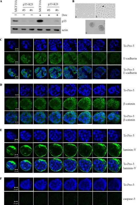
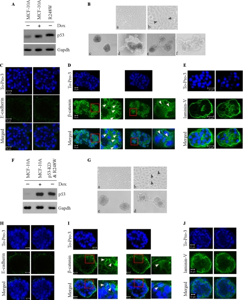
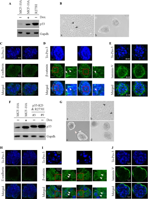
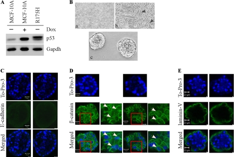
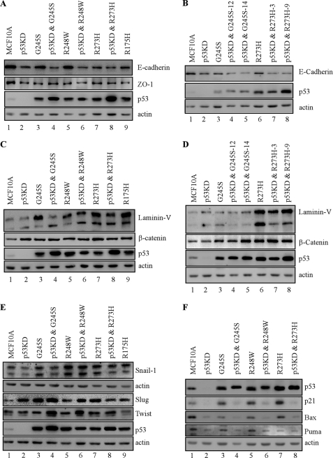
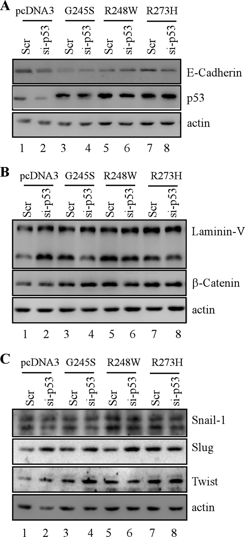
Mutant p53 is not only deficient in tumor suppression but also acquires additional activity, called gain of function. Mutant p53 gain of function is recapitulated in knock-in mice that carry one null allele and one mutant allele of the p53 gene. These knock-in mice develop aggressive tumors compared with p53-null mice. Recently, we and others showed that tumor cells carrying a mutant p53 are addicted to the mutant for cell survival and resistance to DNA damage. To further define mutant p53 gain of function, we used the MCF-10A three-dimensional model of mammary morphogenesis. MCF-10A cells in three-dimensional culture undergo a series of morphological changes and form polarized and growth-arrested spheroids with hollow lumen, which resembles normal glandular architectures in vivo. Here, we found that endogenous wild-type p53 in MCF-10A cells was not required for acinus formation, but knockdown of endogenous wild-type p53 (p53-KD) led to partial clearance of cells in the lumen due to decreased apoptosis. Consistent with this, p53-KD altered expression patterns of the cell adhesion molecule E-cadherin, the cytoskeletal marker β-catenin, and the extracellular matrix protein laminin V. We also found that ectopic expression of the mutant G245S led to a phenotype similar to p53-KD, whereas a combination of ectopic expression of siRNA-resistant G245S with p53-KD led to a less cleared lumen. In contrast, ectopic expression of mutant R248W, R175H, and R273H disrupted normal acinus architectures with filled lumen and led to formation of irregular and multiacinus structures regardless of p53-KD. In addition, these mutants altered normal expression patterns and/or levels of E-cadherin, β-catenin, laminin V, and tight junction marker ZO-1. Furthermore, epithelial-to-mesenchymal transitions (EMT) markers, Snail, Slug, and Twist, were highly induced by mutant p53 and/or p53-KD. Together, we postulate that EMT represents a mutant p53 gain of function and mutant p53 alters cell polarity via EMT.


Figures

FIGURE 1.
FIGURE 1.
MCF-10A cells form acinar-like architectures with hollow lumen. A, selective images of MCF-10A cells in two-dimensional (a, 100×; b, 200×) and three-dimensional (c) cultures. B, schematic diagrams of serial confocal cross-sections (x-y axis) through a hypothetical MCF-10A acinus. The diagrams overlaying each section illustrate the relative position of the optical section with respect to the z axis. C, serial confocal cross-sections of an acinus stained with To-Pro-3 and antibody against E-cadherin. MCF-10A cells were cultured on Matrigel for 20 days and immunostained with antibody against E-cadherin (green). E-cadherin is normally expressed at lateral cell-cell junctions. To-Pro-3 was used for nuclei staining. D, serial confocal cross sections of an acinus stained with To-Pro-3 and antibody against β-catenin. β-Catenin is expressed normally at cell-cell junctions. E, serial confocal cross sections of an acinus stained with To-Pro-3 and antibody against laminin V. Laminin V is normally deposited at the basal surface of the spheroid. F, serial confocal cross-sections of an acinus stained with To-Pro-3 and antibody against active cleaved caspase-3. The experiment was performed as in C except that anti-cleaved caspase-3 was used, and MCF-10A cells were cultured in a three-dimensional culture for 10 day. Active caspase-3 is normally expressed in the lumen of early stage acini.
FIGURE 2.
FIGURE 2.
Knockdown of endogenous wild-type p53 in MCF-10A leads to incomplete clearance of cells in the lumen due to decreased apoptosis. A, generation of MCF-10A cell lines in which endogenous wild-type p53 was stably knocked down (clones #5 and #6). Western blot analysis was prepared using extracts from p53-KD MCF-10A cells that were mock-treated (−) or treated with doxorubicin (Dox; +). The blots were probed with antibodies against p53 and actin, respectively. B, selective images of MCF-10A cells in two-dimensional (a, 100×; b, 200×) and three-dimensional (c) cultures. C, serial confocal cross-sections of an acinus stained with To-Pro-3 and antibody against E-cadherin. D, serial confocal cross-sections of an acinus stained with To-Pro-3 and antibody against β-catenin. E, serial confocal cross-sections of an acinus stained with To-Pro-3 and antibody against laminin V. F, serial confocal cross-sections of an acinus stained with To-Pro-3 and antibody against active cleaved caspase-3.
FIGURE 3.
FIGURE 3.
Mutant G245S has a dominant negative activity toward endogenous wild-type p53 but possesses a weak gain of function in the MCF-10A three-dimensional model. A and F, generation of MCF-10A cell lines in which siRNA-resistant mutant G245S was expressed (A), along with knockdown of endogenous wild-type p53 (F, clones #1 and #12). Western blot analysis was prepared using extracts from MCF-10A cells that were mock-treated (−) or treated with doxorubicin (Dox; +). The blots were probed with antibodies against p53 and GAPDH, respectively. B and G, selective images of MCF-10A cells in two-dimensional (a, 100×; b, 200×) and three-dimensional (c) cultures. C and H, representative confocal images of cross-sections through the middle of acini stained with To-Pro-3 and antibody against E-cadherin. D and I, representative confocal images of cross-sections through the middle of acini stained with To-Pro-3 and antibody against β-catenin. White arrows in D and I indicate the accumulation and translocation of β-catenin in acinar structure. E and J, representative confocal images of cross-sections through the middle of acini stained with To-Pro-3 and antibody against laminin V.
FIGURE 4.
FIGURE 4.
Ectopic expression of mutant R248W disrupts normal acinus architecture with filled lumen regardless of p53-KD. A and F, generation of MCF-10A cell lines in which siRNA-resistant mutant R248W was expressed (A), along with knockdown of endogenous wild-type p53 (F). Western blot analysis was prepared using extracts from MCF-10A cells that were mock-treated (−) or treated with doxorubicin (Dox; +). The blots were probed with antibodies against p53 and GAPDH, respectively. B and G, selective images of MCF-10A cells in two-dimensional (a, 100×; b, 200×) and three-dimensional (B, c–f and G, c–d) cultures. C and H, representative confocal images of cross-sections through the middle of acini stained with To-Pro-3 and antibody against E-cadherin. D and I, representative confocal images of cross-sections through the middle of acini stained with To-Pro-3 and antibody against β-catenin. E and J, representative confocal images of cross-sections through the middle of acini stained with To-Pro-3 and antibody against laminin V.
FIGURE 5.
FIGURE 5.
Ectopic expression of mutant R273H disrupts normal acinar architectures with filled lumen regardless of p53-KD. A and F, generation of MCF-10A cell lines in which siRNA-resistant mutant R273H was expressed (A), along with knockdown of endogenous wild-type p53 (F). Western blot analysis was prepared using extracts from MCF-10A cells that were mock-treated (−) or treated with doxorubicin (Dox; +). The blots were probed with antibodies against p53 and GAPDH, respectively. B and G, selective images of MCF-10A cells in two-dimensional (B, a, 100×; G, a, 100×; b, 200×) and three-dimensional (c and d) cultures. C and H, representative confocal images of cross-sections through the middle of acini stained with To-Pro-3 and antibody against E-cadherin. D and I, representative confocal images of cross-sections through the middle of acini stained with To-Pro-3 and antibody against β-catenin. E and J, representative confocal images of cross-sections through the middle of acini stained with To-Pro-3 and antibody against laminin V.
FIGURE 6.
FIGURE 6.
Ectopic expression of mutant R175H disrupts normal acinus architectures with filled lumen. A, generation of MCF-10A cell lines in which mutant R175H was expressed. Western blot analysis was prepared using extracts from MCF-10A cells that were mock treatment (−) or treatment with doxorubicin (Dox; +). The blots were probed with antibodies against p53 and GAPDH, respectively. B, selective images of MCF-10A cells in two-dimensional (a, 100×; b, 200×) and three-dimensional (c) cultures. C, representative confocal images of cross-sections through the middle of acini stained with To-Pro-3 and antibody against E-cadherin. D, representative confocal images of cross-sections through the middle of acini stained with To-Pro-3 and antibody against β-catenin. White arrows indicate the accumulation and translocation of β-catenin in acinar structure. E, representative confocal images of cross-sections through the middle of acini stained with To-Pro-3 and antibody against laminin V.
FIGURE 7.
FIGURE 7.
Markers for epithelial-to-mesenchymal transitions are regulated upon ectopic expression of siRNA-resistant mutant p53 regardless of stable knockdown of endogenous wild-type p53 in MCF-10A cells. A, C, and E, Western blots were prepared with extracts from parental MCF-10A cells (lane 1), p53-KD MCF-10A cells (lane 2), MCF-10A cells in which a mutant p53 was ectopically expressed (lanes 3, 5, 7, and 9) and MCF-10A cells in which a mutant p53 was ectopically expressed along with knockdown of endogenous wild-type p53 (lanes 4, 6, and 8). The blots were probed with antibodies against E-cadherin (A), ZO-1 (A), p53 (A, C, and E), laminin V (C), β-catenin (C), Snail (E), Slug (E), Twist (E), and actin (A, C, and E). B, the experiments were performed as in A except that two MCF10A cell lines in which mutant G245S or R273H was ectopically expressed along with knockdown of endogenous wild-type p53 were used. D, the experiments were performed as in C except that two MCF10A cell lines in which mutant G245S or R273H was ectopically expressed along with knockdown of endogenous wild-type p53 were used. F, the experiment was performed as in A except that the blots were probed with antibodies against p53, p21, Bax, and Puma, respectively.
FIGURE 8.
FIGURE 8.
EMT markers are regulated upon transient overexpression of siRNA-resistant mutant p53 along with or without transient knockdown of wild-type p53 in MCF-10A cells. Western blots were prepared with extracts from MCF-10A cells in which scramble siRNA (Scr) was co-transfected with pcDNA3 empty vector (lane 1), G245S (lane 3), R248W (lane 5), or R273H (lane 7) and MCF-10A cells in which siRNA against p53 (si-p53) was co-transfected with pcDNA3 empty vector (lane 2), G245S (lane 4), R248W (lane 6), or R273H (lane 8). The blots were probed with antibodies against E-cadherin (A), p53 (A–C), laminin V (B), β-catenin (B), Snail (C), Slug (C), Twist (C), and actin (A–C), respectively.

Similar articles

Cited by

References

    1. Brosh R., Rotter V. (2009) Nat. Rev. Cancer 9, 701–713 - PubMed
    1. Ko L. J., Prives C. (1996) Genes Dev. 10, 1054–1072 - PubMed
    1. Milner J., Medcalf E. A. (1991) Cell 65, 765–774 - PubMed
    1. Willis A., Jung E. J., Wakefield T., Chen X. (2004) Oncogene 23, 2330–2338 - PubMed
    1. Lang G. A., Iwakuma T., Suh Y. A., Liu G., Rao V. A., Parant J. M., Valentin-Vega Y. A., Terzian T., Caldwell L. C., Strong L. C., El-Naggar A. K., Lozano G. (2004) Cell 119, 861–872 - PubMed

Publication types

MeSH terms