. 2011 Apr 22;332(6028):462-5.
doi: 10.1126/science.1199211. Epub 2011 Mar 31.

Protein tyrosine kinase Wee1B is essential for metaphase II exit in mouse oocytes

Affiliations

Protein tyrosine kinase Wee1B is essential for metaphase II exit in mouse oocytes

Jeong Su Oh et al. Science. .

Abstract

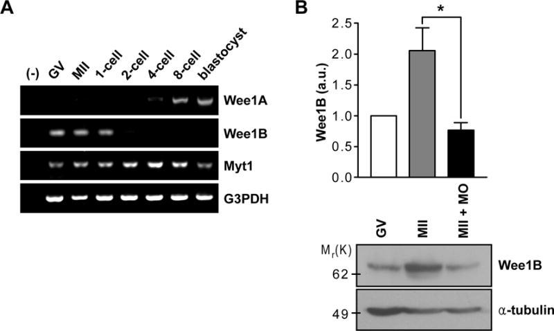
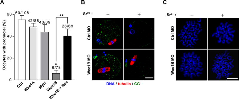
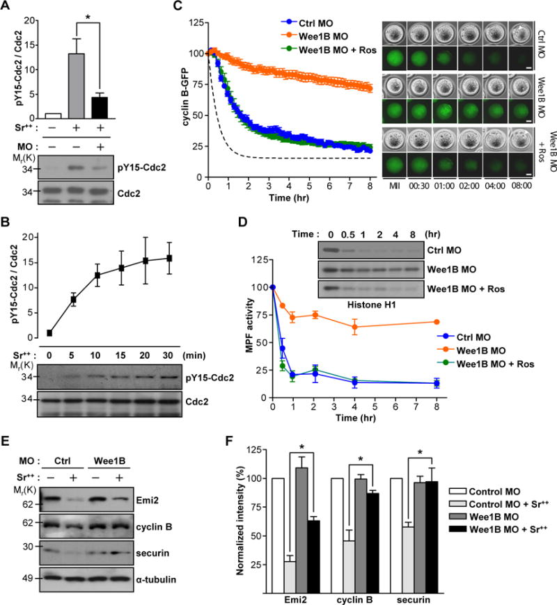
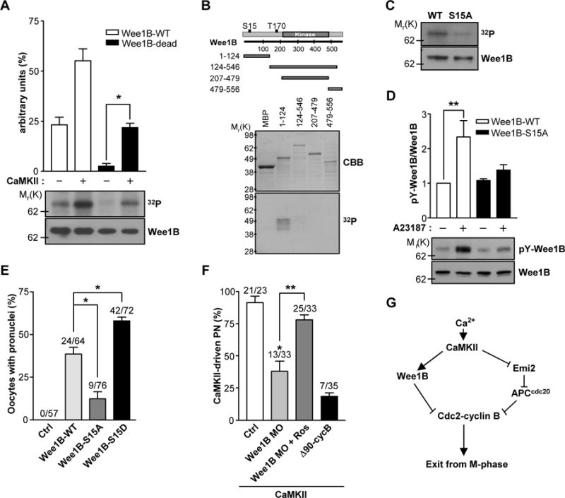
Waves of cyclin synthesis and degradation regulate the activity of Cdc2 protein kinase during the cell cycle. Cdc2 inactivation by Wee1B-mediated phosphorylation is necessary for arrest of the oocyte at G2-prophase, but it is unclear whether this regulation functions later during the metaphase-to-anaphase transition. We show that reactivation of a Wee1B pathway triggers the decrease in Cdc2 activity during egg activation. When Wee1B is down-regulated, oocytes fail to form a pronucleus in response to Ca(2+) signals. Calcium-calmodulin-dependent kinase II (CaMKII) activates Wee1B, and CaMKII-driven exit from metaphase II is inhibited by Wee1B down-regulation, demonstrating that exit from metaphase requires not only a proteolytic degradation of cyclin B but also the inhibitory phosphorylation of Cdc2 by Wee1B.


Figures

Fig.1
Fig.1. Expression of Wee1B during the oocyte-to-embryo transition
(A) RT-PCR of Wee1 kinases in oocyte and preimplantation embryos at different stages of development. G3PDH was used as an internal control. (B) GV and MII oocytes along with Wee1B MO-injected MII oocytes were immunoblotted for Wee1B and α-tubulin. Each lane contains 100 oocytes. Band intensities for Wee1B were quantified and normalized to α-tubulin level. a.u., arbitrary units. *p=0.0283.
Fig.2
Fig.2. Effect of down-regulation of Wee1B during egg activation
(A) GV oocytes injected with indicated MOs were in vitro matured for 20 hours. MII oocytes were activated with strontium with or without roscovitine (Ros) and oocytes with pronuclei were scored after 8 hours. Data are mean ± SEM of three independent experiments. The total number of oocytes used is reported above the bar. *p<0.0001 compared to control, **p<0.005. (B) In vitro matured MII oocytes injected with control (Ctrl) or Wee1B MO at GV stage were stained for DNA, tubulin and cortical granule (CG) before (−) or after (+) activation with strontium for 8 hours. (C) Chromosome spreads of Wee1B knockdown oocytes after strontium activation. Note that sister chromatids remained attached in Wee1B knockdown oocytes, whereas they had undergone disjunction in control oocytes. Bar, 20μm.
Fig.3
Fig.3. Wee1B-dependent inactivation of MPF during egg activation
(A) Non-injected or Wee1B MO injected MII oocytes were activated with strontium for 20 min and subjected to SDS-PAGE followed by immunoblotting for Cdc2 and tyrosine 15 phosphorylation (pY15) of Cdc2. (B) MII oocytes activated with strontium were prepared at different time after activation and subjected to SDS-PAGE followed by immunoblotting for Cdc2 and pY15 Cdc2. Data are the mean ± SEM from three independent experiments. A representative image is shown. (C and D) GV oocytes injected with Wee1B MO and cyclin B-GFP mRNA were in vitro matured and activated with strontium at time t=0. (C) GFP levels were measured every 10 min for 8 hours. Average of MPF activity in control oocytes from (D) is shown with dotted line. Data are mean ± SEM of ten oocytes from two independent experiments. Representative images are shown in right panel. Pronucleus is indicated by arrowheads. Bar, 20μm. (D) Oocyte lysates were incubated with [γ-32P]ATP and either Histone H1 or myelin basic protein (MBP) (see fig.S4). Phosphorylation was detected by SDS-PAGE and autoradiography. Representative images from two independent experiments are shown. (E) In vitro matured MII oocytes injected with control or Wee1B MO at GV stage were incubated with (+) or without (−) strontium for 2 hours. Emi2, cyclin B and securin levels were determined by immunoblots. Each lane contains 100 oocytes. Representative images from three independent experiments are shown. (F) Band intensities of Emi2, cyclin B and securin were quantified and normalized to controls. Data are the mean ± SEM from three independent experiments. *p<0.05.
Fig.4
Fig.4. Phosphorylation of Wee1B by CaMKII
(A) Wee1B was overexpressed in HEK293 cells, immunoprecipitated, and incubated with or without CaMKII in the presence of [γ-32P]ATP for 10 min. The phosphorylation state was detected by SDS-PAGE and autoradiography. The amounts of loaded protein were determined by immunoblot. A representative experiment of the three performed is shown. (B) MBP-tagged truncated forms of Wee1B were generated and expressed in Escherichia coli. After purification by affinity chromatography, fusion proteins were incubated with CaMKII in the presence of [γ-32P]ATP for 10 min. The radiolabeled fusion proteins were detected by autoradiogram. The expression and purification of MBP fusion proteins were monitored by Coomassie brilliant blue (CBB) staining. (C) Immunoprecipitated Wee1B-WT or -S15A mutant were incubated with CaMKII in the presence of [γ-32P]ATP for 10 min, and incorporation of 32P was detected by autoradiography. The amounts of loaded protein were determined by immunoblot. (D) HEK293 cells overexpressing Wee1B-WT or -S15A mutant were treated with 10μM of A23187 for 20 min. The activity of Wee1B was measured by the autophosphorylation of tyrosine residues. A representative image of three independent experiments performed is reported. (E) MII oocytes were injected with mRNAs encoding the indicated Wee1Bs, and oocytes with a pronucleus were counted after 6 hours. (F) MII oocytes injected with Wee1B MO or Δ90cyclin B mRNA were microinjected with constitutively active CaMKII (CA-CaMKII) and pronuclear formation scored after 8 hours. *p<0.005 and **p<0.05 (G) Proposed model of mouse egg activation.

References

    1. Jones KT. Reproduction. 2005 Dec;130:813. - PubMed
    1. Lew DJ, Kornbluth S. Curr Opin Cell Biol. 1996 Dec;8:795. - PubMed
    1. Wasch R, Cross FR. Nature. 2002 Aug 1;418:556. - PubMed
    1. Tsurumi C, Hoffmann S, Geley S, Graeser R, Polanski Z. J Cell Biol. 2004 Dec 20;167:1037. - PMC - PubMed
    1. Yin S, et al. Cell Cycle. 2007 Dec 1;6:2990. - PubMed

Publication types

MeSH terms