. 2011 Nov;45(5):915-22.
doi: 10.1165/rcmb.2010-0113OC. Epub 2011 Mar 31.

Nuclear β-catenin is increased in systemic sclerosis pulmonary fibrosis and promotes lung fibroblast migration and proliferation

Affiliations

Nuclear β-catenin is increased in systemic sclerosis pulmonary fibrosis and promotes lung fibroblast migration and proliferation

Anna P Lam et al. Am J Respir Cell Mol Biol. 2011 Nov.

Abstract

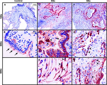
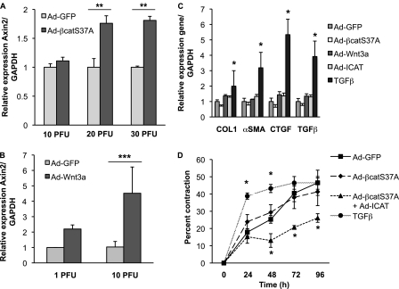
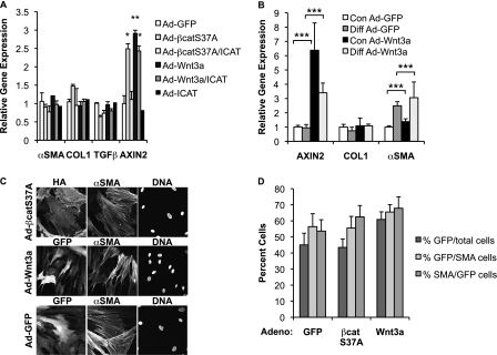
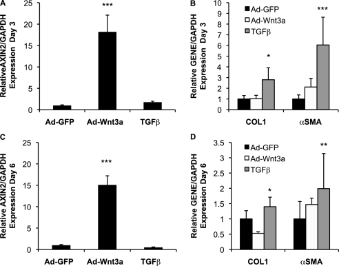
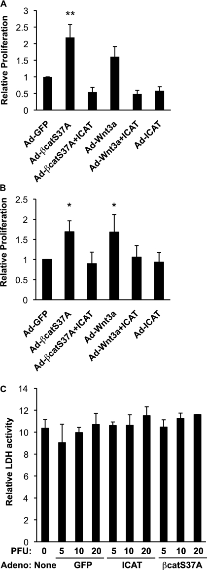
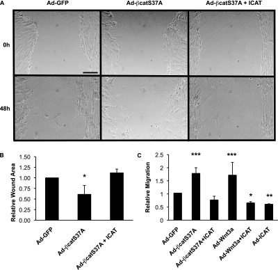
Pulmonary fibrosis is a disease that results in loss of normal lung architecture, but the signaling events that drive tissue destruction are incompletely understood. Wnt/β-catenin signaling is important in normal lung development, but whether abnormal signaling occurs in lung fibrosis due to systemic sclerosis and the consequences of β-catenin signaling toward the fibrogenic phenotype remain poorly defined. In this study, we show nuclear β-catenin accumulation in fibroblastic foci from lungs of patients with systemic sclerosis-associated advanced pulmonary fibrosis. Forced activation of β-catenin signaling in three independently derived sources of normal human lung fibroblasts promotes proliferation and migratory activities but is not sufficient to activate classic markers of fibroblast activation, such as TGF-β, type 1 collagen, α-smooth muscle actin, and connective tissue growth factor. These findings indicate that activation of β-catenin signaling in pulmonary fibroblasts may be a common feature of lung fibrosis, contributing to fibroproliferative and migratory activities associated with the disease.


Figures

Figure 1.
Figure 1.
Nuclear β-catenin staining is observed in systemic sclerosis (SSc)-associated pulmonary fibrosis. (A) Low (100×) and (B) high (600×) magnification images from a control lung show β-catenin staining at the cell–cell contacts of airway epithelial cells (arrows) but no obvious staining in nuclei of alveolar epithelial or interstitial cells. Control lungs are taken from normal donors whose lungs were not used for transplant. (CH) Lung section images from patients with SSc-associated pulmonary fibrosis show β-catenin staining at junctions of airway epithelial cells (D, arrows), in nuclei of alveolar epithelial cells (G, arrowheads), and within cytoplasm and nuclei of cells that appear to be fibroblasts (E and H, arrowheads). Images are representative findings from three control patients and three patients with SSc–interstitial lung disease using Zeiss Axioskop with a CRi Nuance multispectral camera.
Figure 2.
Figure 2.
Overexpression of Wnt and nondegradable β-catenin is insufficient to induce expression of TGF-β–responsive genes in normal human lung fibroblasts. Normal human lung fibroblasts (NHLFs) were infected with Ad-GFP (control) or Ad-S37A–β-catenin (A) or Ad-Wnt3a (B) for 24 hours; total RNA was extracted to quantify expression of AXIN2 mRNA by quantitative PCR (qPCR). Ad-S37A–β-catenin and Ad-Wnt3a are sufficient to promote Wnt/β-catenin signaling in NHLFs 24 hours postinfection. Results are expressed as the mean expression ± SD of AXIN2 relative to glyceraldehyde 3-phosphate dehydrogenase (GAPDH) from three independent experiments (**P < 0.01; ***P < 0.001). (C) NHLFs were treated with Ad-GFP (control), Ad-S37A–β-catenin (20 plaque-forming units (PFU)/cell), Ad-Wnt3a (10 PFU/cell), Ad-ICAT (20 PFU/cell), or TGF-β (5 ng/ml) for 24 hours; total RNA was extracted to quantify expression of type 1 collagen (COL1), α-smooth muscle actin (α-SMA), connective tissue growth factor (CTGF), and TGF-β mRNAs by quantitative PCR. Results are expressed as the mean gene expression ± SD relative to GAPDH from three independent experiments (*P < 0.05). (D) Collagen contraction by NHLFs after treatment with TGF-β (5 ng/ml), Ad-GFP (control), Ad-S37A–β-catenin, or Ad-S37A–β-catenin with Ad-ICAT. Results are expressed as the percentage mean change in matrix diameter size compared with the 0-hour time point ± SD from three independent experiments (*P < 0.05).
Figure 3.
Figure 3.
Wnt/β-catenin signaling is not sufficient to induce expression of TGF-β or its established profibrotic targets in NL-57 and WI-38 human fibroblasts. (A) Lung fibroblasts (NL-57) obtained from a patient without lung disease were treated with Ad-GFP (control), Ad-S37A–β-catenin (20 PFU/cell), Ad-Wnt3a (10 PFU/cell), or Ad-ICAT (20 PFU/cell) for 24 hours; total RNA was extracted to quantify expression of α-SMA, COL1, TGF-β, and AXIN2 mRNAs by quantitative PCR. Results are expressed as the mean gene expression ± SD relative to GAPDH from three independent experiments (*P < 0.05; **P < 0.01). (B) Human embryonic fibroblasts (WI-38) were infected with Ad-GFP (control) or Ad-Wnt3a (10 PFU/cell) after undergoing treatment with control or differentiation media for 3 days; total RNA was extracted to quantify expression of AXIN2, COL1, and α-SMA mRNAs by qPCR. Results are expressed as the mean gene expression ± SD relative to GAPDH from three independent experiments (***P < 0.001). (C) Immunofluorescence of NHLFs infected with Ad-GFP, Ad-Wnt3a-GFP, and Ad-S37A–β-catenin–HA for 24 hours. Immunostaining was performed to confirm the presence of hemagglutinin (HA), GFP, and α-SMA; cell nuclei were stained with Hoechst. (D) Infection efficiency with Ad-GFP, Ad-S37A–β-catenin, and Ad-Wnt3a is routinely > 40% and expressed as GFP- or HA-positive cells divided by the total number of cells (%GFP/total cells). More than 50% of α-SMA–expressing cells are infected, expressed as GFP- or HA-positive cells divided by number of α-SMA–expressing cells (%GFP/SMA). Similarly, more than 50% of infected cells are α-SMA positive (%SMA/GFP). Results are expressed as the percent ± SEM from three independent experiments.
Figure 4.
Figure 4.
Persistent activation of Wnt/β-catenin signaling does not alter COL1 or α-SMA expression. NHLFs were infected with Ad-GFP (control) or Ad-Wnt3a (10 PFU/cell) or treated with TGF-β (5 ng/ml) every 48 hours until RNA was extracted on Days 3 or 6 postinfection. Although persistent infection with Ad-Wnt3a robustly increases AXIN2 expression on Days 3 (A) and 6 (C), this does not alter COL1 or α-SMA gene expression, in contrast to TGF-β (B and D). Results are expressed as the mean gene expression ± SD relative to GAPDH from three independent experiments (*P < 0.05; **P < 0.01; ***P < 0.001).
Figure 5.
Figure 5.
Wnt/β-catenin signaling promotes proliferation and is not required for the survival of normal human lung fibroblasts. (A) Bromodeoxyuridine (BrdU) incorporation assay. Human lung fibroblasts (NL-57) from nondiseased lungs were infected with Ad-GFP, Ad-S37A–β-catenin, Ad-ICAT, and Ad-Wnt3a for 48 hours and then pulse-labeled with BrdU. Results are expressed as mean ± SD relative to control adenovirus from six independent experiments (**P < 0.01). Similar results were observed with NHLFs (Figure E2). (B) Ki67 assay. Human lung fibroblasts (NHLFs) were infected with Ad-GFP, Ad-S37A–β-catenin, Ad-ICAT, and Ad-Wnt3a for 48 hours and processed for immunofluorescence staining with an antibody to Ki67. Results are expressed as mean ± SD relative to control adenovirus from three to five independent experiments (*P < 0.05). (C) Lactate dehydrogenase (LDH) cell viability assay. NHLFs were infected with Ad-GFP, Ad-S37A–β-catenin, and Ad-ICAT for 72 hours, and media was assessed for LDH activity as described in Materials and Methods. Activation of Wnt/β-catenin signaling neither promotes cell death with increasing PFUs of control adenovirus or adenovirus expressing S37A–β-catenin nor is required for cell survival (Ad-ICAT). Results are expressed as mean levels of LDH activity ± SD from at least three independent experiments.
Figure 6.
Figure 6.
Wnt/β-catenin signaling stimulates fibroblast migration. (A) NHLFs were “scratch” wounded, infected with the stated adenoviruses, and photographed at 0-, 24-, and 48-hour time points. Representative images for each infection and 48-hour time point are shown. (B) Quantification of wound size at 48 hours. Results are expressed as mean change ± SD of wound area relative to wound area at 0 time point from four independent experiments (*P < 0.05). (C) Lung fibroblasts (NL-57) obtained from a patient without pulmonary disease were infected with Ad-GFP, Ad-S37A–β-catenin, Ad-ICAT, and Ad-Wnt3a and analyzed in a Boyden chamber assay as described in Materials and Methods. Similar results were observed with NHLFs (Figure E3). Results are expressed as the mean number of cells migrated in each condition relative to control adenovirus ± SD from eight independent experiments (*P < 0.05; **P < 0.01; ***P < 0.001).

References

    1. Selman M, King TE, Pardo A. Idiopathic pulmonary fibrosis: prevailing and evolving hypotheses about its pathogenesis and implications for therapy. Ann Intern Med 2001;134:136–151 - PubMed
    1. Phan SH. The myofibroblast in pulmonary fibrosis. Chest 2002; 122(Suppl)286S–289S - PubMed
    1. Hinz B, Phan SH, Thannickal VJ, Galli A, Bochaton-Piallat ML, Gabbiani G. The myofibroblast: one function, multiple origins. Am J Pathol 2007;170:1807–1816 - PMC - PubMed
    1. Selman M, Pardo A, Kaminski N. Idiopathic pulmonary fibrosis: aberrant recapitulation of developmental programs? PLoS Med 2008;5:e62. - PMC - PubMed
    1. Moon RT, Kohn AD, De Ferrari GV, Kaykas A. Wnt and beta-catenin signalling: diseases and therapies. Nat Rev Genet 2004;5:691–701 - PubMed

Publication types