. 2011 Jun;31(6):1387-96.
doi: 10.1161/ATVBAHA.110.222547. Epub 2011 Mar 31.

Runx2-upregulated receptor activator of nuclear factor κB ligand in calcifying smooth muscle cells promotes migration and osteoclastic differentiation of macrophages

Affiliations

Runx2-upregulated receptor activator of nuclear factor κB ligand in calcifying smooth muscle cells promotes migration and osteoclastic differentiation of macrophages

Chang Hyun Byon et al. Arterioscler Thromb Vasc Biol. 2011 Jun.

Abstract

Objective: Clinical and experimental studies demonstrate the important roles of vascular smooth muscle cells (VSMC) in the pathogenesis of atherosclerosis. We have previously determined that the osteogenic transcription factor Runx2 is essential for VSMC calcification. The present study characterized Runx2-regulated signals and their potential roles in vascular calcification.

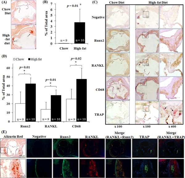
Methods and results: In vivo studies with atherogenic apolipoprotein E(-/-) mice demonstrated that increased oxidative stress was associated with upregulation of Runx2 and receptor activator of nuclear factor κB ligand (RANKL), which colocalized in the calcified atherosclerotic lesions and were juxtaposed to infiltrated macrophages and osteoclast-like cells that are positively stained for an osteoclast marker, tartrate-resistant acid phosphatase. Mechanistic studies using RNA interference, a luciferase reporter system, chromatin immunoprecipitation, and electrophoretic mobility shift assays indicated that Runx2 regulated the expression of RANKL via a direct binding to the 5'-flanking region of the RANKL. Functional characterization revealed that RANKL did not induce VSMC calcification, nor was RANKL required for oxidative stress-induced VSMC calcification. Using a coculture system, we demonstrated that VSMC-expressed RANKL induced migration as well as differentiation of bone marrow-derived macrophages into multinucleated, tartrate-resistant acid phosphatase-positive osteoclast-like cells. These effects were inhibited by the RANKL antagonist osteoprotegerin and with VSMC deficient in Runx2 or RANKL.

Conclusion: We demonstrate that Runx2 directly binds to the promoter and controls the expression of RANKL, which mediates the crosstalk between calcifying VSMC and migration and differentiation of macrophages into osteoclast-like cells in the atherosclerotic lesions. Our studies provide novel mechanistic insights into the regulation and function of VSMC-derived RANKL in the pathogenesis of atherosclerosis and vascular calcification.


Figures

Figure 1
Figure 1. Vascular calcification is associated with increased expression of Runx2 and RANKL as well as macrophage infiltration and TRAP-positive cells
(A) Frozen aortic root sections from chow diet- or high-fat diet-fed ApoE-deficient mice were obtained at the level of the aortic valve leaflets and stained with Alizarin Red. Arrows indicate calcium mineral deposition. (B) Calcification at the aortic root sections was measured using ImageJ software (NIH Bethesda, MD). The percentage of tissue area with calcification was calculated by dividing the calcified area by the total area of the aortic root. Bar values are expressed as the mean ± SD with number of the mice shown above each bar. (C) Frozen aortic root sections were stained with antibodies for Runx2, RANKL, CD68 and secondary antibody alone (negative control) and histochemically stained for TRAP. Immunopositive areas shown in boxes in low magnification (× 100, left panels) are shown at higher magnification (× 400, right panels). Arrows indicate TRAP-positive cells. (D) Runx2-, RANKL-, and CD68-immunopositive areas in the aortic root sections. The immunopositive areas were quantified as a percent of total aortic root area in each section. Bar values are expressed as mean ± SD with number of the mice shown above each bar. (E) Immunofluorescent staining of Runx2, RANKL, and TRAP in frozen aortic root sections from high-fat diet-fed ApoE-deficient mice. Negative control shows autofluorescence (green). Boxes in the low magnification image (upper panels) are shown in higher magnification in the lower panels. Alizarin red staining showing on left panel is to indicate the calcified areas.
Figure 2
Figure 2. Oxidative stress induces the expression of RANKL in VSMC during calcification
(A) VSMC were exposed to control or 0.05 to 0.4 mM H2O2 in osteogenic media for 3 weeks with media changes every 2–3 days. In vitro VSMC calcification was determined by Alizarin Red staining. Representative images of stained dishes (upper) and microscopic views (× 40, lower) from 4 independent experiments are shown. (B) Expression of RANKL in VSMC exposed to 0.05 to 0.4 mM H2O2 in osteogenic media for 3 weeks was determined by quantitative real-time PCR (n=4, *p <0.05, **p <0.01 and ***p<0.005 compared with control conditions). (C) RANKL protein was measured in media and cell lysates from cells under oxidative stress for 2 weeks. Results from 2 independent ELISA assays performed in triplicate are shown (*p<0.05 compared with control condition in media or cell lysates). D) Expression of Runx2 and RANKL during VSMC calcification was determined by RT-PCR. VSMC were exposed to 0.4 mM H2O2 in osteogenic media for up to 21 days. Representative RT-PCR results of 3 independent experiments are shown. (E). RANKL promoter activity in VSMC under oxidative stress was determined using the Dual-Luciferase Reporter Assay System with six deletion mutants of RANKL promoter-reporters RL(FL), RL(−700), RL(−550), RL(−400), RL(−200), RL(−150), RL(−50) and a plasmid expressing Renilla Luciferase as an internal control reporter. Results shown are relative luciferase activities in each of the conditions compared with that of the RL(FL) under control condition, which was assigned a value of 1.0. Results from 3 independent experiments performed in duplicate are shown (* p < 0.05 vs. RL(FL) control).
Figure 3
Figure 3. Runx2 regulates RANKL transcription
(A) Putative transcription factors which bind to 5'-flanking and promoter regions of murine RANKL (AF332141) were identified through the TFSEARCH engine (http://www.cbrc.jp/research/db/TFSEARCH.html). Three Runx2-binding sites were identified within the −400 to −200 bp region and one additional binding site was found near −200 bp. The putative Runx2 binding sites (Runx2-1, Runx2-2, Runx2-3, and Runx2-4) and primers for ChIP assay are indicated. (B) ChIP assay with Runx2 immunoprecipitates (anti-Runx2; Santa Cruz) and PCR using primer sets for whole, R1&2, and R3&4. Representative pictures of 3 independent experiments are shown. (C) ChIP assay with PCR primer sets R1, R2, R3, and R4. Representative pictures of 3 independent experiments are shown. (D) EMSA was performed using four probes (P1, P2, P3, and P4) containing the putative Runx2 binding elements. Probes carrying mutations of the Runx2-binding sites (P1m, P2m, P3m, and P4m) were used as negative controls. Representative EMSA pictures of 3 independent experiments are shown. (E) Competitive EMSA was performed using 100-fold molar excess amounts of unlabeled probes. Probes carrying mutation of the Runx2-binding sites were used as negative controls. Representative EMSA pictures of three independent experiments are shown. (F) Specific antibody for Runx2 was used to detect supershift in Runx2 binding complex. A representative EMSA picture of 2 independent experiments are shown. (G) VSMC with stable Runx2 knockdown by shRNA against Runx2 (Lenti-shRunx2) or control lentiviruses encoding GFP (Lenti-GFP) were exposed to 0.4 mM H2O2 for 2 weeks. Expression of Runx2 protein was determined by Western blot analysis. A representative blot of 2 independent experiments is shown. (H) Expression of RANKL transcript in control or Runx2 knockdown VSMC in G was determined by RT-PCR. Representative results from two independent experiments are shown. (I) VSMC were transduced with Ad-Runx2 or control virus (Ad-GFP) and cultured in osteogenic media for 2 weeks. Expression of RANKL transcript was determined by RT-PCR. Representative RT-PCR results of 2 independent experiments are shown.
Figure 4
Figure 4. RANKL does not induce VSMC calcification and is not required for oxidative stress-induced VSMC calcification
(A) RANKL (100 ng/ml) and/or OPG (50 ng/ml) were added to VSMC with or without the addition of 0.4 mM H2O2 in osteogenic media for 3 weeks with media changes every 2–3 days. In vitro VSMC calcification was determined by von Kossa staining. Representative images of stained dishes (upper) and microscopic views (× 100, lower) from 3 independent experiments are shown. (B) VSMC were cultured in DMEM with 10% FBS and 10 mM beta-glycerophosphate with or without the addition of RANKL (100 ng/ml) for 10 days. Calcification was quantified in cell lysates by the o-cresolphthalein complexone method. Results shown are from 3 independent experiments (NS, no statistically significant difference). (C) VSMC from WT or RANKL-deficient mice were treated with 0.4 mM H2O2 in osteogenic media for 3 weeks. Representative images of von Kossa stained dishes (upper) and microscopic views (× 100, lower) from 2 independent experiments are shown. (D) In separate experiments, VSMC from WT or RANKL-deficient mice were treated with 0.4 mM H2O2 in osteogenic media for 1 or 2 weeks; and calcium mineral was quantified by the o-cresolphthalein complexone method. Results from 2 independent experiments performed in triplicates are shown (*p <0.05 compared with control conditions, no statistically significant differences were found between WT and RANKL−/− VSMC).
Figure 5
Figure 5. Oxidative stress-stimulated VSMC promote BMM migration in a Runx2/RANKL-dependent manner
A) After VSMC were cultured in osteogenic media for 2 weeks with or without 0.4 mM H2O2, cells were coated on the lower side of Transwell filter, and DiI-labeled BMMs were added to the upper chambers. After 24 hours, migrated BMMs were measured using a microplate fluorescence reader. Results from 3 independent experiments performed in duplicate are shown (p = NS for unstimulated WT VSMC vs. serum-free, p = 0.01 for oxidative stress-stimulated WT VSMC vs. serum-free). B) WT VSMC were pre-incubated in osteogenic media for 2 weeks with or without 0.4 mM H2O2, then coated on the lower side of Transwell. Effects of conditioned media from the pre-incubation or addition of OPG (50ng/ml) to the upper side of the Transwell on macrophage migration was determined. Results from 3 independent experiments performed in duplicate are shown (*p = 0.005 for oxidative stress-stimulated VSMC vs. serum-free control). Addition of OPG abolished the effects.
Figure 6
Figure 6. Oxidative stress-stimulated VSMC promote osteoclastic differentiation of BMM in a Runx2/RANKL-dependent manner
BMMs were plated on top of VSMC that were pre-incubated in osteogenic media for 2 weeks with or without 0.4 mM H2O2; cultured in α-MEM containing 10% Hi-FBS/10 ng/ml M-CSF with or without RANKL (100 ng/ml); and stained for TRAP 1 week later. (A) Representative images of stained dishes (left) and microscopic views (× 100, right) from 4 independent experiments performed in duplicate. (B) Effect of OPG (500 ng/ml) on the co-culture. Representative images of TRAP stained dishes (upper) and microscopic views (× 100, lower) from 2 independent experiments performed in duplicate are shown. (C) Runx2 KO VSMC were co-cultured with BMM for 1 week. Representative images of TRAP stained dishes (left) and microscopic views (× 100, right) from 3 independent experiments performed in duplicate are shown.

Comment in

  • Vascular calcification: harder than it looks.
    Kovacic JC, Randolph GJ. Kovacic JC, et al. Arterioscler Thromb Vasc Biol. 2011 Jun;31(6):1249-50. doi: 10.1161/ATVBAHA.111.227868. Arterioscler Thromb Vasc Biol. 2011. PMID: 21593455 Free PMC article. No abstract available.

Similar articles

Cited by

References

    1. Demer LL. Effect of calcification on in vivo mechanical response of rabbit arteries to balloon dilation. Circulation. 1991;83:2083–2093. - PubMed
    1. Steitz SA, Speer MY, Curinga G, Yang HY, Haynes P, Aebersold R, Schinke T, Karsenty G, Giachelli CM. Smooth muscle cell phenotypic transition associated with calcification: upregulation of Cbfa1 and downregulation of smooth muscle lineage markers. Circ Res. 2001;89:1147–1154. - PubMed
    1. Tintut Y, Parhami F, Bostrom K, Jackson SM, Demer LL. cAMP stimulates osteoblast-like differentiation of calcifying vascular cells. Potential signaling pathway for vascular calcification. J Biol Chem. 1998;273:7547–7553. - PubMed
    1. Canfield AE, Doherty MJ, Wood AC, Farrington C, Ashton B, Begum N, Harvey B, Poole A, Grant ME, Boot-Handford RP. Role of pericytes in vascular calcification: a review. Z Kardiol. 2000;89(Suppl 2):20–27. - PubMed
    1. Hayashi K, Nakamura S, Nishida W, Sobue K. Bone morphogenetic protein-induced MSX1 and MSX2 inhibit myocardin-dependent smooth muscle gene transcription. Mol Cell Biol. 2006;26:9456–9470. - PMC - PubMed

Publication types

MeSH terms