Comparative Study
. 2011 Apr;96(4):558-66.
doi: 10.3324/haematol.2010.033167.

Methylation profiling of mediastinal gray zone lymphoma reveals a distinctive signature with elements shared by classical Hodgkin's lymphoma and primary mediastinal large B-cell lymphoma

Affiliations
Comparative Study

Methylation profiling of mediastinal gray zone lymphoma reveals a distinctive signature with elements shared by classical Hodgkin's lymphoma and primary mediastinal large B-cell lymphoma

Franziska C Eberle et al. Haematologica. 2011 Apr.

Abstract

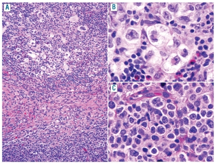
Background: Mediastinal gray zone lymphoma is a newly recognized entity with transitional morphological and immunophenotypic features between the nodular sclerosis subtype of Hodgkin's lymphoma and primary mediastinal large B-cell lymphoma. Diagnostic criteria for mediastinal gray zone lymphoma are still challenging, and the optimal therapy is as yet undetermined. Epigenetic changes have been implicated in the loss of the B-cell program in classical Hodgkin's lymphoma, and might provide a basis for the immunophenotypic alterations seen in mediastinal gray zone lymphoma.

Design and methods: We performed a large-scale DNA methylation analysis of microdissected tumor cells to investigate the biological underpinnings of mediastinal gray zone lymphoma and its association with the related entities classical Hodgkin's lymphoma and primary mediastinal large B-cell lymphoma, making comparisons with the presumptively less related diffuse large B-cell lymphoma.

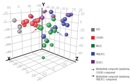
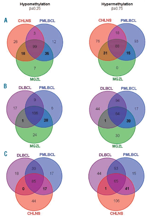
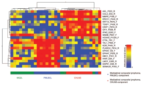
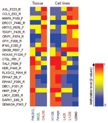
Results: Principal component analysis demonstrated that mediastinal gray zone lymphoma has a distinct epigenetic profile intermediate between classical Hodgkin's lymphoma and primary mediastinal large B-cell lymphoma but remarkably different from that of diffuse large B-cell lymphoma. Analysis of common hypo- and hypermethylated CpG targets in mediastinal gray zone lymphoma, classical Hodgkin's lymphoma, primary mediastinal large B-cell lymphoma and diffuse large B-cell lymphoma was performed and confirmed the findings of the principal component analysis. Based on the epigenetic profiles we were able to establish class prediction models utilizing genes such as HOXA5, MMP9, EPHA7 and DAPK1 which could distinguish between mediastinal gray zone lymphoma, classical Hodgkin's lymphoma and primary mediastinal large B-cell lymphoma with a final combined prediction of 100%.

Conclusions: Our data confirm a close relationship between mediastinal gray zone lymphoma and both classical Hodgkin's lymphoma and primary mediastinal large B-cell lymphoma. However, important differences were observed as well, allowing a clear distinction from both parent entities. Thus, mediastinal gray zone lymphoma cannot be assigned to either classical Hodgkin's lymphoma or primary mediastinal large B-cell lymphoma, validating the decision to create an intermediate category in the World Health Organization classification.


Figures

Figure 1.
Figure 1.
Distinct epigenetic profile of MGZL as assessed by principal component analysis. The methylation data for 1421 CpG targets from all studied tissue samples were subjected to principal component analysis and projected onto the first three principal components. MGZL appears to have a distinct epigenetic profile intermediate between CHLNS and PMLBCL, but clearly different from that of DLBCL. One of the lymphomas studied was a composite lymphoma, comprising two distinct components of CHLNS and PMLBCL in the same biopsy which were microdissected separately. Both elements of the composite lymphoma clustered with cases of MGZL, but the two components also demonstrated a particularly close association with cases of CHLNS or PMLB-CL, respectively.
Figure 2.
Figure 2.
Histological features of mediastinal composite lymphoma showing two different histological components in the same biopsy: one characteristic of CHLNS and another of PMLBCL. (A) CHLNS-component with Hodgkin and Reed-Sternberg cells in an inflammatory background with numerous eosinophils (top) separated by fibrous collagen bands from the PMLBCL-component composed of tumor cells with abundant pale cytoplasm (bottom) (original magnification: x10). (B) High magnification of the CHLNS-component showing characteristic lacunar cells (original magnification: x40). (C) High magnification of the PMLBCL-component showing sheets of relatively monomorphic large cells (original magnification: x40).
Figure 3.
Figure 3.
CpG target hypomethylation and hypermethylation in MGZL, CHLNS, PMLBCL and DLBCL. The Venn diagrams show the numbers of common hypomethylated (β≤0.25) targets (left hand panels) or common hypermethylated (β≥0.75) targets (right hand panels) in the indicated lymphoma entities. Comparison of MGZL with CHLNS and PMLBCL (A) or DLBCL and PMLBCL (B) and comparison of CHLNS with DLBCL and PMLBCL (C). (A) MGZL shares a number of unique common hypo- and hypermethylated targets with CHLNS and PMLBCL (numbers in bold). (B) In contrast, only one hypomethylated and one hypermethylated target are shared between MGZL and DLBCL, exclusive of those shared by PMLBCL. Several hypomethylated and hypermethylated targets are shared in common by MGZL and PMLBCL but not shared by DLBCL, 28 and 39 respectively (numbers in bold). (C) Some overlap of target methylation is present between CHLNS and PMLBCL (17 hypomethylated CpG, 41 hypermethylated CpG) but almost none between CHLNS and DLBCL (numbers in bold).
Figure 4.
Figure 4.
Hierarchical cluster analysis of 22 differentially methylated targets in MGZL, CHLNS and PMLBCL. Columns represent individual cases of microdissected tumor samples, rows represent the indicated CpG sites (selected targets of prediction models; P<0.01, one-way ANOVA; Δβ≥ 0.30 between any two sample conditions; see Design and Methods section for details). The color represents the methylation level (β-values) from 0 (0% methylation, blue) to 1 (100% methylation, red) as given in the vertical color bar. The horizontal color bar indicates the tumor samples. The two components of a single mediastinal composite lymphoma were analyzed separately. Whereas the PMLBCL-component was closest to the profile of MGZL, the CHLNS-component clustered with the cases of CHLNS. PMLB-CL and CHLNS are readily distinguished, while MGZL shows overlap with both groups.
Figure 5.
Figure 5.
Epigenetic characterization of lymphoma cell lines using the identified 22 differentially methylated targets of the prediction models. Columns represent the indicated lymphoma tissue samples or the indicated lymphoma cell lines. Rows represent the 22 selected CpGs based on Figure 4 (selected targets of prediction models; see Design and Methods section for details). The color represents the methylation level (mean of β-values of tissue samples and β-values of individual cell lines) as given in the vertical color bar (0, 0% methylation, blue; 1, 100% methylation, red). The analysis of the 22 selected CpG loci reveals great similarity of the methylation pattern of the PMLBCL-cell lines (blue) and the Hodgkin-cell lines (red) to their tissue counterparts. The pattern of U2940, a cell line derived from a patient with a DLBCL sequential to CHL, was most similar to the tissue methylation profile of CHLNS.

Comment in

  • Mediastinal gray zone lymphoma.
    Quintanilla-Martinez L, Fend F. Quintanilla-Martinez L, et al. Haematologica. 2011 Apr;96(4):496-9. doi: 10.3324/haematol.2011.043026. Haematologica. 2011. PMID: 21454881 Free PMC article. No abstract available.

References

    1. Rudiger T, Jaffe ES, Delsol G, deWolf-Peeters C, Gascoyne RD, Georgii A, et al. Workshop report on Hodgkin’s disease and related diseases (‘grey zone’ lymphoma) Ann Oncol. 1998;9 (Suppl 5):S31–8. - PubMed
    1. Kuppers R. The biology of Hodgkin’s lymphoma. Nature Rev. 2009;9(1):15–27. - PubMed
    1. Jaffe ES, Wilson WH. Gray zone, synchronous, and metachronous lymphomas: diseases at the interface of non-Hodgkin’s lymphomas and Hodgkin’s Lymphoma. In: Mauch PM, Armitage JO, Coiffier B, Dalla-Favera R, Harris NL, editors. Non-Hodgkin’s Lymphoma. Philadelphia, PA: Lippincott, Williams, and Wilkins; 2004. pp. 69–80.
    1. Traverse-Glehen A, Pittaluga S, Gaulard P, Sorbara L, Alonso MA, Raffeld M, et al. Mediastinal gray zone lymphoma: the missing link between classic Hodgkin’s lymphoma and mediastinal large B-cell lymphoma. Am J Surg Pathol. 2005;29(11):1411–21. - PubMed
    1. Eberle FC, Mani H, Jaffe ES. Histopathology of Hodgkin’s lymphoma. Cancer J. 2009;15(2):129–37. - PubMed

Publication types

MeSH terms