Review
. 2011 Apr;10(4):261-75.
doi: 10.1038/nrd3428.

Targeting G-quadruplexes in gene promoters: a novel anticancer strategy?

Affiliations
Review

Targeting G-quadruplexes in gene promoters: a novel anticancer strategy?

Shankar Balasubramanian et al. Nat Rev Drug Discov. 2011 Apr.

Abstract

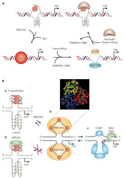
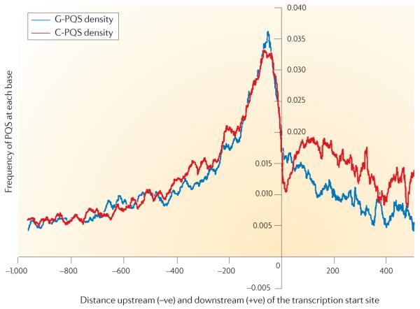
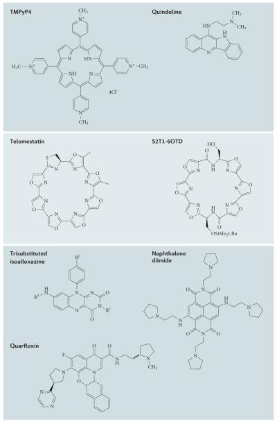
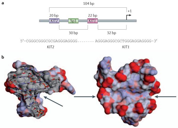
G-quadruplexes are four-stranded DNA structures that are over-represented in gene promoter regions and are viewed as emerging therapeutic targets in oncology, as transcriptional repression of oncogenes through stabilization of these structures could be a novel anticancer strategy. Many gene promoter G-quadruplexes have physicochemical properties and structural characteristics that might make them druggable, and their structural diversity suggests that a high degree of selectivity might be possible. Here, we describe the evidence for G-quadruplexes in gene promoters and discuss their potential as therapeutic targets, as well as progress in the development of strategies to harness this potential through intervention with small-molecule ligands.


Figures

Figure 1
Figure 1. G-quadruplexes in promoter regions: MYC as an example
A | A proposed model of MYC transcriptional regulation that involves the resolution of the G-quadruplex by NM23H2 (from the non-metastasis 23 (NM23) protein family) for duplex DNA formation and subsequent transcriptional activation by transcription factor Sp1. The binding of heterogeneous nuclear ribonucleoprotein K (HNRNPK) and cellular nucleic acid binding protein (CNBP) to single-stranded DNA induced by negative supercoiling also leads to activation of MYC transcription. The stabilization of the G-quadruplex by nucleolin results in negative regulation of MYC transcription. B | This diagram shows the involvement of NM23H2 and a G-quadruplex-interactive compound in modulating the activation and silencing of the nuclease hypersensitive element III1 (NHE III1) in the MYC promoter. Ba shows the G-quadruplex/i-motif form of the NHE III1, which is the silencer element. Bb shows a molecular model of the purine-rich strand unwound from the G-quadruplex form, which is superimposed on an NM23H2 trimer. Ba to Bc via Bb illustrates the remodelling of the G-quadruplex/i-motif complex by NM23H2, in which a stepwise unfolding of the secondary DNA structure is proposed to take place. Part A reproduced, with permission, from REF. © (2010) International Union of Pure and Applied Chemistry. Part B is modified, with permission, from REF. © (2010) Annual Reviews. RNA Pol II, RNA polymerase II.
Figure 2
Figure 2. Identification of putative G-quadruplex-forming regions in gene promoters using computational analysis
The density plot shows the frequency (that is, the probability) of each nucleo tide upstream (−ve) or downstream (+ve) of the transcription start site being part of a putative G-quadruplex-forming sequence (PQS), based on the Quadparser algorithm. The data has been averaged over all human protein coding genes in the genome. The blue and red plots reflect the presence of a G-quadruplex motif (G-PQS) or the C-rich complement of a G-quadruplex motif (C-PQS), respectively. The downstream data are related to the spliced sequence (that is, exons), and there is clearly some evidence of strand bias, in contrast with the symmetry between the blue and red plots in the upstream sequence. Figure is modified, with permission, from REF. © (2008) Oxford University Press.
Figure 3
Figure 3. Structures of selected G-quadruplex ligands
Each of these small molecules has shown a G-quadruplex-associated biological activity in one or more studies.
Figure 4
Figure 4. G-quadruplexes in the promoter of the gene coding for KIT
a | The minimal promoter structure of the KIT gene showing the spatial relationship between G-quadruplexes KIT1 and KIT2 and a transcription factor Sp1 binding site. Shown in the inset are the sequences of KIT1 and KIT2. b | The two orthogonal views of the averaged nuclear magnetic resonance structure of the KIT1 quadruplex are shown, with the solvent-accessible surface coloured by charge. The arrows indicate the large irregular cleft in the structure, which has not been found in any other G-quadruplex structure determined to date. bp, base pair.
Figure 5
Figure 5. Properties of quarfloxin
a | Origin and lead optimization of quarfloxin (CX-3543). The design of the fluoroquinolone QQ58 (REF. 103) was based on the principle that nor-floxacin and A-62176 inhibited gyrase and topoisomerase II by interacting with the enzyme–duplex DNA complex, and this scaffold was amenable to G-quadruplex interaction by extending the tetracyclic nucleus to a larger and approximately planar system that would interact more favourably with the tetrad in the G-quadruplex than with duplex DNA. Cylene Pharmaceuticals then optimized the planar, presumably G-tetrad-interacting moiety and identified groove-binding arms that would both prevent topoisomerase poisoning and increase selectivity between different G-quadruplexes. b | Diagram showing the proposed mechanism of action of quarfloxin specifically involving the MYC G-quadruplex,. The other target proteins of nucleolin are mentioned in the main text. Quarfloxin is preferentially concentrated in the nucleolus. Binding of quarfloxin to the non-template G-quadruplexes in the recombinant DNA displaces nucleolin, which is relocated to the nucleoplasm, where it has been shown to bind to the MYC G-quadruplex to inhibit MYC expression. RNA Pol I, RNA polymerase I.

References

    1. Kohn KW. Beyond DNA cross-linking: history and prospects of DNA-targeted cancer treatment — fifteenth Bruce F. Cain Memorial Award Lecture. Cancer Res. 1996;56:5533–5546. - PubMed
    1. Roche VF. In: Foye’s Principles of Medicinal Chemistry. Lemke TL, Williams DA, Roche VF, Zito SW, editors. Lippincott Williams & Wilkins; Baltimore: 2008. pp. 1147–1192.
    1. Sen D, Gilbert W. Formation of parallel four-stranded complexes by guanine-rich motifs in DNA and its implications for meiosis. Nature. 1988;334:364–366. - PubMed
    1. Sundquist WI, Klug A. Telomeric DNA dimerizes by formation of guanine tetrads between hairpin loops. Nature. 1989;342:825–829. - PubMed
    1. Zahler AM, Williamson JR, Cech TR, Prescott DM. Inhibition of telomerase by G-quartet DNA structures. Nature. 1991;350:718–720. - PubMed

Publication types

Substances